




No CrossRef data available.
Article contents
Biology of Thelazia callipaeda (Spirurida, Thelaziidae) eyeworms in naturally infected definitive hosts
Published online by Cambridge University Press: 05 October 2004
Abstract
Thelazia callipaeda (Spirurida, Thelaziidae) eyeworm causes ocular infection in carnivores and humans in the Far East; this infection has been recently reported also in Europe – northern and southern Italy – in dogs, cats and foxes. The natural vector/s of T. callipaeda is/are unknown and the development of the nematode in its definitive hosts is limited to an experimental trial on dogs. To contribute new insights into the development of T. callipaeda in the definitive host in field conditions, eyeworms were collected from naturally infected dogs from an area with a high prevalence of infection (up to 60·14%) in the Basilicata region of southern Italy, from January 2002 to December 2003. Conjunctival secretions were also collected and examined for the presence of immature stages. The presence of blastomerized eggs throughout the period – except for the months from May to November – indicates a seasonality in the reproductive activity of T. callipaeda, coinciding with the presence/absence of the vector. In fact, 1st-stage larvae were found in the lachrymal secretions of dogs in summer (June–July 2002 and 2003), ready to be ingested by flies feeding about the eyes. The evidence of 4th-stage larvae in March 2002 and April, July and October 2003 may be accounted for by the presence of flies that act as intermediate hosts of T. callipaeda from early spring to early autumn. The presence of immature stages in October indicates an overlapping generation of nematodes and a 2nd cycle of vector infection. This basic knowledge of the development of T. callipaeda will hopefully help future epidemiological studies to identify the intermediate hosts and define the likely risk for vectors in field conditions.
Information
- Type
- Research Article
- Information
- Copyright
- © 2004 Cambridge University Press
INTRODUCTION
Among the numerous species of Thelazia causing ocular infections in animals, Thelazia callipaeda Railliet & Henry 1910 and Thelazia californiensis Price 1930 are parasites found in dogs, cats, foxes, rabbits and humans. The adult stages are found in the eyes of animals, precisely under the lid and nictitating membrane, and in the conjunctival sacs, inducing epiphora, conjunctivitis, ocular discharge and chemosis sometimes resulting in corneal ulcer.
Female worms produce a large number of 1st-stage larvae (L1) that are deposited in the lachrymal secretions of the hosts and ingested by secretophagous dipteran insects. L1 develop in the flies to the 3rd infectious stage larva (L3) and leaves the vector when the latter feeds on the ocular secretions of animals (Skrjabin, Sobolev & Ivashkin, 1967).
The nature of the natural vectors of T. callipaeda remain uncertain although there is some evidence that Musca domestica (Diptera, Muscidae) and, experimentally, Amiota variegata and Amiota okadai (Diptera, Drosophilidae) are involved (Kozlov, 1962; Shi et al. 1988; Wang et al. 2003).
While T. californiensis is mainly confined to the western USA (Skrjabin et al. 1967; Soulsby, 1982), T. callipaeda has been reported in humans and dogs in the Russian Federation and the Far East (Bhaibulaya, Prasertsilpa & Vajrasthira, 1970; Hong, Lee & Kim, 1988; Kosin, Kosman & Depary, 1989; Hong et al. 1995). In particular, in the past 20 years the number of reports of human thelaziosis in the Far East has increased (Kosin et al. 1989).
In Europe, after the first report in dogs from northern Italy (Rossi & Bertaglia, 1989), the presence of T. callipaeda and the prevalence of infection in dogs, cats and foxes have been studied in northern and southern Italy (Otranto et al. 2003a). These findings indicate that the occurrence of canine thelaziosis is not occasional but represents an infection that is spreading throughout Italy.
Apart from the general morphological descriptions of Skrjabin et al. (1967) and Soulsby (1982), morphological findings have been provided by Bhaibulaya et al. (1970), Hong et al. (1988), Rossi & Bertaglia (1989) and Otranto et al. (2003b). As far as the development of T. callipaeda in dogs is concerned, very few data are available and, what data there are, were obtained only under experimental conditions (Kozlov, 1962) (Fig. 1). No studies are available on the seasonal development of different instars of T. callipaeda in the definitive host under natural conditions. Therefore, the aim of this paper is to contribute basic knowledge regarding the development of T. callipaeda in naturally infected dogs throughout 2 infection seasons, in order to provide new insights into the reproductive activity and likely risk for vectors in field conditions.

Fig. 1. Schematic representation of the life-cycle and timing of Thelazia callipaeda in the definitive and intermediate host (by Kozlov, 1962).
MATERIALS AND METHODS
Study area
The survey was carried out from January 2002 to December 2003. The dogs were from a mountainous area comprising twelve municipalities in the Basilicata region of southern Italy (altitude: from 548 m to 1367 m above sea level; latitude 39° and 41° North, longitude 15° and 16° East).
Collection procedures
The dogs (n=31) were restrained with a muzzle (no anaesthetic was used) and the conjunctival secretions were collected monthly by instillation of 15 ml of physiological saline (0·9% NaCl) and transferred to Petri dishes. The nematodes and the collected rinsing solution were labelled and delivered to the Parasitology Laboratory of the School of Veterinary Medicine of the University of Bari.
The eyeworms were fixed in a solution of amman (lactic acid 1[ratio ]5, carbolic acid 1[ratio ]5, glycerine 2[ratio ]5, distilled water 1[ratio ]5) and Methylene blue (1[ratio ]50) and examined under light microscopy (Leica Microsystem DMLS) at different magnifications up to ×1000. The identification process was carried out following the morphological descriptions of Skrjabin et al. (1967) and Bhaibulaya et al. (1970). All the most important morphological characteristics of T. callipaeda were evaluated for adult forms as reported in a previous paper (Otranto et al. 2003b).
The rinsing solutions containing the conjunctival secretions were examined under a stereomicroscope (LEICA MSV) for the presence of immature eyeworms after centrifugation at 1550 g for 30 sec to concentrate the immature stages, when present.
Study of the biology
Several morphological features and the reproductive status were examined to evaluate the developmental stages of the worms collected throughout the observation period (Table 1). In particular, the reproductive status of the females was determined by the presence/absence of eggs, presence/absence of blastomerization, and presence and location of larvae in the uterus and close to the opening of the vulva. The mature stages of the males were identified based on the presence of spicules. Measurements and pictures were taken using a Leica Microsystem DC300 – Software Leica QWIN.
Table 1. Morphological features and measurements used to determine the developmental stage of Thelazia callipaeda according to Kozlov (1962) and Skrjabin et al. (1967) (BC: Buccal Capsule; ND: Not Described.)

Statistical analysis
A comparison was made for parasite sex in each month of collection using the Chi square test. The number of non-blastomerized, blastomerized and larvated eggs calculated on the total number of females examined in the different months throughout all the observation period, were analysed by Chi-square for linear trends in proportions, to highlight significant variations over time (EPI.INFO 6.0). Differences were considered to be significant when P[les ]0·05.
RESULTS
A total of 318 (mean=13·21±10·57, range 9 to 58 nematodes) comprising 219 females, 94 males and 5 immature stages (Table 2) were directly collected with sterile cotton swabs from the eyes of naturally infected dogs (Fig. 2). On the whole the female/male ratio was 2·3[ratio ]1 although no significance was noticed for this value during the different months of the year.

Fig. 2. Eye of a dog with heavy Thelazia callipaeda infection.
Table 2. Number of Thelazia callipaeda specimens collected from the eyes of the dogs from January 2002 to December 2003 broken down according to month of collection and developmental stage (F, female; M, male; L, 4th larval stage. The number of females with non-blastomerized (nBE), blastomerized (BE) and/or larvated eggs (LE) is also reported.)

All the adult nematodes were identified as T. callipaeda by the position of the vulva located anterior to the oesophagus–intestinal junction in the females and the presence of 5 pairs of postcloacal papillae in the males.
In the females, non-larvated or larvated eggs along the uteri were present during the period. Eggs with blastomerization were present during the entire observation period, with the exception of late spring to early autumn (i.e. May–November 2002 and 2003) (Table 2). In Fig. 3 the percentage of non-larvated (i.e. blastomerized or non-blastomerized) and larvated eggs are shown over the time of observation. Non-blastomerized eggs were linearly present over the whole period (χ2 for trend=1·713, P>0·05), whereas blastomerized and larvated eggs showed significant variations over time (χ2 for trend=4·022, P<0·05 and χ2 for trend=10·811, P<0·01, respectively).

Fig. 3. Percentages over the observation period of non-blastomerized (nBE), blastomerized (BE) and larvated (LE) eggs (showed in lines only) over the total number of Thelazia callipaeda females. BE are shown in bars and line whereas BE and LE are shown in lines.
Five immature worms were collected from the infected dogs: 2 in March 2002, 1 in April 2003, 1 in July 2003 and the last in October 2003 (Table 2). The immature stages ranged in length from 3·88 to 7·66 mm and in width from 0·15 to 0·25 mm, and no genitalia were detected. All the immature nematodes were identified as 4th-stage larvae (L4).
First-stage eyeworms (Fig. 4) were present in the lachrymal secretions of 2 dogs (n=23) in June–July 2002 and 2 dogs (n=40) in June–July 2003.

Fig. 4. Light micrograph of Thelazia callipaeda. First-stage larva outside the nematode presenting a shell membrane (SM). Magnification ×200.
DISCUSSION
This paper represents the first biological survey on T. callipaeda in naturally infected dogs over a period of 2 years. The life-cycle of eyeworms in naturally infected dogs is still completely unknown, probably due to the enormous difficulties associated with approaching such a study. In particular, when studying the development of eyeworms in field conditions (mainly linked to the intermediate host), one major difficulty lies in collecting a sufficient number of specimens throughout the observation period. In fact such research requires the minimizing of biases due to the environment, the vectorial infection rate and the parasite population in each dog and, where possible, using animals living in similar habitats. The above constraints are even greater when collecting T. callipaeda eyeworms, since they affect dogs kept as pets. This is not the case for other eyeworms affecting routinely slaughtered livestock (e.g. Thelazia gulosa, Thelazia rhodesi and Thelazia skrjabini in cattle, and Thelazia lacrymalis in equids), from which it is easier to collect nematodes. This study was possible only because it was carried out in an area where T. callipaeda is endemic, with the highest prevalence of infection in dogs (60·14%) ever reported (Otranto et al. 2003a).
Based on published reports concerning the immature stages of T. callipaeda (Kozlov, 1962), several morphological features were considered here as key to investigating the development of this nematode. Sexually mature males and females (with larvated eggs close to the vulvar opening) were detected throughout the observation period. The presence of larvated eggs in spring, summer and early autumn might be explained by the release of larvated eggs into the lachrymal secretions of dogs, possibly coinciding with the vector/s activity. This is supported by the finding during summer of L1 in the animals' lachrymal secretions, which are then ingested by flies feeding about the eyes. These results agree with those of Kozlov (1962), who found sexually mature T. callipaeda in experimentally infected dogs throughout 1 year, and also with data available for T. lacrymalis harbouring larvae in uteri (during 5 years of infection) with no differences between seasons (Lyons et al. 2000). The presence of L1 in uteri in winter might erroneously appear to be irrelevant to the maintenance of eyeworm life-cycles due to the likely absence of the vector/s at low temperatures. Nevertheless, the presence of larvae in uteri during the colder months could be related to the overwintering of the adult female T. callipaeda. Overwintering could be of great importance for the maintenance of T. callipaeda infections in a specific area, with the release of larvae in the following spring and summer seasons to perpetuate the biological cycle as soon as the vector/s appear/s. It has actually been experimentally demonstrated that T. callipaeda survives at least 9 months in definitive hosts with overlapping generations (Kozlov, 1962; Anderson, 2000).
The finding of blastomerized eggs throughout the observation period, with exception of the months from May to November 2002–2003, might be accounted for by a seasonality in the reproductive activity of T. callipaeda coinciding with the presence of the vector. In late autumn to late spring the vector presumably is not present. In that period, the occurrence of blastomerized eggs and larvae close to the vulvar opening indicates the potential of T. callipaeda overwintering females to infect the vector with L1 as soon as the flies appear. The blastomerized eggs very quickly produce other larvated eggs to increase the infection rate of the vectors. The vector should be present from spring to late autumn. No blastomerization was observed in the same period, probably because the mature females are striving to release L1 to maintain the biological cycle. This is confirmed by the fact that in June–July L1 were detected in both uteri and the lachrymal secretions.
The presence of L4 in March 2002 and April, July and October 2003 may be accounted for by the presence of flies acting as intermediate hosts of T. callipaeda from very early spring to early autumn; in fact, the 3rd moult of T. callipaeda from L3 to L4 takes place 7–9 days after the flies transmit L3 to dogs (Kozlov, 1962). This assumption is supported by the activity of some species of flies (e.g. Musca autumnalis and Musca larvipara) which act as intermediate hosts of bovine eyeworms (Otranto et al. 2003c). Musca domestica, which some authors consider to be a vector of T. callipaeda (Shi et al. 1988), is also active in southern Italy from early spring to autumn (Giangaspero, 1997).
Another possible explanation for the presence of immature stages in early spring is that flies infected the previous summer are re-activated in early spring (after hibernation) and transmit T. callipaeda in the L3 stage to dogs. The hibernation of flies infected by bovine Thelazia has already been demonstrated in face flies (Chirico, 1994). The presence of L4 in October might indicate a 2nd peak of fly infection during the summer season.
The intermediate host of canine eyeworms in natural conditions is currently unknown and data on fluctuations in the temporal pattern of parasitization by T. callipaeda in insects are unavailable. Nevertheless, high values of parasitism by T. gulosa have been detected in flies in late spring/early summer and late summer (Geden & Stoffolano, 1981; Otranto et al. 2003c). The data presented herein are consistent with these findings, since T. callipaeda L1 were detected in ocular secretions of infected animals in June–July 2002 and 2003 and, therefore, those are most likely the periods with the highest rate of parasitism in the intermediate hosts. This assumption is supported by findings of high infection rates by T. callipaeda in dogs in October–December in the same area as this study (Otranto et al. 2003a).
These results on the biology of T. callipaeda provide useful information for further epidemiological studies to identify the insects that may act as intermediate hosts and to define the likely risk for vectors in field conditions. The identification of the vectors of canine eyeworms is of paramount importance, not only to enhance insight into the biology and parasitic behaviour of T. callipaeda but also for prophylaxis of the disease. If the vector of T. callipaeda is identified in the field it might be possible to prevent the spread of this disease, not only in dogs but especially in disadvantaged socio-economic settings where humans, often children and the elderly, are infected.
This study was supported by the Foreign Affairs Ministries of Italy and the People's Republic of China (represented by the Universities of Bari and Lanzhou, respectively) for the period 2002–2005. The authors are grateful to Nicola Leone and Michele Garaguso for their assistance in the field and to all the inhabitants of the test sites for kindly permitting the clinical examinations. The authors thank Gioia Capelli (Faculty of Veterinary Medicine, University of Padova, Italy) for her assistance in the statistical work and 2 anonymous reviewers who provided helpful comments on the manuscript.
References
REFERENCES
ANDERSON, R. C. (
2000).
Nematode Parasites of Vertebrates. Their Development and Transmission, pp.
428–430. 2nd Edn.
CABI Publishing,
Guilford, UK.
BHAIBULAYA, M.,
PRASERTSILPA, S. &
VAJRASTHIRA, S. (
1970).
Thelazia callipaeda Railliet and Henry, 1910, in man and dog in Thailand.
American Journal of Tropical Medicine and Hygiene
19,
476–479.CrossRefGoogle Scholar
CHIRICO, J. (
1994).
Prehibernating Musca autumnalis (Diptera: Muscidae) an overwintering host for parasitic nematodes.
Veterinary Parasitology
52,
279–284.CrossRefGoogle Scholar
GEDEN, C. J. &
STOFFOLANO, J. G. (
1981).
Geographic range and temporal patterns of parasitization of Musca autumnalis De Geer by Thelazia spp. in Massachusetts with observation on Musca domestica as an unsuitable host.
Journal of Medical Entomology
18,
449–456.Google Scholar
GIANGASPERO, A. (
1997).
Le mosche di interesse veterinario. I Muscidae. Guida alla conoscenza e al riconoscimento.
Edagricole,
Bologna,
Italy.
HONG, S. T.,
LEE, S. H. &
KIM, S. I. (
1988).
A human case of Thelazia callipaeda infection with references to its internal structures.
Korean Journal of Parasitology
2,
137–139. (In Korean.)CrossRefGoogle Scholar
HONG, S. T.,
PARK, Y. K.,
LEE, S. K.,
YOO, J. H.,
CHUNG, Y. H. &
HONG, S. J. (
1995).
Two human cases of Thelazia callipaeda infection in Korea.
Korean Journal of Parasitology
2,
139–144. (In Korean.)CrossRefGoogle Scholar
KOSIN, E.,
KOSMAN, M. L. &
DEPARY, A. A. (
1989).
First case of human thelaziasis in Indonesia.
Southeast Asiatic Journal of Tropical Medicine and Public Health
20,
233–236.Google Scholar
KOZLOV, D. P. (
1962).
The life cycle of nematode Thelazia callipaeda parasitic in the eye of the man and carnivores.
Doklady Akademy Nauk SSSR
142,
732–733. (In Russian.)Google Scholar
LYONS, E. T.,
SWERCZEK, T. W.,
TOLLIVER, S. C.,
BAIR, H. D.,
DRUDGE, J. H. &
ENNIS, L. E. (
2000).
Prevalence of selected species of internal parasites in equids at necropsy in central Kentucky.
Veterinary Parasitology
92,
51–62.CrossRefGoogle Scholar
OTRANTO, D.,
FERROGLIO, E.,
LIA, R.,
TRAVERSA, D. &
ROSSI, L. (
2003 a).
Current status and epidemiological observations of Thelazia callipaeda (Spirurida, Thelaziidae) in dogs, cats and foxes in Italy: a ‘coincidence’ or a parasitic disease of the Old Continent?
Veterinary Parasitology
116,
315–325.Google Scholar
OTRANTO, D.,
LIA, R. P.,
TRAVERSA, D. &
GIANNETTO, S. (
2003 b).
Thelazia callipaeda (Spirurida, Thelaziidae) of carnivores and humans: morphological study by light and scanning electron microscopy.
Parassitologia
45,
125–133.Google Scholar
OTRANTO, D.,
TARSITANO, E.,
TRAVERSA, D.,
DE LUCA, F. &
GIANGASPERO, A. (
2003 c).
Molecular epidemiological survey on the vectors of Thelazia gulosa, Thelazia rhodesi and Thelazia skrjabini (Spirurida: Thelaziidae).
Parasitology
127,
365–373.Google Scholar
ROSSI, L. &
BERTAGLIA, P. (
1989).
Presence of Thelazia callipaeda Railliet & Henry, 1910, in Piedmont, Italy.
Parassitologia
31,
167–172.Google Scholar
SHI, Y. E.,
HAN, J. J.,
YANG, W. Y. &
WEI, D. X. (
1988).
Thelazia callipaeda (Nematoda: Spirurida): transmission by flies from dogs to children in Hubei, China.
Transactions of the Royal Society of Tropical Medicine and Hygiene
82,
627.CrossRefGoogle Scholar
SKRJABIN, K. I.,
SOBOLEV, A. A. &
IVASHKIN, V. M. (
1967).
Principles of Nematology. Vol. IX. Spirurata of Animals and Man and the Disease Caused by Them. Part. 4: Thelazioidea, pp.
26–36.,
Izdatel'sto Akademii Nauk SSSR,
Moskow. Published by Israel Program for Scientific Translations,
Washington, USA.
SOULSBY, E. J. L. (
1982).
Helminths, Arthropods and Protozoa of Domesticated Animals, 7th Edn. pp.
289–290.
Bailliere Tindall,
Oxford,
UK.
WANG, Z. X.,
HU, Y.,
SHEN, J. L.,
WANG, K. C.,
WANG, H. Y.,
JIANG, B. L.,
ZHAO, P.,
WANG, Z. C.,
DING, W.,
WANG, F. &
XIA, X. F. (
2003).
Longitudinal investigation and experimental studies on thelaziasis and the intermediate host of Thelazia callipaeda in Guanghua county of Hubei province.
Zhonghua Liu Xing Bing Xue Za Zhi
24,
588–590. (In Chinese.)Google Scholar

Fig. 1. Schematic representation of the life-cycle and timing of Thelazia callipaeda in the definitive and intermediate host (by Kozlov, 1962).
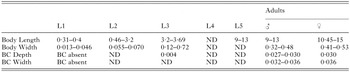
Table 1. Morphological features and measurements used to determine the developmental stage of Thelazia callipaeda according to Kozlov (1962) and Skrjabin et al. (1967)
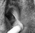
Fig. 2. Eye of a dog with heavy Thelazia callipaeda infection.

Table 2. Number of Thelazia callipaeda specimens collected from the eyes of the dogs from January 2002 to December 2003 broken down according to month of collection and developmental stage
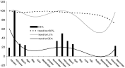
Fig. 3. Percentages over the observation period of non-blastomerized (nBE), blastomerized (BE) and larvated (LE) eggs (showed in lines only) over the total number of Thelazia callipaeda females. BE are shown in bars and line whereas BE and LE are shown in lines.
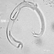
Fig. 4. Light micrograph of Thelazia callipaeda. First-stage larva outside the nematode presenting a shell membrane (SM). Magnification ×200.
 You have
Access
- 101
- Cited by