The concept of developmental programming
The developmental origins of adult disease hypothesis had its origin in epidemiological data collated by Professor David Barker and Clive Osmond at the MRC Environmental Epidemiological Unit, Southampton University. They identified different geographical areas with differing rates of neonatal, post-neonatal and infant mortality, and correlated these with differing rates of mortality from cardiovascular disease in the same regions some 50 to 60 years later, i.e. when contemporaries of those who died as infants were well into middle age (Barker and Osmond, Reference Barker and Osmond1986). This simple, perhaps serendipitous, but astute observation sparked the ‘foetal/developmental origins of adult disease’ paradigm that has become applied to many different areas of science (Langley-Evans, Reference Langley-Evans2004; Rich-Edwards, Reference Rich-Edwards2004). Since low birth weight was known to be a major contributor to neonatal mortality, this later became the focus for many subsequent studies and, for historical records, was the best proxy of foetal growth. However, growth is a continuum from conception to death with birth being one (major) step in that pathway and many recent human and animal studies have indicated that ‘developmental programming’, i.e. an increased tendency towards a less-than-optimal adult phenotype, may operate despite no obvious change in birth weight. To illustrate the difficulty, consider the low birth weight infant that more than likely will demonstrate a degree of early growth acceleration (Cameron et al., Reference Cameron, Preece and Cole2005) – is low weight per se the inducing factor or is early growth acceleration more important? Early statistical models tended to adjust for current weight (at a fixed time point reflecting the rate of growth to that point in time) but this was criticised (Lucas et al., Reference Lucas, Fewtrell and Cole1999; Huxley et al., Reference Huxley, Neil and Collins2002). Indeed, early growth rate per se or ‘centile crossing’ is now thought to be more important for inducing later disease than simply being born small at term, although clearly the two interact (Eriksson et al., Reference Eriksson, Forsen, Tuomilehto, Osmond and Barker2000; Barker et al., Reference Barker, Osmond, Forsen, Kajantie and Eriksson2005). For example, risk of hypertension is potentiated in individuals born relatively small but who subsequently put on excess weight-for-height. The economic consequences of cardiovascular disease in the UK are vast, estimated at £26 billion per year or £434 per capita (http://www.heartstats.org/uploads/documents%5C2007.chapter13.pdf) and thus any potential early influence that may increase the proportion of individuals with cardiovascular morbidity will have a significant economic impact on the UK’s health budget.
Conceptual framework of developmental programming
For addressing potential mechanistic pathways that may explain a proportion of the variation in adult disease phenotype, one has to turn to animal models, being largely free from methodological bias and socio-economic confounding. A number of topical reviews have been published recently that adequately summarise the current state of animal research with regard to the developmental origins hypothesis (Armitage et al., Reference Armitage, Khan, Taylor, Nathanielsz and Poston2004; Langley-Evans, Reference Langley-Evans2004; McMillen and Robinson, Reference McMillen and Robinson2005; Sinclair et al., Reference Sinclair, Lea, Rees and Young2007) and this will not be covered in detail here. Briefly, the evidence from both small and large animal models suggests that there are two, not independent, pathways towards an increased susceptibility for non-communicable disease: low birth weight per se (Barker and Osmond, Reference Barker and Osmond1988) and relative postnatal (neonatal–adolescent) growth acceleration (Barker et al., Reference Barker, Osmond, Forsen, Kajantie and Eriksson2005).
Given the plethora of varied animal studies in multiple species around the world that have now lent biological support to the concept of developmental programming, it is important to have a working conceptual framework that can, at the very least, integrate the basic scientific data. The original hypothesis, postulated ~5 years after the first epidemiological data were published, was ‘The Thrifty Phenotype’ hypothesis (Hales and Barker, Reference Hales and Barker2001). Here the concept is that the developing foetus responds to a ‘poor’, e.g. nutritionally poor, prenatal environment by engaging mechanisms of developmental plasticity to ensure immediate survival to term. The gene/proteome responds to environmental cues, for example, variation in nutrient availability, in different ways, giving rise to variation in postnatal phenotype. The trade-off is that whilst the resulting offspring may be better adapted to a nutritionally poor postnatal environment, it will theoretically be less well adapted to a nutritionally abundant postnatal environment. Many studies support this hypothesis (Gardner et al., Reference Gardner, Bell and Symonds2007) and to a certain extent the hypothesis is akin to Popkins ‘nutrition transition’, i.e. a global change in nutrient intake from low- to high-energy foodstuffs, but at the level of tissues and cells rather than whole populations (Popkin, Reference Popkin2006). A case-in-point may currently be happening in the Indian sub-continent in which the incidence of coronary heart disease and Type 2 diabetes is increasing exponentially, together with their gross domestic product (Yajnik, Reference Yajnik2004). However, the hypothesis fails to account for the increased mortality in the many already industrialised, Westernised cultures. As such, the recent ‘predictive adaptive response’ theory arose (Gluckman and Hanson, Reference Gluckman and Hanson2004). Here, developmental plasticity ‘conditions’ a growing foetus in expectation of its future environment; i.e. to develop under conditions of nutritional excess and live as an adult in a nutritionally abundant environment would confer no greater risk on adult health than the population average. Whilst this hypothesis has successfully stimulated research that incorporates an evolutionary perspective into the developmental programming arena (Kuzawa, Reference Kuzawa2005), unfortunately, it is difficult to reconcile with existing data in human and animal models (Rodrigues et al., Reference Rodrigues, Robinson and Gray-Donald1999; Catalano et al., Reference Catalano, Kirwan, Haugel-de Mouzon and King2003).
One alternative and attractive model was proposed by Jackson (Reference Jackson1996). Here an individual’s metabolic competence, i.e. ability to withstand infection, reproduce and engage in physically active behaviours, was considered to be ‘optimal’ or 100% around 20 to 21 years of age (in humans at least) and then to decline gradually into senescence, at which ill-health becomes more likely (Figure 1). Transposed into the developmental programming theatre there are two elements to consider: (1) does a poor prenatal diet reduce an individual’s maximal functional capacity or ‘reserve functional capacity at a given age? and (2) is the rate of decline in metabolic performance increased? Thus individuals born small or showing early growth acceleration may not fully realise their genetic/metabolic potential when in the prime of their lives (hypothesis 1) and/or may exhibit an increased age-related decline in performance (hypothesis 2). Effectively, the relative risk of succumbing to pathophysiological processes is increased after suffering a poor developmental environment. There are much data in the literature to support such a hypothesis (Ozanne and Hales, Reference Ozanne and Hales2004). To what extent can such a conceptual framework be practically applied to developmental programming of the reproductive axis? There are two questions to ask: does a poor developmental environment
Development of functional capacity with age. An individual’s phenotypic competence, i.e. ability to withstand infection, reproduce and engage in physically active behaviours is considered to be ‘optimal’ or 100% around 20 to 21 years of age (in the human at least) and then to decline gradually into senescence. At this time a threshold is reached where their metabolic reserve is reduced and ill-health is more likely to supervene. Developmental programming may reduce the maximal functional capacity achieved, thereby also reducing the ‘metabolic reserve’ and/or increase the rate of decline of functional capacity with age. Redrawn with permission from Jackson (Reference Jackson1996).

1. reduce an individual’s capacity to reproduce and bear forth offspring? and/or
2. advance reproductive senescence?
Cumulatively, these may be considered as an early life influence on fertility.
Effects on fertility in humans
Sinclair et al. (Reference Sinclair, Lea, Rees and Young2007) considered the effects of malnutrition during pregnancy on offspring fertility in an evolutionary context and concluded that whilst teleologically it may make sense for a poly-ovular species faced with a long-term decline in nutrient provision to reduce their ovulation rate in order to moderate litter size, failure to ovulate would be unsustainable in the longer term. Mechanisms underlying this process may, therefore, be ‘protected’ from nutritional programming. Consequently, the effects of maternal undernutrition during pregnancy may be more apparent in poly-ovular than mono-ovular species, and more so in altricial mammals whose investment in reproductive effort (i.e. annual fecundity rate) is estimated to be 10 times that of the human (Phelan and Rose, Reference Phelan and Rose2005).
There is clearly a lack of convincing evidence from human epidemiological studies to indicate that offspring fertility is adversely affected by maternal undernutrition during pregnancy. For example, investigations into the long-term consequences of the war-time famine in the Netherlands revealed that undernutrition during gestation can lead to a number of disease states in later life, the nature and severity of which are dependent on the stage of gestation at the time of insult (Painter et al., Reference Painter, Roseboom and Bleker2005). However, the subsequent fertility of offspring (including age at first pregnancy, completed family size or inter-pregnancy interval) from this cohort was unaffected and was not associated with birth weight (Lumey, Reference Lumey1998). The authors did note, however, that there was an increase in the risk of still-birth and perinatal death among the offspring of women prenatally exposed to the famine in the third trimester of pregnancy (Lumey and Stein, Reference Lumey and Stein1997). Similarly, there is only scant evidence at present to support the notion that foetal growth restriction can advance the onset of reproductive senescence or menopause in women (Cresswell et al., Reference Cresswell, Egger, Fall, Osmond, Fraser and Barker1997). A separate study with 323 Australian female twin pairs failed to establish a relationship between birth weight and age at menopause (Treloar et al., Reference Treloar, Sadrzadeh, Do, Martin and Lambalk2000). Early postnatal influences, however, may have some bearing on fertility, ovarian ageing and reproductive senescence. Hardy and Kuh (Reference Hardy and Kuh2002) found no relationship between birth weight and onset of menopause, but women who were of relatively low weight at 2 years and/or who had received formula milk rather than breast milk had an earlier menopause. Childhood exposure to the 1944 to 1945 Dutch famine, between the ages of 3 and 12, led to only modest effects on subsequent reproductive function in women (Elias et al., Reference Elias, van Noord, Peeters, den Tonkelaar and Grobbee2005). For example, mean age at menarche and proportion childless were not affected by childhood famine, but the likelihood of giving birth to a first or second child was reduced, and the incidence of surgical menopause increased.
As outlined earlier, a common feature of in utero growth restriction is the association with a period of accelerated or compensatory growth that normally follows during early childhood. This phenomenon has been proposed to underlie many of the adverse effects on cardiovascular function and metabolism observed during adulthood (Singhal and Lucas, Reference Singhal and Lucas2004). It certainly complicates interpretation of the effects of nutrient restriction during pregnancy per se on physiological function in offspring. Moreover, the impact of catch-up growth on fertility in humans is largely unknown, but a key feature of ‘catch-up growth’ is that the rate of weight gain during childhood appears to be particularly important in the aetiology of central adiposity (Wells, Reference Wells2007), which, in turn, is thought to be causally linked to metabolic syndrome (Despres and Lemieux, Reference Despres and Lemieux2006). This is significant because it is quite probable that the increased prevalence of anovulatory adolescent girls (40% v. 4%), and the reduced ovulation rate in ovulatory girls (1.4 v. 1.9), all of whom were born small for gestational age (SGA) (Ibanez et al., Reference Ibanez, Potau, Ferrer, Rodriguez-Hierro, Marcos and de Zegher2002b), may be a consequence of deranged metabolism associated with central adiposity (Ibanez et al., Reference Ibanez, Potau, Ferrer, Rodriguez-Hierro, Marcos and de Zegher2002a) and not as a direct consequence of in utero nutrient restriction. Characteristic features of the SGA-related anovulation reported by Ibáňez and colleagues, in addition to abdominal obesity, were hyperinsulinaemia, sub-clinical hyperandrogenism and dyslipidaemia, all of which were corrected following 3 months of metformin (an insulin-sensitising drug) therapy. Importantly, metformin treatment restored ovulatory activity in 70% of subjects within 11 weeks of treatment, indicating that the reproductive pathology was linked to metabolism and was reversible.
Effects on fertility in farm animals
Sheep
Early studies in sheep indicated that plane of nutrition during the rearing period (0 to 12 months) has long-term effects on ovulation rate and, ultimately, litter size in adult ewes (Gunn, Reference Gunn1983). The permanent reduction in adult ovulation rate was also accompanied by a reduction in adult body weight. Although reproductive data in that study were adjusted for body condition score at mating, there was no information on body fat distribution in these animals, and it is not certain if this may have been altered along with body mass. The study could also provide no insight, within the 12-month rearing period, regarding the developmental window most sensitive to nutritional programming, although a re-analysis of the data of Allden (Reference Allden1979) indicated that the first 2 months of life may be most critical in determining adult ovulation rate and litter size (Gunn, Reference Gunn1983). A subsequent study by Rhind et al. (Reference Rhind, Elston, Jones, Rees, McMillen and Gunn1998), working with 499 Brecon Cheviot sheep, confirmed the importance of female offspring nutritional status during lactation on their fertility (e.g. incidence of multiple births); indeed, undernutrition of the female lambs during the first months of life resulted in a reduction in their lifetime reproductive performance, irrespective of nutrition during adult life. Whether such effects may be related to the concomitant effects on postnatal growth rate are unknown.
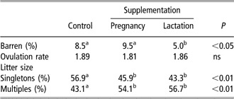
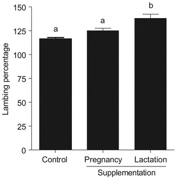
Attention in sheep has since focused on the effects of nutritional programming during foetal development. A comparatively recent study by Rae et al. (Reference Rae, Kyle, Miller, Hammond, Brooks and Rhind2002) reported a significant effect of global nutrient restriction (by 50%) during the first 95 days of gestation on ovulation rate (determined by laparoscopic examination) at 20 months of age in female offspring. No effects on conception rates or litter size were reported. However, working with a flock of 450 Scottish Blackface ewes, managed in a two-pasture hill system, Gunn et al. (Reference Gunn, Sim and Hunter1995) assessed the effects of supplementing ewes during either the final 100 days of pregnancy or the first 100 days of lactation on offspring fecundity over three successive parities. The effects of supplementation were compared to naturally grazed controls (Table 1). Although ovulation rate was not affected, the proportion of multiples was greater in offspring from ewes supplemented during either pregnancy or lactation. Supplementation during lactation had the additional benefit of reducing the proportion of barren offspring. These data, however, do not accurately represent the complete picture. Supplementation also affected mortality in offspring (although not significantly so), so that the proportion of female offspring that died during the experimental period was 17.8%, 17.4% and 13.4%. A simple re-analysis of the data over the three parities, looking at the effects of supplementation on mean lambing percentage, and taking into consideration female offspring mortality, barrenness and litter size, is presented in Figure 2. This analysis confirms that the greatest effect of supplementation, on mean lambing percentage, was during lactation, and so confirms the earlier observations made by this group (Gunn, Reference Gunn1983) and others (Rhind et al., Reference Rhind, Elston, Jones, Rees, McMillen and Gunn1998). Whilst this analysis provides no mechanistic insight into this phenomenon, it does set in a farming-system context the practical significance of these findings.
The effect of supplementary feeding during pregnancy or lactation on offspring fecundity over three successive parities (adapted from Gunn et al., Reference Gunn, Sim and Hunter1995)

Within a row, values with different superscript letters are significantly different.
Re-analysis of the data of Gunn et al. (Reference Gunn, Sim and Hunter1995) who assessed the effects of supplementary feeding during pregnancy or lactation on offspring fecundity over three successive parities. The data are lambing percentages averaged over the three parities that account for offspring survival, barrenness and litter size.

Cattle
In a study to assess the effects of dam nutrition on the reproductive performance of crossbred beef-heifer calves in Nebraska, Martin et al. (Reference Martin, Vonnahme, Adams, Lardy and Funston2007) assessed the effects of late gestation-protein supplementation, and early lactation hay-feeding v. lush pasture grazing in a 2 × 2 factorial design. Protein supplementation during late gestation led to higher pregnancy rates in heifer offspring with a greater proportion of these animals calving in the first 3 weeks of the first calving season. This improvement in fertility was associated with increased heifer weight gains up to weaning and increased weight at the beginning of the breeding season. As with Gunn et al. (Reference Gunn, Sim and Hunter1995), this study in pasture-fed beef cattle sheds no light on the underlying mechanisms, but does provide some insight into the practical significance of the early nutritional environment on offspring fecundity. Once again, however, the improvement in fertility of female offspring from protein-supplemented dams could be a secondary effect of enhanced early growth.
In multiparous dairy cows, late embryo and early foetal development coincides with the significant competitive demand of a pronounced lactation. This has led some investigators to assess the effects of the early nutritional environment on subsequent reproductive success in dairy heifers. Working with data from two genetic lines of dairy cow, Pryce et al. (Reference Pryce, Simm and Robinson2002) analysed the effects of maternal nutritional status on the reproductive performance of 988 daughters as maiden heifers. They found no difference in the reproductive performance of maiden heifers whether their dam was a primiparous heifer or a multiparous cow. Similarly, there was no relationship between the authors’ relatively crude estimates of maternal nutritional status (i.e. dam body condition score, dry matter intake and yields of milk fat and protein) and daughter reproductive performance. More recently, Swali and Wathes (Reference Swali and Wathes2006) assessed the relationship between daughter birth weight (low, average and high) and subsequent reproductive success as nulliparous heifers and primiparous cows. There was no evidence of catch-up growth in the low birth-weight calves whose subsequent reproductive performance was not adversely affected. On the contrary, although not statistically significant, there was some indication that cumulative conception rates were greater for the low birth weight group compared to the other two birth-weight categories. Furthermore, a significantly higher proportion (0.88 v. 0.50) of low rather than high birth weight offspring displayed a normal pattern of cyclical activity. Collectively, these observations suggest that, at present, there is little evidence to indicate that cow reproductive performance is adversely affected to any significant degree by maternal nutritional status during pregnancy.
Effects on foetal hypothalamic–pituitary–gonadal axis development and function in farm animals
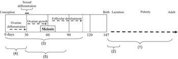
Whilst the picture appears uncertain with regard to a specific deleterious effect of foetal undernutrition on adult fertility in the human and in farm animals, there is some evidence to suggest that foetal undernutrition may indeed have an effect on the development of the female reproductive axis (summarised in Figure 3). A number of studies have shown that the timing of the period of undernutrition during gestation appears important with respect to ‘programming’ of the reproductive axis (Gunn et al., Reference Gunn, Sim and Hunter1995; Rae et al., Reference Rae, Kyle, Miller, Hammond, Brooks and Rhind2002). For example, as previously stated, ovulation rate is reduced in adult female sheep that have suffered foetal undernutrition for the first 110 days of pregnancy, most likely through a direct effect on folliculogenesis during intrauterine development (Rae et al., Reference Rae, Palassio, Kyle, Brooks, Lea, Miller and Rhind2001). Interestingly, limiting the period of undernutrition to an early, middle or late window (each 1 to 2 months duration) during this first 110 days also reduced the number of follicles developing beyond the primordial stage regardless of when the nutritional restriction was applied (Rae et al., Reference Rae, Palassio, Kyle, Brooks, Lea, Miller and Rhind2001). Since these periods of nutrient restriction were calculated to correspond with foetal ovarian differentiation, meiosis I and folliculogenesis, respectively, it follows that different developmental mechanisms are sensitive to maternal undernutrition. In support, further studies following the same nutritional regimen as Rae et al. (Reference Rae, Palassio, Kyle, Brooks, Lea, Miller and Rhind2001 and Reference Rae, Kyle, Miller, Hammond, Brooks and Rhind2002) have shown that underfeeding from 0 to 110 days or from 65 to 110 days alters the expression of ovarian genes that regulate apoptosis, whereas undernutrition during the first 30 days of gestation reduces germ cell proliferation at day 65 but increased granulosa cell proliferation at day 110 (Lea et al., Reference Lea, Andrade, Rae, Hannah, Kyle, Murray, Rhind and Miller2006). Thus it is likely that both of these mechanisms contribute to the impact of maternal undernutrition on folliculogenesis in the female foetal ovary. Interestingly, in a separate study, Murdoch and colleagues reported that underfeeding pregnant sheep from day 28 to day 78 of pregnancy increased the incidence of DNA damage in foetal ovaries and that this was associated with altered ovarian developmental gene expression (Murdoch et al., Reference Murdoch, Van Kirk, Vonnahme and Ford2003).
A reduced plane of nutrition from 0 to 12 months reduces ovulation rate and lifter size (1; Gunn, Reference Gunn1983; Gunn et al., 1995), but the first 2 months of life are most critical for this effect (2; aix’ see Figure 2). Foetal undernutrition during the first 95 days may also reduce the subsequent adults’ ovulatory function (3; Rae et al., Reference Rae, Palassio, Kyle, Brooks, Lea, Miller and Rhind2001 and Reference Rae, Kyle, Miller, Hammond, Brooks and Rhind2002), most likely through direct effects on folliculogenesis (3; Rae et al., Reference Rae, Palassio, Kyle, Brooks, Lea, Miller and Rhind2001). Undernutrition restricted to only the first 30 days of gestation in sheep may have a similar effect when examined at 65 or 110 days in the sheep (4; Lea et al., Reference Lea, Andrade, Rae, Hannah, Kyle, Murray, Rhind and Miller2006). Increased ovarian DNA damage has also been observed following early (d28) to mid (d80) undernutrition in sheep (5; Murdoch et al., Reference Murdoch, Van Kirk, Vonnahme and Ford2003).

To date, there has been little evidence to show an effect of prenatal undernutrition on the endocrine regulation of the hypothalamic–pituitary–gonadal (HPG) axis. For example, given the anatomical effects described above on folliculogenesis, no corresponding functional effects on FSH and LH profiles or response to exogenous GnRH in the adult female offspring have been described. In contrast, however, employing a very different nutritional paradigm, that of the overnourished, adolescent sheep (Wallace et al., Reference Wallace, Aitken and Cheyne1996), alterations in the HPG axis have been reported (Da Silva et al., Reference Da Silva, Aitken, Rhind, Racey and Wallace2001, Reference Da Silva, Aitken, Rhind, Racey and Wallace2002 and Reference Da Silva, Aitken, Rhind, Racey and Wallace2003). This model is characterised by reduced placental size, resulting in low birth weight lambs that were effectively undernourished during gestation due to altered nutrient partitioning. Growth-restricted female foetuses from this model exhibited higher pituitary luteinising hormone-β mRNA expression and fewer ovarian follicles than controls (Da Silva et al., Reference Da Silva, Aitken, Rhind, Racey and Wallace2002). In terms of an impact on the offspring, however, there were no reported effects on the females but male lambs exhibited reduced testosterone and a delay in puberty.
In conclusion, taken together, there is little strong evidence to indicate that offspring fertility in the human and in farm animal species has been overtly affected by their prenatal nutritional exposure. Thus in response to the first question posed earlier, the answer would seem to be no; an individual’s capacity to reproduce and bear forth offspring is largely unaffected by variations in nutritional exposure within the ‘normal physiological range’. To date, it would also appear premature to suggest that reproductive lifespan is affected, i.e. reproductive senescence is also resistant to early environmental perturbation. Nevertheless, mechanistic studies have illustrated the exquisite sensitivity of the developing female gonad to intrauterine nutritional perturbation and, clearly, further work is required to determine more precisely the long-term consequences of nutritional perturbations during pregnancy on adult reproductive well-being. In particular, the potential for early postnatal growth acceleration to confound the effects of low birth weight per se with regard to effects on fertility has yet to be fully tested in many of the large animal models and human studies outlined in this review. In terms of programming of reproductive dysfunction in the human, there is undoubtedly a paucity of supporting data; however, ‘absence of evidence’ is not ‘evidence of absence’.