First described 4 decades ago, the so-called “one-and-a-half” ventricular repair caters for the situation in which one of the ventricles retains its capacity to support the systemic circulation, despite the heart being unable to function in biventricular fashion. In the surgical repair, the other ventricle, which struggles to support the pulmonary circulation, is off-loaded by surgical creation of a shunt. The off-loaded ventricle then continues to pump the pulmonary circulation, with the procedure avoiding the well-known negative shortcomings of the Fontan procedure. Reference Billingsley, Laks and Boyce1–Reference Chowdhury, Airan and Talwar20 The off-loaded ventricle, almost always morphologically right, is usually either hypoplastic or dysfunctional. Reference Billingsley, Laks and Boyce1–Reference Van Arsdell18 The concept of continuing to provide some degree of kinetic energy to the pulmonary circulation is enticing. Reference Billingsley, Laks and Boyce1–Reference Wright, Knight and Thomas14 The size of the off-loaded ventricle permitting such an approach is a continuum, ranging from those that are clearly unsuitable for normal use, to those that are nearly normal. The anatomic settings, furthermore, are manifold. In reality, however, although described as the “one-and-a-half ventricle repair”, the procedure has little to do with half of a given ventricle. It is better described as providing the means of supporting one and a half circulations.
As yet, we have been unable to find specific criterions to determine when the procedure should be adopted in preference to biventricular or functionally univentricular repairs, nor whether the morphologic and physiologic characteristics are more favourable when compared to the alternative procedures. Reference Hanley, Sade and Blackstone6,Reference Hanley9 As far as we can establish, the issue of qualitative assessment of the underperforming side of the heart has still to be addressed. Little attention has been directed, furthermore, to the difficulties that might be encountered during any additionally required intracardiac repairs. The safe upper limits of pulmonary arterial pressure and pulmonary vascular resistance have also yet to be established. At present, all too frequently, the surgeon is left to make subjective decisions. Reference Hanley, Sade and Blackstone6,Reference Hanley9 At All India Institute of Medical Sciences, New Delhi, we have long shown our interest in supporting one and half circulations. In 2001, we did attempt to establish morphological and functional criterions for selection of candidates. Reference Chowdhury, Airan and Sharma19 Then, in 2005, we reported our long-term results and concerns. Reference Chowdhury, Airan and Talwar20 Our ongoing experience shows that problems continue to accrue. In this review, therefore, based on all these uncertainties, we present our ongoing appreciation of the surgical value of the pulsatile superior cavopulmonary connection as a definitive palliative operation, this being the procedure that provides the means for one of the ventricles to support half of the pulmonary circulation.
Methods
We performed a narrative review of the extant literature for described instances of construction of a pulsatile cavopulmonary shunt so as to produce one and a half circulations. We then evaluated all clinical studies describing its principles, indications, techniques, and outcomes. We collated and synthesised, in particular, the role of atrial septal fenestration, the issues relating to the need for ligation of the azygos vein, problems relating to free pulmonary regurgitation and pulsatile superior caval venous flow, the potential for growth of the unloaded ventricle, and long-term functional results. The search engines employed were PubMed, Google Scholar, Cochrane Database for Systematic Reviews, Cochrane Central Register of Controlled Trials, Ovid Medline, ACP Journal Club, Ovid EMBASE, and Database of Abstracts of Review of Effectiveness in all languages.
Methodological index for studies scale was used for critical appraisal of study quality using PRISMA 2020 guidelines. Reference Page, McKenzie and Bossuyt21 Three authors independently graded the quality of the included studies using the methods established by Sundemo and colleagues. Reference Sundemo, Hamrin Senorski and Karlsson22 Cconsensus discussions were carried out to resolve any disagreements.
Our approach has permitted us to make an unbiased individualised review of 201 investigations of potential interest. Reference Billingsley, Laks and Boyce1–Reference Konstantinov and Alexi-Meskishvili28,E1–E173 Based on their contained evidences (Tables S1-S3), we have attempted to establish criterions for selection of candidates when compared to either biventricular or functionally univentricular repairs.
Historical background
In medicine, an operation or syndrome is often not named after those who first described it. This is certainly the case for the superior cavopulmonary connection. The concept of unloading the venous return from the upper body, leaving the right ventricle supporting half the pulmonary circulation, was developed independently by many surgeons. Reference Carlon, Mondini and de Marchi23–Reference Konstantinov and Alexi-Meskishvili28,E145–E164 Carlon, for example, had advocated the concept in Padova by 1950, Reference Page, McKenzie and Bossuyt21 predating by some years the accounts provided by Glenn and his associates. Reference Glenn and Patino24–Reference Glenn26 The first successful bidirectional cavopulmonary shunt was reported in 1961, again in Italy. Reference Dogliotti, Actis-Dato, Venere and Tarquini27 The excellent historical account provided by Konstantinov and Alekshi-Meskishvili Reference Konstantinov and Alexi-Meskishvili28 has listed the others who have contributed to its subsequent evolution. Reference Konstantinov and Alexi-Meskishvili28
Concerns for performing the superior cavopulmonary connection
Despite the successful application of unloading by means of the superior cavopulmonary connection in many different settings, several anatomical and pathophysiological issues remain either poorly appreciated or ignored. Reference Cabrelle, Castaldi and Vedovelli5,Reference Hanley, Sade and Blackstone6,Reference Hanley9,Reference Van Arsdell18–Reference Chowdhury, Airan and Talwar20,E1–E14 One such concern is its safety in the setting of elevated pulmonary vascular resistance. Reference Chowdhury, Airan and Talwar20,E15-E18 A second concern is found in patients with the tendency of developing heart block, such as those with congenitally corrected transposition. Reference Barron7,E19 A third concern is the creation of free pulmonary regurgitation. This may increase right ventricular diastolic dimensions, but may compromise the functional efficiency of the subpulmonary ventricle due to retrograde flow from the superior caval vein. Reference Hoashi, Kitano and Kagisaki8,Reference Stellin, Vida and Milanesi11,Reference Van Arsdell18,E9–E27 A fourth identified concern is the advisability of creating an atrial septal fenestration when the tricuspid valve is between one-third and one-half of normal in the setting of postoperative right ventricular dysfunction. Reference Hoashi, Kitano and Kagisaki8,Reference Stellin, Vida and Milanesi11,Reference Van Arsdell18,E9–E27 A fifth concern is the consequences of leaving open the azygos vein. Reference Chowdhury, Airan and Sharma19,Reference Chowdhury, Airan and Talwar20,E7,E8,E20–E31 A sixth concern is the issue of whether concomitantly to band the right pulmonary artery, thus preventing the superior caval venous syndrome. Reference Chowdhury, Airan and Sharma19,Reference Chowdhury, Airan and Talwar20,E7, E17,E30 A seventh concern is found in those requiring intricate intracardiac repair with prolonged cardiopulmonary bypass time so as to commit only the inferior caval venous blood to the right ventricle. The additional procedures currently have unacceptably high perioperative complications and mortality. Reference Chowdhury, Airan and Sharma19,Reference Chowdhury, Airan and Talwar20,E8 Perhaps the greatest concern, nonetheless, is that unloading of the venous return from upper body may not provide the same functional capacity as a true biventricular repair. Reference Chowdhury, Airan and Talwar20,E1-E20,E28
Criterions for selection of patients
Based on our extensive review, we identified at least 8 settings in which the pulsatile superior cavopulmonary connection has been used extensively:
-
To simplify complex surgical procedures. Reference Cabrelle, Castaldi and Vedovelli5,Reference Patino, Glenn and Guilfoil15,E7,E8,E10,E26,E28,E29,E31–E34
-
To limit the risk from repair of a straddling right atrioventricular valve when attempting ventricular septation.E7,E10,E33–E36
-
To prolong conduit survival by reducing venous ventricular preload in patients undergoing Rastelli-type repairs.E6–E8
-
To augment the flow of blood to the lungs prior to definitive repair of congenitally corrected transposition. Reference Barron7,Reference Van Arsdell18,E2,E11,E19,E25,E37–E39
-
To simplify full atrial redirection in cases of an abnormally located ventricular mass. Reference Barron7,E11
-
To decrease right ventricular preload in the setting of Ebstein’s malformation.E7,E10,E19,E23,E24,E40–E43
-
As an interstage procedure prior to attempting biventricular repair.E44–E47
-
During conversion from the Fontan circulation in individuals with pulmonary arteriovenous malformations, recurrent intractable arrhythmias, and protein-losing enteropathy.E33-35,E48–E53
In addition to the settings listed above, there are, of course, multiple additional circumstances in which the surgeon may opt to provide one and half circulations. The circumstances are to be found cited in our electronic references.E1–E172
Examples of the use of the superior cavopulmonary connections
In individuals with pulmonary atresia with intact ventricular septum
Due to anatomic heterogeneity, patients with pulmonary atresia and intact ventricular septum undergo a variety of individualised approaches. The best patients are candidates for either biventricular surgical repair or repair by means of laser perforation of the imperforate valve and interventional balloon dilation. Reference Anderson, Anderson and Zuberbuchler2–Reference Anderson and Ho4,Reference Daubeney, Delany and Anderson15,E54–E57 At the other end of the spectrum, patients are found with muscular overgrowth of the apical ventricular component and a hypoplastic ventricular inlet. In this category, most are candidates only for conversion to the Fontan circulation.E112
Following preliminary systemic-to-pulmonary arterial shunting and pulmonary valvotomy, multi-institutional studies have demonstrated that approximately one-tenth of these patients remain in a “grey area”. Because it is well accepted that the right ventricle in this malformation is intrinsically tripartite, and only become bipartite or unipartite from muscular overgrowth and cavitary obliteration, investigators have attempted right ventricular overhaul in selected cases as an intermediate step towards either the biventricular or a one-and-a-half ventricular repair. Data provided by the Congenital Heart Surgeons Society, and the group working in Melbourne, indicate that creation of the superior cavopulmonary connection is an effective alternative to attempting “right ventricular overhaul” as a step towards biventricular repair. Reference Anderson, Anderson and Zuberbuchler2–Reference Anderson and Ho4,Reference Hanley, Sade and Blackstone6,Reference Hoashi, Kitano and Kagisaki8,E15,E54–E57
In the setting of straddling and overriding tricuspid valves
Depending on the degree of overriding of the atrioventricular junction, the morphologically right ventricle will be more or less incomplete. With minimal override in this setting, there has been a renaissance of interest in biventricular repair. Reference Anderson and Ho3–Reference Barron7,Reference Hanley9,E31,E35,E36 Between the extremes of complete biventricular repair and conversion to Fontan circulation, some individuals benefit from creation of the superior cavopulmonary connection.
Transposition with ventricular septal defect and hypoplastic right ventricle
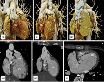
This subset of patients (Fig. 1) may be treated initially by balloon atrial septostomy and banding of the pulmonary trunk. Creation of the superior cavopulmonary connection can then be performed as an adjunct to the arterial switch, with subsequent closure of the ventricular septal defect, when the pulmonary vasculature has adequately matured. Reference Chowdhury, George and Sankhyan12,Reference Chowdhury, Airan and Sharma19,Reference Chowdhury, Airan and Talwar20,E27–E30
Volume rendered images (a–c), oblique coronal images (d and e), and oblique axial image (f) show a small right ventricle (RV) in the setting of transposition. Note is made of pulmonary valvar (PV) stenosis and a subaortic ventricular septal defect (*). [A = aorta; LV = left ventricle; P = pulmonary trunk; LA = left atrium].

Transposition with ventricular septal defect and hypoplastic left ventricle
The cavopulmonary anastomosis is part of the treatment of early atrial septostomy followed by delayed atrial switching. Reference Chowdhury, Airan and Talwar20,E2,E6,E24,E27,E28 This is one of the rare instances where it is the morphologically left ventricle that is unloaded so as to support only half of the pulmonary circulation.
Congenitally corrected transposition with or without a ventricular septal defect and pulmonary stenosis or pulmonary atresia
Many such individuals have morphologically right ventricles of borderline size. In this setting, the cavopulmonary connection has been performed as an adjunct to the “double-switch” procedure, or to delay the Rastelli procedure in which the pulmonary ventricle would be made anatomically and functionally smaller by the intracardiac baffle and ventriculotomy. Reference Malhotra, Patel and Pandya13,Reference Chowdhury, Airan and Sharma19,Reference Chowdhury, Airan and Talwar20,E2,E5,E6,E13,E18,E19,E37,E38,E58
Double outlet right ventricle with remote interventricular communication
In a small subset of patients with double outlet right ventricle, the defect is remote from either outlet.E29,E35,E39,E59,E82 In order to avoid compromising a portion of right ventricular volume by placement of a large intraventricular tunnel, some have been treated by creation of the cavopulmonary connection. An analogous situation may be seen in patients with transposition and a ventricular septal defect remote from the outlet, in whom the classic techniques for repair are untenable. Reference Chowdhury, Airan and Talwar20,E26,E27
Individuals with supero-inferior ventricles
In many of these patients, the superiorly positioned morphologically right ventricle is grossly hypoplastic. Depending on the type of atrioventricular and ventriculoarterial connections, some patients have been converted to the Fontan circulation, whereas others have been treated with either a biventricular repair or creation of a superior cavopulmonary connection. Reference Anderson, Anderson and Zuberbuchler2–Reference Anderson and Ho4,Reference Van Arsdell18,E25,E38
Right ventricular hypoplasia
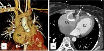
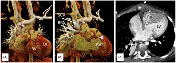
Hypoplasia of the subpulmonary right ventricle is occasionally encountered in those with ventricular or atrioventricular septal defects, tetralogy of Fallot, right ventricular outflow tract obstruction, and transposition with large ventricular septal defects. Although a standard biventricular repair may be possible, many of these individuals have been palliated by creation of a superior cavopulmonary connection (Figs. 2, 3). Reference Chowdhury, Airan and Sharma19,Reference Chowdhury, Airan and Talwar20,E60–E65
The volume-rendered image (a), and the oblique axial image (b) show a hypoplastic tricuspid valve (curved arrow) and a hypoplastic right ventricle (RV). [A = aorta; LV = left ventricle; P = pulmonary trunk; RA = right atrium].

Volume rendered images (a and b) and oblique axial image (c) shows features of tetralogy of Fallot with hypoplasia of the morphologically right ventricle (RV). Note the patent cavopulmonary shunt. [A = aorta; LV = left ventricle; P = pulmonary trunk; * = subaortic ventricular septal defect; arrowhead = infundibular stenosis].

Individuals with isomeric atrial appendages
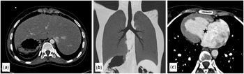
A feature of many of these individuals is the presence of bilateral superior caval veins, often with a common atrium and other complex cardiac anomalies (Fig. 4). In these settings, the superior cavopulmonary connection avoids the need to create a complex intra-atrial baffle, thus simplifying the operation. Reference Chowdhury, Airan and Talwar20,E20,E27
The axial image (a), the coronal minimum intensity projection image (b), and the oblique axial image (c) show the features of right isomerism, in patient with absence of the spleen, a transverse midline liver, bilateral morphologically right bronchuses, and a right-sided heart with atrioventricular septal defect (star).

Those with Ebstein’s malformation
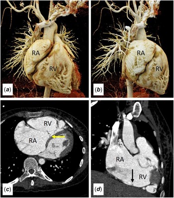
The indications for creation of a superior cavopulmonary connection in individuals with Ebstein’s malformation are complex and somewhat controversial. Having the right ventricle support only half the pulmonary circulation has generally been used when the ventricle itself is severely dilated, dysplastic, or severely dysfunctional (Fig. 5). Reference Wright, Knight and Thomas14,Reference Chowdhury, Airan and Sharma19,Reference Chowdhury, Airan and Talwar20,E10,E19,E23,E41,E66,E67 Those with a small or hypoplastic right ventricle are more likely to go down the route of a Starnes procedure and functionally univentricular palliation.
The volume-rendered images (a and b), the oblique axial image (c), and the oblique coronal image (d) show apical displacement of septal leaflet (yellow arrow) and posterior leaflet (black arrow) of the tricuspid valve in a patient with Ebstein’s malformation. Note the grossly dilated right atrium (RA). [RV = right ventricle].

Univentricular connection to a dominant left ventricle
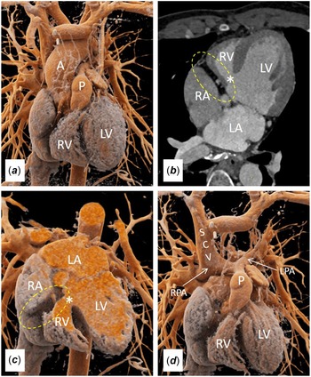
In this setting, the incomplete right ventricle supports the pulmonary trunk when the ventriculo-arterial connections are concordant, which is usually the case in the setting of tricuspid atresia. Reference Chowdhury, Airan and Sharma19,Reference Chowdhury, Airan and Talwar20,E18,E68 The atriopulmonary variation of the Fontan procedure, although not now performed, produced the closest anatomical situation of a true “one-and-a-half ventricle” arrangement.E7,E10,E69,E70 Such patients were on occasion, palliated by creation of the superior cavopulmonary connection (Fig 6).
The volume rendered images (a, c) and the oblique axial image (b) show absence of communication (dotted yellow arrow) between the right atrium (RA) and the incomplete right ventricle (RV), with fat noted within the right atrioventricular groove. This is classical tricuspid atresia. Note the ventricular septal defect (*) and the hypoplastic and incomplete morphologically right ventricle (RV). The volume rendered image (d, with aorta digitally subtracted) shows the superior cavopulmonary connection, connecting the superior caval vein (SCV) to the right pulmonary artery (RPA). [A = aorta; P = pulmonary trunk; LA = left atrium; LPA = left pulmonary artery].

Special circumstances
In addition to the settings listed above, there are multiple additional circumstances in which the surgeon may opt to construct the superior cavopulmonary connection. E1–E172
-
Patients with cardiac Ewing’s sarcoma undergoing tumour debulking, patients undergoing resection of a metastatic tumour to the right ventricle along with lobectomy for lung cancer, tricuspid valve endocarditis, and post infarction right ventricle.E70–E73
-
Patients with Uhl’s anomaly and right ventricular endomyocardial fibrosis.E70–E79
-
Patients undergoing the Rastelli procedure with a large intracardiac baffle. E7,E8,E10,E47,E80
-
Atrial redirection with systemic atrioventricular valvular regurgitation.E25,E80
-
Creation of the cavopulmonary connection in the setting of acute postoperative right ventricular dysfunction following an attempted biventricular repair. Reference Chowdhury, Airan and Sharma19,Reference Chowdhury, Airan and Talwar20,E70–E80
-
Conversion from the Fontan circulation in those with pulmonary arteriovenous malformations, Fontan conduit obstruction, recurrent intractable arrhythmias, and protein-losing enteropathy. Reference Chowdhury, George and Sankhyan12,E37,E51,E81
-
As an interstage procedure before biventricular repair to minimise the risk of left ventricular outflow tract obstruction, heart block, iatrogenic atrioventricular valvular regurgitation, and the requirement of a right ventricle-to-pulmonary artery conduit. Reference Hoashi, Kitano and Kagisaki8,E7,E10,E44–E47
The ability of the subpulmonary ventricle to support the pulmonary circulation subsequent to the creation of the cavopulmonary connection
The safe limits of right ventricular size and function are currently unknown. Different institutions follow different protocols for selection. Reference Chowdhury, Airan and Sharma19,Reference Chowdhury, Airan and Talwar20,E20,E27,E28 A simple means of quantitating the ability of the morphologically right ventricle to handle volume is to measure the size of tricuspid annulus with this measurement then plotted on a nomogram to obtain a z value. Reference Chowdhury, Airan and Sharma19,Reference Chowdhury, Airan and Talwar20,E28,E86–E90 . Intraoperative assessment can be done with direct visualisation and measurement with Hegar’s dilator. Contrast-enhanced and multidetector computed-tomographic angiocardiography in angled oblique views, or cardiac velocity-encoded magnetic resonance imaging, now provide the best comprehensive assessment of ventricular dimensions. Reference Chowdhury, Airan and Sharma19,Reference Chowdhury, Airan and Talwar20,E91–E101 Successful creation of the superior cavopulmonary connection has been achieved when predicted ventricular volumes have been as small as 30%. Reference Wright, Knight and Thomas14,E5,E6,E61 An alternative approach is to combine tricuspid valve Z-scores and pulmonary ventricular volume (Table S1). Reference Chowdhury, Airan and Talwar20,E18,E27 A combination of parameters, therefore, namely, tricuspid Z-values, tricuspid/ mitral valve diameter, right ventricular morphology, right ventricular diastolic volumes, presence or absence of right ventricular-coronary artery fistulae, and the degree of right ventricular endocardial fibroelastosis are usually taken into consideration for the decision-making of construction of a pulsatile superior cavopulmonary connection as opposed to opting for a biventricular or functionally univentricular type of repair.E101,E168–E172
In those with pulmonary atresia and intact interventricular septum, high mortalities ensued when biventricular repair was attempted with tricuspid z-values of less than minus three.E4 Conversely, good outcomes were achieved with z-values greater than minus three.E4,E102 Our own criterions are shown in Table S2. We agree with others, nonetheless, that absolute sizes of the right ventricle and the tricuspid valve may not be the only factors for operative success. Reference Chowdhury, Airan and Sharma19,Reference Chowdhury, Airan and Talwar20,E101–E110 Right ventricular compliance, tricuspid regurgitation, relative pulmonary arterial hypoplasia, and pulmonary vascular resistance are important additional considerations in preoperative candidate selection. Reference Chowdhury, Airan and Sharma19,Reference Chowdhury, Airan and Talwar20
Functional analysis can be achieved echocardiographically or angiographically, permitting grading as normal, or mildly, moderately, or severely decreased. Reference Chowdhury, Airan and Sharma19,Reference Chowdhury, Airan and Talwar20 Visual inspection of the mural thickness and diastolic filling is also made. Reference Hanley, Sade and Blackstone6,Reference Chowdhury, Airan and Sharma19,Reference Chowdhury, Airan and Talwar20,E4,E11,E18,E29,E42,E92–E102,E111 Patients with poorly functioning right ventricles are also considered for the superior cavopulmonary connection when there is dilation to 120%. or more and at least a moderate reduction in ventricular function. Reference Hanley, Sade and Blackstone6,Reference Chowdhury, Airan and Sharma19,E4,E11–E14,E18,E29,E42,E92–E102,E111 Recently, velocity-encoded MRI has been used more accurately to quantitate right ventricular systolic and diastolic function and volume. Reference Chowdhury, Airan and Sharma19,Reference Chowdhury, Airan and Talwar20,E91-E106
A third important determinant of the ability of the ventricle properly to function is the pulmonary afterload. In this regard, we took several physiologic criterions into consideration when contemplating creation of a superior cavopulmonary connection, specifically;
-
Mean pulmonary arterial pressure less than 15 mm Hg, or less than 20 mm Hg with a net left-to-right shunt.
-
Indexed pulmonary vascular resistance less than 3.0 Woods units/m2 and a preoperative Mayo index less than 4.0.E173
-
Systemic ventricle end-diastolic pressure less than 12 mm Hg and ejection fraction greater than 0.45.
-
Probability of residual pulmonary arterial hypertension, when two-ventricle repair with atrial pop-off was considered a better option. Reference Chowdhury, Airan and Sharma19,Reference Chowdhury, Airan and Talwar20
Results
Early surgical outcomes have been good, or even excellent for most individuals undergoing conversion to one and a half circulations. The overall reported operative mortalities prior to 2000 were around 20%. After 2000, the overall risk of surgical repair has been cited to be between 3% and 8.5% Reference Van Arsdell18–Reference Chowdhury, Airan and Talwar20,E24,E27,E30–E35,E47,E50,E101 There are multiple reported causes of death. Reference Van Arsdell18–Reference Chowdhury, Airan and Talwar20,E24,E27,E30–E35,E47,E50,E101 Despite appropriate selection of patients, nonetheless, complications related to a pulsatile superior cavoatrial junction have been reported in almost one-tenth of operations, with a small risk of anastomotic takedown. Reference Chowdhury, Airan and Talwar20,E18 In contrast, there have been no reported instances of plastic bronchitis, protein losing enteropathy or liver cirrhosis. Supraventricular arrhythmias, however, have been reported in one-eighth to one-third of instances. Reference Chowdhury, Airan and Talwar20,E5–E14,E27–E30 Data are limited on long-term outcomes according to pathologic substrates but over seven-tenths of patients are reported to be faring well after 20 years in most long-term studies. Reference Cabrelle, Castaldi and Vedovelli5,Reference Hanley, Sade and Blackstone6,Reference Wright, Knight and Thomas14,Reference Toh, Kotani and Akagi17–Reference Chowdhury, Airan and Talwar20,E1–E172
Evidence supporting the generally held view that a pulsatile superior cavopulmonary anastomosis is superior to a Fontan procedure is limited. Higher functional capacity has been demonstrated by some investigators. Reference Cabrelle, Castaldi and Vedovelli5,Reference Chowdhury, Airan and Sharma19,Reference Chowdhury, Airan and Talwar20,E18,E28,E112 Other investigations, in contrast, have documented restrictive right ventricular physiology. They have also demonstrated functional capacity and health-related quality of life scores similar or worse than those with a Fontan circulation. Reference Cabrelle, Castaldi and Vedovelli5,49,E9,E50,E81,E107,E144,E9,E167 The finding of a trend towards worse survival in those with one and a half circulations as revealed by multi-institutional studies is intriguing. It may be related to adverse effects on the coronary perfusion at the time of right ventricular decompression, or unfavourable consequences of pulmonary regurgitation and right atrial hypertension. Reference Hanley, Sade and Blackstone6,Reference Daubeney, Delany and Anderson15,E167–E172 There are isolated reports of sufficient growth of the unloaded ventricle subsequent to creation of the cavopulmonary connection to permit conversion to biventricular repair. Reference Hoashi, Kitano and Kagisaki8,E44,E46,E167 Up to half of long-term survivors show some degree of growth, albeit insufficient for biventricular conversion. Reference Chowdhury, Airan and Sharma19,Reference Chowdhury, Airan and Talwar20,E7,E9,E10,E15,E17,E18,E28,E44–E47,E50,E49,E78,E81,E107,E115,E144 Successful conversion of the Fontan circulation to one and a half circulations is also reported, and the reverse as an interstage procedure prior to biventricular repair. Reference Hoashi, Kitano and Kagisaki8,E44-E46 The superior cavopulmonary connection has also been used as a salvage procedure following failed biventricular repair. Reference Van Arsdell18–Reference Chowdhury, Airan and Talwar20,E8,E9,E11,E18,E27,E37,E38,E44–E46,E69–79,E82,E84
Discussion
Some readers might find disturbing over the use of "so-called" in our title. Our purpose in producing our review is in no way meant to be a criticism of the value of the surgical procedure that is dominated by the construction of a bidirectional cavopulmonary anastomosis. On the contrary, we hope that our review, based on our extensive experience, points to the importance of the procedure. Our use of “so-called” is meant to do no more than emphasise that the procedure has nothing to do with “half a ventricle”. In fact, as was pointed out to use recently by Dr Wayne Tworetsky, from Boston, the more appropriate term for the procedure is construction of one and a half circulations. It is difficult to appreciate how a heart could exist with “half a ventricle”. The early surgical outcomes of the procedure, however, it is described, have been good, or even excellent.
Thus, numerous combinations of lesions can be palliated by off-loading the venous return from the upper half of the body by performing a superior cavopulmonary connection and thus leaving a ventricle, usually the morphologically right ventricle, to support only half the pulmonary circulation. The procedure has acceptable surgical mortality, morbidity, and functional results. Reference Hanley, Sade and Blackstone6–Reference Stellin, Vida and Milanesi11 We emphasise again, nonetheless, that the approach has little to do with “half a ventricle”. Indeed, construction of the cavopulmonary connection had been used to “rescue” an incomplete right ventricle subsequent to its incorporation as part of the Fontan procedure. The atrioventricular connection, although part of the evolution of the Fontan concept, is no longer used in this fashion. The global experiences we have reviewed, nonetheless, do little to provide understanding of the role of “half a ventricle”. They do, in contrast, emphasise the problems that continue to be faced when opting to use a ventricle to support half of the pulmonary circulation.
Nothing in the literature, for example, as far as we can establish, has addressed the issue of making a qualitative assessment of the right side of the heart. Similarly ignored is the “difficulty factor” encountered when performing intricate intracardiac repairs to divert inferior caval venous blood. Also in need of attention are the safe upper limits of pulmonary arterial pressure and pulmonary vascular resistance. At present, the surgeon is called upon to make subjective decisions regarding the optimal approach. Reference Hanley, Sade and Blackstone6,Reference Barron7,Reference Hanley9,E21 We know that we can successfully create the situation in which a ventricle supports only half the pulmonary circulation, but as yet there is no certainty as to who the candidates should be. We also do not know, currently, whether supporting half the pulmonary circulation produces benefits over and above those conferred by the Fontan procedure. We were unable to find data to allow us to make a rational decision as to when the size and function of the subpulmonary ventricle reaches the point at which the benefit accrues by avoiding the Fontan operation. Reference Hanley, Sade and Blackstone6,Reference Barron7,Reference Hanley9,,E21 Our own reported experiences suggest that these criterions will be similar to those required for creation of the Fontan circulation. Reference Chowdhury, Airan and Sharma19,Reference Chowdhury, Airan and Talwar20,E14 Over and above these concerns relating to the underlying pathophysiology, the procedure also introduces specific issues of surgical concern (Table S3).
Atrial septal fenestration
Fenestration of the atrial septum obviously permits right-to-left shunting. It also increases left ventricular preload and cardiac output, albeit with mild desaturation. Reference Chowdhury, Airan and Sharma19,Reference Chowdhury, Airan and Talwar20 The indications and long-term results for fenestration are limited and conflicting.E18,E47,E50 At AIIMS, we currently fenestrate in patients with tricuspid valves between one-third and one-half of normal, or in the setting of right ventricular dysfunction and post-operative supraventricular arrhythmias. This strategy has improved our clinical outcomes, with none of our long-term survivors developing significant desaturation. Reference Chowdhury, Airan and Sharma19,Reference Chowdhury, Airan and Talwar20 On the downside, we did encounter perioperative or late supraventricular arrhythmias in around one-sixth of our cohort. Reference Chowdhury, Airan and Sharma19,Reference Chowdhury, Airan and Talwar20
Pulmonary regurgitation
This feature removes from the operation part of its physiologic rationale, since the ventricular preload is no longer reduced. Reference Chowdhury, Airan and Sharma19,Reference Chowdhury, Airan and Talwar20,E7,E10 In our experience, nonetheless, patients with only mild pulmonary regurgitation have run a smooth postoperative course. Reference Chowdhury, Airan and Sharma19,Reference Chowdhury, Airan and Talwar20 Should mild to moderate right ventricular outflow tract obstruction be present, it is better left intact, as opposed to courting the development of free pulmonary regurgitation. In the setting of congenitally corrected transposition, creation of the superior cavopulmonary connection is particularly attractive for “pressure unloading” of the pulmonary ventricle.E37,E111
Should the azygos vein be ligated?
A patent azygos or hemiazygos vein was thought advantageous in patients with superior caval venous hypertension. This notion was also pertinent for the right atrium in cases of severe right ventricular dysfunction, or during exercise when the hypoplastic right heart was incapable of handling the increased systemic venous return. Reference Cabrelle, Castaldi and Vedovelli5,E7,E8,E27–E30 Our practice is to ligate the azygos vein in all our patients, with no observed adverse haemodynamic consequences. Reference Chowdhury, Airan and Sharma19,Reference Chowdhury, Airan and Talwar20
Complications related to the superior caval vein
Due to the reversed pulsatile flow in the superior cavopulmonary anastomosis, an increased incidence of postoperative chylothorax, pleural effusions, early morning periorbital oedema, and left superior caval venous aneurysm have been reported by some investigators. Reference Chowdhury, Airan and Sharma19,Reference Chowdhury, Airan and Talwar20,E17,E18,E112–E115 Banding of the right pulmonary artery at its junction with the pulmonary trunk has been advocated to prevent this complication. Reference Chowdhury, Airan and Sharma19,Reference Chowdhury, Airan and Talwar20,E7,E17,E30 This procedure, however, has its own complication, namely development of pulmonary arteriovenous malformations on the banded side.E112–E116 We have undertaken such unilateral banding with satisfactory results. Reference Chowdhury, Airan and Sharma19,Reference Chowdhury, Airan and Talwar20
Growth and function of the subpulmonary ventricle
Previous reports have largely concentrated on establishing the anatomical criterions for using the subpulmonary ventricle to support only half the pulmonary circulation. Reference Billingsley, Laks and Boyce1,Reference Hanley, Sade and Blackstone6,Reference Hanley9,Reference Stellin, Vida and Milanesi11,Reference Van Arsdell18,E7,E20,E30 Within the ranges of size is a bandwidth of ventricles that are capable of dealing with inferior caval venous blood alone. The criterions for opting for this intermediate procedure need to be clarified. Reference Hanley, Sade and Blackstone6,Reference Hanley9 In our earlier publication, we demonstrated that individuals with severe pulmonary ventricular hypoplasia with tricuspid valve Z score lower than -4.8 and mild pulmonary ventricular hypoplasia with tricuspid valve Z score higher than -1.5 would predicate a Fontan type or a two ventricle repair, respectively. Reference Chowdhury, Airan and Sharma19,Reference Chowdhury, Airan and Talwar20 Even when used to support only half the circulation, the hypoplastic pulmonary ventricle needs to be functioning reasonably well. Our experience indicates that patients with borderline ventricular function may better be suited for a functionally univentricular repair. Reference Chowdhury, Airan and Sharma19,Reference Chowdhury, Airan and Talwar20
There are some reports of adequate growth of the right ventricle subsequent to creation of the superior cavopulmonary connection, in some instances permitting conversion to biventricular repair in the setting of pulmonary atresia with intact ventricular septum. Reference Hoashi, Kitano and Kagisaki8,E44,E46 In our own experience, just less than half of survivors demonstrated significant growth of the tricuspid valve and right ventricle. Reference Chowdhury, Airan and Sharma19,Reference Chowdhury, Airan and Talwar20 At a median follow-up of almost nine years in several series, none had qualified for conversion to biventricular repair Reference Chowdhury, Airan and Sharma19,Reference Chowdhury, Airan and Talwar20,E7,E10,E28,E44–E47,E107,E112,E117,E135,E167
Failure of the procedure, with conversion to a functionally univentricular pathway
As a treatment of elevated right atrial pressure and/or supraventricular arrhythmias in a failing superior cavopulmonary connection, some investigators have employed conversion to total cavopulmonary connection, with cryoablation of the atrial wall.E9,E11,E37,E58,E136,E137 Such conversion is suggested to reduce atrial arrhythmias. E9
Conversion of the Fontan circulation to the one and a half circulation
Patients initially palliated with the Fontan procedure have been successfully converted to the one and half circulation with acceptable outcomes. The primary indications for such conversion include exercise intolerance, protein-losing enteropathy, pulmonary arteriovenous malformations, intractable arrhythmias, and right atrioventricular conduit obstruction. Concomitant arrhythmia surgery is recommended as a routine in all such patients. Reference Chowdhury, George and Sankhyan12,E82–E84,
Conversion of the one and a half to biventricular circulations
We found one such example, reported four years after the primary repair in a child with unbalanced atrioventricular septal defect. Reference Hoashi, Kitano and Kagisaki8,E44,E46 The development of massive venovenous collaterals between the superior and inferior caval venous compartments had facilitated the growth of the right ventricle.
The one and a half circulation as a salvage procedure
Results for the procedure used as a salvage operation for pulmonary ventricular dysfunction have been unsatisfactory. Between 1990 and 2003, we performed such salvage operations in 5 patients, but three of the patients died. Reference Chowdhury, Airan and Sharma19,Reference Chowdhury, Airan and Talwar20 . Between 2003 and 2021, we have now attempted 17 more salvage repairs, but now without mortality. Reference Chowdhury, Airan and Sharma19,Reference Chowdhury, Airan and Talwar20
Supraventricular arrhythmias
These arrhythmias are reported in between one-eighth and one-third of patients, albeit without having undue impact. Reference Billingsley, Laks and Boyce1,Reference Chowdhury, Airan and Sharma19,Reference Chowdhury, Airan and Talwar20,E1,E5-E14,E27-E30. As a treatment of elevated right atrial pressure, or supraventricular arrhythmias subsequent to pulsatile superior cavopulmonary connection, we performed atrial septal fenestration. In patients with failing pulsatile cavopulmonary connection or high right atrial pressure, some investigators have converted them to either the superior cavopulmonary connection or the Fontan circulation.E7,E9,E50 We had previously reported perioperative and late postoperative supraventricular arrhythmias in around one-sixth of our cohort after median follow-up of almost nine years. The risk of such arrhythmias was greater in those with isomeric appendages, Ebstein’s anomaly, and systemic ventricular dysfunction. Reference Chowdhury, Airan and Sharma19,Reference Chowdhury, Airan and Talwar20
Can the procedure restore ventriculo-arterial coupling?
The optimal haemodynamic of the normal circulation involves a high pulmonary arterial pressure and a lower caval venous pressure. The paradox of the Fontan circulation is that it produces caval and pulmonary arterial hypertension. Since the systemic venous circulation is in series with the pulmonary circulation, these patients develop chronic hepatic congestion with ascites, pulmonary arteriovenous malformations, plastic bronchitis, arrhythmias, thromboembolism, protein-losing enteropathy, plastic bronchitis, liver cirrhosis, and ventricular failure. Reference Chowdhury, Airan and Sharma19,Reference Chowdhury, Airan and Talwar20 In this setting, use of a subpulmonary ventricle to support half of the pulmonary circulation can theoretically restore ventriculo arterial coupling, thus maintaining a low pressure in the inferior caval venous district. Indeed, several investigators, including ourselves, have demonstrated reversal of the “Fontan paradox” subsequent to construction of the superior cavopulmonary connection. Reference Chowdhury, Airan and Sharma19,Reference Chowdhury, Airan and Talwar20 The procedure presumably provides kinetic energy and pulsatility to pulmonary blood flow, while reducing the volume load of the ventricle. Reference Billingsley, Laks and Boyce1,Reference Hoashi, Kitano and Kagisaki8,Reference Hanley9 Chronic atrial arrhythmias, cyanosis, or protein-losing enteropathies have not been identified in the short-term and long-term follow-up of those with one-and-a-half circulations. Reference Chowdhury, Airan and Sharma19,Reference Chowdhury, Airan and Talwar20,E125–E130,E165
Future directions
Our review shows that it is safe to produce one and a half circulations, with this arrangement providing good results at mid- and long-term follow-up. As yet, however, we do not know for whom the procedure should optimally be performed. We still need to know whether creation of one and a half circulations provides better definitive palliation when the alternatives are the Fontan procedure, or even biventricular repair. The physiologic and morphologic criterions for optimal use remain to be defined. Properly to test the hypothesis that one and a half circulations produces superior outcomes in selected patients, when compared to biventricular or functionally univentricular repairs, will require multi-institutional prospective and randomised trials.
Supplementary material
The supplementary material for this article can be found at https://doi.org/10.1017/S1047951123001646.
Acknowledgements
None.
Financial support
This research received no specific grant from any funding agency, commercial, or not-for-profit sectors.
Competing interests
The author(s) declared no potential conflicts of interest with respect to the research, authorship, and/or publication of the article.
Ethical standard
The authors assert that all procedures contributing to this study comply with the ethical standards of the relevant national guidelines on human experimentation and with the Helsinki declaration of 1975, as revised in 2008.