INTRODUCTION
Ventral prefrontal cortex (VPFC), a key regulatory component of the corticolimbic neural system involved in emotional and motivated behaviors, is implicated in bipolar disorder (BD) (Blumberg et al., Reference Blumberg, Charney and Krystal2002). Neuroimaging studies of adults with BD provide evidence for abnormal VPFC morphology and functioning. The VPFC undergoes structural and functional maturation during adolescence and early adulthood (Giedd et al., Reference Giedd, Blumenthal, Jeffries, Castellanos, Liu, Zijdenbos, Paus, Evans and Rapoport1999; Gogtay et al., Reference Gogtay, Giedd, Lusk, Hayashi, Greenstein, Vaituzis, Nugent, Herman, Clasen, Toga, Rapoport and Thompson2004; Sowell et al., Reference Sowell, Thompson, Holmes, Jernigan and Toga1999), coinciding with a peak for acute BD episodes. Abnormal VPFC morphology and function in BD, combined with the intersection of VPFC developmental maturation and the onset of BD, indicate that abnormalities in VPFC neurodevelopment may play a role in the behavioral expression of BD in adolescence and young adulthood. VPFC disturbances may not be expressed fully until the region passes through its course of programmed development in late adolescence/early adulthood, when characteristics of the adult phenotype emerge (Blumberg et al., Reference Blumberg, Kaufman, Martin, Charney, Krystal and Peterson2004).
The progressive course of PFC neurodevelopment may contribute to differing results obtained in the morphometric study of the region in diverse age groups of those with BD. Gray matter volume decreases in multiple VPFC subregions, including subgenual anterior cingulate cortex (ACC), orbitofrontal cortex, and inferior frontal cortex, are consistently reported in adults with BD (Blumberg et al., Reference Blumberg, Krystal, Bansal, Martin, Dziura, Durkin, Martin, Gerard, Charney and Peterson2006; Drevets et al., Reference Drevets, Price, Simpson, Todd, Reich, Vannier and Raichle1997; Lopez-Larson et al., Reference Lopez-Larson, DelBello, Zimmerman, Schwiers and Strakowski2002; Lyoo et al., Reference Lyoo, Kim, Stoll, Demopulos, Parow, Dager, Friedman, Dunner and Renshaw2004). In contrast, studies of adolescents with BD have yielded inconsistent findings with reports of volume decreases in sub- and pregenual ACC and orbitofrontal cortex (Kaur et al., Reference Kaur, Sassi, Axelson, Nicoletti, Brambilla, Monkul, Hatch, Keshavan, Ryan, Birmaher and Soares2005; Wilke et al., Reference Wilke, Kowatch, DelBello, Mills and Holland2004) or failures to detect differences (Adler et al., Reference Adler, DelBello, Jarvis, Levine, Adams and Strakowski2007; Chang et al., Reference Chang, Barnea-Goraly, Karchemskiy, Simeonova, Barnes, Ketter and Reiss2005; Dickstein et al., Reference Dickstein, Milham, Nugent, Drevets, Charney, Pine and Leibenluft2005; Frazier et al., Reference Frazier, Breeze, Makris, Giuliano, Herbert, Seidman, Biederman, Hodge, Dieterich, Gerstein, Kennedy, Rauch, Cohen and Caviness2005; Sanches et al., Reference Sanches, Sassi, Axelson, Nicoletti, Brambilla, Hatch, Keshavan, Ryan, Birmaher and Soares2005). VPFC volume decreases reported by Blumberg et al. (Reference Blumberg, Krystal, Bansal, Martin, Dziura, Durkin, Martin, Gerard, Charney and Peterson2006) were statistically significant in young adults, but not adolescents, with BD. Based on these cross-sectional results, the authors suggested that VPFC volume abnormalities may progress over the course of adolescence and therefore be less likely to emerge as significantly divergent until late adolescence or early adulthood. We conducted a prospective longitudinal study of brain volume in adolescents and young adults with BD in order to address this issue empirically. We hypothesized that adolescents/young adults with BD would show greater volume loss in VPFC over time relative to healthy comparison participants (HC).
MATERIALS AND METHODS
Participants
Participants consisted of 10 adolescent/young adult outpatients with BD I (five females) and 8 adolescent/young adult HCs (six females), as described previously (Blumberg et al., Reference Blumberg, Fredericks, Wang, Kalmar, Spencer, Papademetris, Pittman, Martin, Peterson, Fulbright and Krystal2005). The HCs did not meet criteria for Diagnostic and Statistical Manual, 4th edition (DSM-IV) Axis I diagnoses (American Psychiatric Association, 2000) and had no family history of DSM-IV Axis I diagnoses in their first-degree relatives. Participants with BD were referred from the Yale School of Medicine Medical Center, the Veterans Affairs Connecticut Healthcare System, and the greater New Haven, Connecticut community, and the HCs were recruited from the community. At the time of initial study participation, individuals with BD were between 10 and 21 years of age and HCs were between 11 and 19 years of age. Follow-up study participation occurred on average 2.28 ± 0.55 SD years after baseline participation. Participants were without a history of other neurological disorders, loss of consciousness for longer than 5 min, or significant medical illness, with the exception of one BD participant with treated hypothyroidism.
The revised Schedule for Affective Disorders and Schizophrenia for School-Age Children-Present and Lifetime Version (Kaufman et al., Reference Kaufman, Birmaher, Brent, Rao, Flynn, Moreci, Williamson and Ryan1997) was administered to the participants and a parent/guardian for participants 18 years of age and younger by a board-certified child psychiatrist or a psychologist expert in childhood mood disorders. For those participants older than 18 years, the Structured Clinical Interview for DSM-IV Axis I Disorders (Version 2.0) (First et al., Reference First, Spitzer, Gibbon and Williams1995) was administered by a board-certified adult psychiatrist. These interviews were administered for each participant at both time points. Final DSM-IV diagnoses were established by the consensus of clinical and structured interviews. At the time of the baseline neuroimaging scan, five participants with BD were unmedicated, six met criteria for rapid cycling, nine were in the midst of a mood episode, and one was euthymic. At the time of the second neuroimaging session, seven participants with BD were unmedicated, seven met criteria for rapid cycling, and three were euthymic. Psychotropic medications at the first scan included mood stabilizers (n = 2), anticonvulsants (n = 3), antidepressants (n = 2) (both tricyclics and selective serotonergic reuptake inhibitors), atypical antipsychotics (n = 2), stimulants (n = 1), levothyroxine (n = 1), and clonidine (n = 1). Psychotropic medications at the second time point included mood stabilizers (n = 1), anticonvulsants (n = 2), antidepressants (n = 2) (both tricyclics and selective serotonergic reuptake inhibitors), atypical antipsychotics (n = 2), stimulants (n = 2), and atomoxetine (n = 1). All five participants who were not taking psychotropic medication at the time of both scans had been exposed to lithium, valproate, or other psychotropic medications. Comorbidities included attention-deficit/hyperactivity disorder (n = 1), oppositional defiant disorder (n = 2), social phobia (n = 1), post-traumatic stress disorder (n = 1), learning disorder not otherwise specified (n = 2), and developmental coordination disorder (n = 1). One participant had a history of alcohol dependence and hallucinogen and marijuana abuse in remission for more than 1 year prior to baseline scan. All BD participants reported positive family history, within their first- or second-degree relatives, of a mood spectrum disorder, including BD, major depressive disorder, or alcohol abuse. For further detail on sample characterization, see Blumberg et al. (Reference Blumberg, Fredericks, Wang, Kalmar, Spencer, Papademetris, Pittman, Martin, Peterson, Fulbright and Krystal2005).
Following a complete description of the research, written informed consent was obtained from parents/guardians and participants older than 18 years. Written informed assent was obtained from minors. This research was approved by the human investigation committees of the Yale School of Medicine and the Department of Veterans Affairs.
Magnetic Resonance Image Acquisition and Processing
Magnetic resonance imaging scans at baseline and follow-up time points were obtained using the same 1.5-T scanner (GE Signa; General Electric, Milwaukee, WI). Head positioning was standardized using canthomeatal landmarks. Images were obtained using a three-dimensional sagittal spoiled gradient echo sequence (repetition time, 24 ms; echo time, 5 ms; flip angle, 45°; frequency encoding, superior/inferior; no wrap; 256 × 192 matrix; field of view, 30 × 30 cm2; two excitations; slice thickness, 1.2 mm; and 124 contiguous slices).
Morphometric measures were computed using the Yale Bioimage Suite software package (http://www.bioimagesuite.org/) running on Linux workstations. We employed a nonrigid registration method that relied on the normalized mutual information metric. The transformation was parameterized using a Free-Form Deformation Model (Papademetris et al., Reference Papademetris, Jackowski, Schultz, Staib and Duncan2004; Rueckert et al., Reference Rueckert, Sonoda, Hayes, Hill, Leach and Hawkes1999). Using this nonrigid registration technique, the follow-up scans for each participant were pairwise registered to the baseline scans. The resulting transformations were then used to produce Jacobian maps of local expansion/contraction that were generated based on the determinant of the Jacobian of the displacement field generated by each transformation (Staib et al., Reference Staib, Jackowski and Papademetris2006). The Jacobian maps had a resolution of 1.2 × 1.2 × 1.2 mm3 where each voxel has a value representing the local volume change required to map an individual participant’s baseline time point to his/her follow-up time point (i.e., 1 = no volume change, >1 = follow-up time point was larger than baseline time point, and <1 = baseline time point was larger than follow-up time point). The Jacobian maps were checked to ensure that the transformations were all free of singularities (i.e., |J| < 0). Next, the baseline images for each participant were normalized to the Colin Brain template which is in Montreal Neurological Institute (MNI) space using the same nonlinear registration algorithm, and the individual Jacobian maps were then warped to the space of the MNI template using the second registration. Jacobians were computed at each voxel throughout the brain, enabling exploration of differences in volume change over time in regions not hypothesized a priori.
Data Analysis
Two-tailed two-sample t tests (BD vs. HC) were performed using BioImage Suite with Jacobian values as the dependent variables. Findings of volume changes in the brain over time were considered significant at p < .05 uncorrected and clusters >50 contiguous voxels (at resolution 2 × 2 × 2 mm3). Post hoc exploratory analyses were performed to determine the potential effects of any demographic factors with large effect sizes, as well as presence or absence of psychotropic medication at time of scanning, on the dependent measures. These exploratory analyses [including analyses covarying for any demographic factors with large effect sizes and an analysis of variance comparing those BD participants who were medicated at either time point (n = 5) to those participants with BD who were unmedicated at both time points (n = 5)] were conducted using the signal extracted from each participant’s normalized Jacobian maps from the maximum point of the prefrontal regions where volume change over time differed between the two groups.
RESULTS
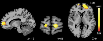
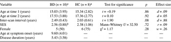
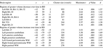
The groups did not differ significantly in age at the baseline or follow-up scans, time between scans, gender, or socioeconomic status (Table 1). Volume decreases from baseline to follow-up were greater in the BD group, relative to the HCs, in bilateral PFC as displayed in orthogonal views in Figure 1. While HCs showed approximately a 0.66% relative volume decrease in this region over time, the BD group showed 3.59% volume decrease in this region. The left PFC cluster included superior and medial frontal gyri [Brodmann’s area (BA) 10] and extended to rostral ACC (BA 32) and ventrally to BA 11. The right PFC cluster was in the medial frontal gyrus (BA 10). Differences were also observed in other areas not hypothesized a priori, and description for all significant clusters is provided in Table 2. Post hoc analyses revealed that among the BD participants, no significant effects were detected with regard to the presence or absence of psychotropic medication. While inter-scan interval had a large effect size, on average the inter-scan interval differed by ½ year between groups, and analysis using this factor as a covariate resulted in the group difference in volume change over time in the prefrontal regions maintaining statistical significance.
Regions of volume loss in prefrontal cortex over time. Sagittal (left), coronal (middle), and axial (right) images show regions of statistically significant volume loss over 2 years in the VPFC and rostral PFC in adolescents/young adults with BD, compared to HCs (p < .05). The color bar represents the range of p values from red (p < .05) to yellow (p < .005).

Demographic characteristics of the samples

Notes
SES, socioeconomic status.
a Values given as mean (SD), excluding gender given as whole number (percentage).
b n = 9.
Regions where volume change over time differed between groups

Notes
x, y, z coordinates of the maximum point within Talairach space. t Statistic for maximum point. BA, Brodmann’s area; WM, white matter.
DISCUSSION
We observed greater volume reductions over time in VPFC and rostral PFC bilaterally in adolescents/young adults with BD relative to HCs, extending our previous cross-sectional findings in providing evidence for progressive PFC abnormalities during adolescence in the disorder (Blumberg et al., Reference Blumberg, Krystal, Bansal, Martin, Dziura, Durkin, Martin, Gerard, Charney and Peterson2006). To the best of our knowledge, this work, together with our report of persistent amygdala abnormalities over the adolescent developmental epoch (Blumberg et al., Reference Blumberg, Fredericks, Wang, Kalmar, Spencer, Papademetris, Pittman, Martin, Peterson, Fulbright and Krystal2005), represents the first comparison of volume change over time in adolescents with BD, relative to HCs. While amygdala volume reductions are present early in the disorder and remain stable over this 2-year interval, changes in PFC volume progress over this time period, suggesting that subcortical and cortical disturbances in BD may emerge at different points in development. Other longitudinal neuroimaging work in BD includes the study of an adolescent sample before and after disease onset by Gogtay et al. (Reference Gogtay, Ordonez, Herman, Hayashi, Greenstein, Vaituzis, Lenane, Clasen, Sharp, Giedd, Jung, Nugent, Toga, Leibenluft, Thompson and Rapoport2007) and longitudinal study of an adolescent/young adult BD sample by Farrow et al. (Reference Farrow, Whitford, Williams, Gomes and Harris2005) that did not incorporate repeat neuroimaging of their HCs. ACC volume loss is consistently reported in these three longitudinal imaging efforts and suggests that the region may be particularly important for the emergence of BD symptomatology in adolescence. Disparities in regions of relative change reported herein and by Gogtay et al. (Reference Gogtay, Ordonez, Herman, Hayashi, Greenstein, Vaituzis, Lenane, Clasen, Sharp, Giedd, Jung, Nugent, Toga, Leibenluft, Thompson and Rapoport2007) may be related to sampling or methodological differences, such as whether baseline scanning occurred before or after illness onset and the degree of psychosis present in the two samples. While we employed a whole-brain analysis, Gogtay et al. (Reference Gogtay, Ordonez, Herman, Hayashi, Greenstein, Vaituzis, Lenane, Clasen, Sharp, Giedd, Jung, Nugent, Toga, Leibenluft, Thompson and Rapoport2007) studied the cortical surface. Volume changes reported by Gogtay et al. (2007) may relate more to the onset of mania, while the present findings may convey an interaction between disease progression and neurodevelopmental changes of adolescence. As it is possible that the volume changes detected in our BD group may relate to progressive changes in the disease over time, future longitudinal neuroimaging studies that quantify changes in degree of symptomatology would be of interest.
The progressive course of PFC neurodevelopment may also contribute to divergent results obtained in functional study of the VPFC in diverse age groups of those with BD. Deficits in VPFC activation during Stroop performance have been observed repeatedly in adults with BD (Blumberg et al., Reference Blumberg, Leung, Skudlarski, Lacadie, Fredericks, Harris, Charney, Gore, Krystal and Peterson2003a; Kronhaus et al., Reference Kronhaus, Lawrence, Williams, Frangou, Brammer, Williams, Andrew and Phillips2006; Malhi et al., Reference Malhi, Lagopoulos, Sachdev, Ivanovski and Shnier2005), but these deficits were not detected during a study of Stroop performance in adolescents with BD (Blumberg et al., Reference Blumberg, Martin, Kaufman, Leung, Skudlarski, Lacadie, Fulbright, Gore, Charney, Krystal and Peterson2003b). In the latter study, age-related increases in VPFC activation were observed in healthy adolescents but not in adolescents with BD, supporting the presence of abnormalities in VPFC functional maturation over the course of adolescence in BD. Some recent studies have reported VPFC dysfunction in adolescents with BD, including VPFC deficits during commission errors on a stop signal task (Leibenluft et al., Reference Leibenluft, Rich, Vinton, Nelson, Fromm, Berghorst, Joshi, Robb, Schachar, Dickstein, McClure and Pine2007), as well as both increases and decreases in VPFC activation relative to controls during the performance of emotional processing tasks (Pavuluri et al., Reference Pavuluri, O’Connor, Harral and Sweeney2007; Rich et al., Reference Rich, Vinton, Roberson-Nay, Hommer, Berghorst, McClure, Fromm, Pine and Leibenluft2006). These recent study designs may have been especially sensitive to VPFC dysfunction that emerges earlier in adolescence, while other aspects of VPFC functional maturation may progress during adolescence and be difficult to detect prior to late adolescence/early adulthood.
The PFC region of greater volume reduction in adolescents/young adults with BD reported herein extended to include a rostral/frontopolar region, consistent with functional abnormalities reported in rostral PFC in adults with BD performing both emotional and cognitive tasks (Blumberg et al., Reference Blumberg, Stern, Ricketts, Martinez, de Asis, White, Epstein, Isenberg, McBride, Kemperman, Emmerich, Dhawan, Eidelberg, Kocsis and Silbersweig1999, Reference Blumberg, Leung, Skudlarski, Lacadie, Fredericks, Harris, Charney, Gore, Krystal and Peterson2003a; Rubinsztein et al., Reference Rubinsztein, Fletcher, Rogers, Ho, Aigbirhio, Paykel, Robbins and Sahakian2001) and the involvement of rostral PFC in reward processing (Rogers et al., Reference Rogers, Owen, Middleton, Williams, Pickard, Sahakian and Robbins1999) and emotional regulation (Bermpohl et al., Reference Bermpohl, Pascual-Leone, Amedi, Merabet, Fregni, Gaab, Alsop, Schlaug and Northoff2006), functions impaired in adults with BD. The loss of PFC volume over time in adolescents with BD we observed is similar to the phenomena reported in adolescents with schizophrenia. However, the patterns of localized volume loss differed between the disorders with significant decreases in dorsolateral PFC regions found in schizophrenia (Thompson et al., Reference Thompson, Vidal, Giedd, Gochman, Blumenthal, Nicolson, Toga and Rapoport2001), suggesting that the regional localization of volume loss over time may contribute to the differing phenotypes. While the pathophysiological mechanisms that may contribute to the progressive prefrontal abnormalities we observed cannot be determined based on this work, it is possible that similar to what is postulated in schizophrenia, this volume loss represents an acceleration of normal regressive processes in gray matter during this developmental stage (Weinberger, Reference Weinberger1987).
The VPFC is a key component of a larger corticolimbic neural system that is implicated in BD. Consistent with the involvement of other brain regions in BD, we report herein other regions not hypothesized a priori where volume change over time differed between individuals with BD and HCs. The observed greater volume increases over time in posterior and cerebellar regions in the adolescents/young adults with BD relative to the HCs are consistent with a report of volume increases in these regions in first-episode patients with BD (Adler et al., Reference Adler, DelBello, Jarvis, Levine, Adams and Strakowski2007). Furthermore, our report of greater volume increases over time in periventricular white matter is supported by findings of increased white matter in BD (Nugent et al., Reference Nugent, Milham, Bain, Mah, Cannon, Marrett, Zarate, Pine, Price and Drevets2006). The specificity to BD of the regional developmental abnormalities reported herein should be evaluated in future studies via direct comparison with other diagnostic groups.
These findings are preliminary, and further investigation is required due to limitations including the modest sample size, a wide age range, an inter-scan interval that varied between groups, and possible confounding factors such as medication exposure. We did not detect significant effects of presence of medication or inter-scan interval. However, our ability to detect effects could have been limited by inadequate power, and future, within-subject, longitudinal imaging investigations in adolescents with BD with larger sample sizes and increased power to address associations with clinical factors would be important.
ACKNOWLEDGMENTS
This article is dedicated to Ms. Kathleen Colonese whose devotion toward advancing BD research and aiding those suffering from psychiatric illnesses helped make this longitudinal study possible. The authors thank Karen Martin, RTRMR, Terry Hickey, RTRMR, and Hedy Sarofin, RTRMR, for their technical expertise and the research subjects for their participation. Funding sources: This work was supported by grants from the National Institute of Mental Health R01MH69747 (H.P.B.), R01MH070902 (H.P.B.), T32MH14276 (J.H.K.), National Institutes of Health (NIH)/National Institute of Neurological Disorders and Stroke R01NS35193 (J.S.D., L.H.S., R.T.C., and X.P.), the Department of Veterans Affairs Research Enhancement Award Program (H.P.B.), the National Alliance for Research in Schizophrenia and Depression (H.P.B. and J.H.K.), the Attias Family Foundation (H.P.B.), Marcia Simon Kaplan (J.H.K.), the Ethel F. Donaghue Women’s Investigator Program at Yale (H.P.B.), and the Klingenstein Foundation (J.H.K.). BioImage Suite was supported in part by a grant from the NIH/National Institute of Biomedical Imaging and Bioengineering R01EB006494. This publication was also made possible by Clinical and Translational Science Award Grant Number UL1 RR024139 from the National Center for Research Resources (NCRR), a component of the NIH, and NIH roadmap for Medical Research. Its contents are solely the responsibility of the authors and do not necessarily represent the official view of NCRR or NIH.