Implications
The composition of muscle fiber types is an important factor which can affect the meat quality and numerous studies have evidenced that microRNAs (miRNAs) have the ability to influence the formation and transition of muscle fiber types. In the present study, porcine Sox6 was identified to be a target of miR-499-5p. There was an inverse correlation between miR-499-5p and Sox6 expression in extensor digitorum longus and Soleus muscles of pigs. We identified miR-499-5p may facilitate the oxidative myofibers formation by downregulating pSox6 expression. This work may lead to new strategies for improving pork quality.
Introduction
Meat quality, which is a vital economic trait in domestic animals, is mainly decided by muscle color, pH value, water holding capacity, tenderness, intermuscular fat and flavor substance content, etc. The composition of muscle fiber types has an important influence on meat quality (Karlsson et al., Reference Karlsson, Klont and Fernandez1999). According to the polymorphisms of myosin heavy chain (MyHC), muscle fibers are generally divided into four different types, namely type I with MyHC I, type IIa with MyHC IIa, type IIx with MyHC IIx and type IIb with MyHC IIb (Pette and Staron, Reference Pette and Staron2000). The type I and IIa fibers are slow and fast oxidative types, respectively. The type IIx and IIb fibers are intermediate and fast glycotic fibers, respectively. Compared with type IIb fiber, type I fiber has higher mitochondria and myoglobin content, lower myosin ATPase activity level and glycolytic capacity. Whereas type IIb fiber, which has a larger fiber size, contains higher glycogen and exhibits more rapid contraction speed. Accordingly, a high type I and type IIa fibers (oxidative myofibers) content contributes more to intramuscular fat, flavor, water holding capacity, tenderness and can reduce the formation of pale, soft and exudative pork effectively (Choi and Kim, Reference Choi and Kim2009; Lee et al., Reference Lee, Joo and Ryu2010). However, regulators and accurate pathways that greatly impact the procedure of myofiber-type specification are still little studied.
The composition of muscle fiber types can be affected by a cadre of transcription factors. For example, Sox6, an important member of the Sox (Sry-related high mobility group (HMG) box) family of transcription factors, is highly expressed in heart and skeletal muscle (Wen et al., Reference Wen, Chen, Chen, Yu, Luo and Huang2016). In zebrafish and mice, Sox6 acts as a repressor of fetal slow-twitch-specific gene expression, thus decreasing the slow-twitch fibers differentiation (Hagiwara et al., Reference Hagiwara, Ma and Ly2005 and Reference Hagiwara, Yeh and Liu2007; von Hofsten et al., Reference von Hofsten, Elworthy, Gilchrist, Smith, Wardle and Ingham2008). Quiat et al. (Reference Quiat, Voelker, Pei, Grishin, Grange, Bassel-Duby and Olson2011) demonstrated that Sox6 is required for fast myofiber maintenance in adult mice muscle, and the lack of Sox6 can result in a conversion to slow myofiber phenotype. Thus it can be seen that Sox6 plays a critical role during skeletal muscle fiber-type convertion.
MicroRNAs (miRNAs) are a class of small non-coding RNAs of about 22 nucleotides that negatively regulate gene expression at the posttranscriptional level via binding to the target messenger RNA’s (mRNA) 3'-untranslated region (3'UTR) (Gladka et al., Reference Gladka, da Costa Martins and De Windt2012). A fair amount of studies have proved that miRNAs play a crucial role in muscle development, including differentiation and fiber-type transition. It was reported that knockout of microRNA-208b (miR-208b) and microRNA-499-5p (miR-499-5p) in mice exhibited a striking slow-to-fast switch in the muscle fiber types (van Rooij et al., Reference van Rooij, Quiat, Johnson, Sutherland, Qi, Richardson, Kelm and Olson2009). In skeletal muscle of Nile Tilapia, miR-499-5p and Sox6 are highly expressed in red and white muscle, respectively, indicating that miR-499-5p may promote the formation of red muscle via reducing Sox6 expression (Nachtigall et al., 2015).
Although it has been proven that Sox6 is a direct miR-499-5p target in rat (McCarthy et al., Reference McCarthy, Esser, Peterson and Dupont-Versteegden2009; Jia et al., Reference Jia, Wang, Shi, Liu, Wang, Tian, Lu, Chen, Ma and Zhou2016), mouse (Bell et al., Reference Bell, Buvoli and Leinwand2010; Li et al., Reference Li, Wang, Jia, Cui, Zhang, Wang, Chen, Ma and Zhou2013) and human (Hosoda et al., Reference Hosoda, Zheng, Cabral-da-Silva, Sanada, Ide-Iwata, Ogórek, Ferreira-Martins, Arranto, D’Amario, del Monte, Urbanek, D’Alessandro, Michler, Anversa, Rota, Kajstura and Leri2011), the relationship between them and their impacts on the myofiber specification in pigs are still fully unknown. So, in this report, we tried to verify the relations between miR-499-5p and pSox6 in vivo and in vitro. At the same time, the role of miR-499-5p in the regulation of porcine myofiber specification has also been characterized.
Material and methods
Ethics statement
All animal procedures were performed in accordance with protocols approved by the Animal Care Advisory Committee of Sichuan Agricultural University.
Cloning of porcine Sox6 3'UTR
In our previous study, we cloned the full coding sequence (CDS) of pSox6 (Wen et al., Reference Wen, Chen, Chen, Yu, Luo and Huang2016). To obtain the 3'UTR of pSox6, forward primer 5'-CCAACCAAGAGAAGCAACC-3' and reverse primer 5'-ATGAGACACGGCAGTTCAG-3' were designed according to the identified pSox6 CDS (GenBank accession no. KF933861) and the predicted pSox6 gene (GenBank accession no. XM_013994541), respectively. The 3'UTR of pSox6 was PCR-amplified from the total RNA isolated from the longissimus lumborum (LL) muscle of a 3-day-old male Duroc×Landrace×Yorkshire (DLY) pig using the above-mentioned primers. The purified PCR product was cloned into pMD19-T vector (TaKaRa, Dalian, China) and sequenced, resulting in pMD19-T-pSox6-3'UTR.
Plasmid construction
For pSox6 3'UTR reporter assay, a 276-bp fragment encompassing the miR-499-5p binding site was PCR-amplified using the specific primers (forward primer 5'-GGGCTCGAGGGAGTGCTTGTTTGCTGAATTAAAATAC-3' and reverse primer 5'-AATGCGGCCGCGGGTTGAGATAAGTTTTCAAGTC-3') and the plasmid pMD19-T-pSox6-3'UTR as a template. The forward and reverse primers introduced Xho I and Not I sites, respectively. After digestion with Xho I and Not I, the PCR product was cloned into psiCHECK-2 dual-luciferase reporter vector (Promega, Madison, WI, USA) to produce psiCHECK-2-pSox6-3'UTR-wt. The mutant pSox6 3'UTR reporter, designated as psiCHECK-2-pSox6-3'UTR-mu, was created by mutating the seed region of the predicted ssc-miR-499-5p site (GUCUUAA to CAGAAUU) by nested PCR.
Luciferase reporter assay
The human embryonic kidney (HEK) 293 T cell line was from the American Type Culture Collection (Rockville, MD, USA). Luciferase reporter assay was performed as previously described (Huang et al., Reference Huang, Chen, Yu, He and Chen2012). Briefly, the HEK 293 T cells were plated into a 24-well plate and cultured in Dulbecco modified Eagle medium (DMEM) (Invitrogen, Carlsbad, CA, USA) containing 10% fetal bovine serum (FBS) (Invitrogen) at 37°C, 5% CO2 in a humidified atmosphere; 24 h later, cells were cotransfected with 0.5 μg of either the psiCHECK-2-pSox6-3'UTR-wt, psiCHECK-2-pSox6-3'UTR-mu or empty plasmid psiCHECK-2, and 50 nM of either miRNA mimics Negative Control or miR-499-5p mimics (GenePharma, Shanghai, China) using Lipofectamine 3000 (Invitrogen) according to the manufacturer’s protocols. After 2 days, the transfected cells were lysed in passive lysis buffer (Promega, Madison, WI, USA) and Firefly and Renilla luciferase activities were measured with a GloMax 20/20 Luminometer (Promega) using Dual-Luciferase Reporter Assay System (Promega) according to the instructions.
Muscle sample collection
Three 10-week-old female DLY pigs (BW 31 to 31.6 kg) were slaughtered in a humane manner according to protocols approved by the Animal Care Advisory Committee of Sichuan Agricultural University. The extensor digitorum longus (EDL) muscle and Soleus (SOL) muscle were removed and immediately snap frozen in liquid nitrogen before being stored at −80°C for RNA isolation.
Cell isolation and culture
Porcine satellite cells were isolated from LL muscle of 3-day-old male DLY pigs. The pig was slaughtered according to protocols approved by the Animal Care Advisory Committee of Sichuan Agricultural University, and the carcass was carefully rinsed with 75% ethanol solution. Then, about 2 g of LL muscle was excised (make sure the fascia was removed) and washed twice in cold PBS (pH 7.4) under sterile conditions. After that, the muscle was cut into small pieces with scissors and digested with 0.2% collagenase type II (Sigma, St. Louis, MO, USA) at 37°C in a water bath. An hour later, the cell suspension was centrifuged at 1500 r.p.m. for 10 min. After the supernatant was removed, PBS was added into the precipitation and repeated the centrifugal process again. Then removed the PBS, and added 0.25% trypsin (about three to four times of the precipitation) to continue digesting for 20 min. After the digestion, the cell suspension was filtered through 200 and 400 mesh in turn, and the collected cells were then purified by differential adhesion method. The cell suspension were plated on 2% gelatin coated-flask and cultured in DMEM/F12 (1 : 1) medium (Gibco, Grand Island, NY, USA) supplemented with 20% FBS (Gibco), and 1% antibiotics (100 U/ml penicillin and 100 μg/l streptomycin) (Invitrogen). Cells were maintained at 37°C in a saturated humidly atmosphere containing 95% air and 5% CO2, with the medium being renewed every 2 days. The cells were identified by immunofiuorescence with PAX7 antibody (Developmental Studies Hybridoma Bank, Iowa city, IA, USA).
Transfection
Porcine satellite cells were seeded in a 12-well plate at 3×10⁴ cells per well and induced differentiation with DMEM/F12/2% horse serum when the cell density reached ~80%; 24 h later, the cells were transfected respectively with 100 nM miR-499-5p mimics, 100 nM miRNA mimics Negative Control, 200 nM miR-499-5p inhibitor or 200 nM miRNA inhibitor Negative Control (GenePharma) using Lipofectamine 3000 (Invitrogen) following the manufacturer’s instructions.
RNA isolation and reverse transcription
Total RNA was extracted from the collected tissue samples and adherent cultured porcine satellite cells using RNAiso Plus reagent (TaKaRa) according to the manufacturer’s directions. The concentrations of total RNA were determined spectrophotometrically using a Beckman Coulter DU800 (Beckman Coulter, Fullerton, CA, USA). Reverse transcription that for the quantitation of mRNA expression was done with 500 ng total RNA from each sample using a PrimeScript RT reagent Kit with gDNA Eraser (TaKaRa) according to the manufacturer’s instructions. While the reverse transcription for detection of miRNA expression was done with 10 ng of total RNA usting a TaqMan MicroRNA Reverse Transcription kit (Applied Biosystems, Foster, CA, USA).
Real-time quantitative polymerase chain reaction
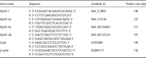
Real-time quantitative PCR was performed in a 7900HT real-time PCR system (384-cell standard block) (Applied Biosystems) using SYBR Select Master Mix (Applied Biosystems) and TaqMan MicroRNA Assay kit (Applied Biosystems) for the detection of mRNA and miRNA, respectively. The gene-specific primers used are listed in the Table 1. The PCR cycling conditions for mRNA detection were 45 cycles at 95°C for 15 s and 60°C for 30 s. Melting curve analysis was performed at the end of each PCR run to confirm amplicon specificity. The PCR cycling conditions for miRNA detection were 40 cycles at 95°C for 15 s and 60°C for 1 min. Relative mRNA and miRNA expression was quantified using the comparative Ct method with β-actin and U6 snRNA (Applied Biosystems) as an endogenous control, respectively (Livak and Schmittgen Reference Livak and Schmittgen2001).
Primers used for real-time quantitative PCR.

Statistical analysis
Data expressed as mean±SE were compared by one-way ANOVA followed by Tukey’s tests (SPSS Inc., Chicago, IL, USA). P<0.05 was considered to be statistical significant.
Results
Cloning and sequence analysis of porcine Sox6 3'UTR
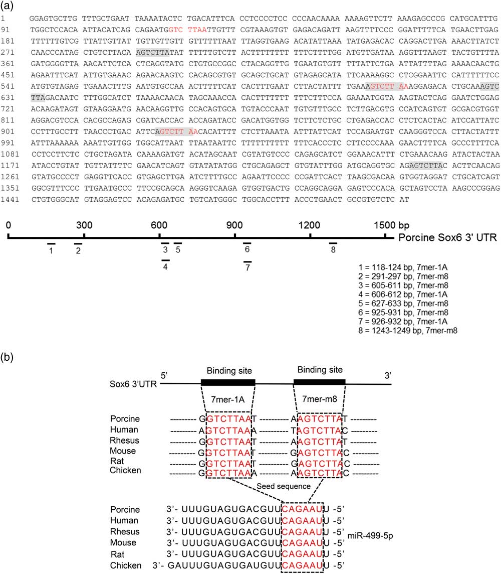
A length of 2012-bp mRNA was amplified from skeletal muscle of a DLY pig and successfully inserted into pMD19-T vector. The resulting plasmid pMD19-T-pSox6-3'UTR was confirmed by colony PCR and DNA sequencing. DNA sequencing showed that the amplified fragment contains a 500-bp pSox6 CDS and a 1512-bp pSox6 3'UTR. This nucleotide sequence has been deposited in GenBank under the accession number KJ123704. Eight potential binding sites for miR-499-5p with three for 7mer-1A and five for 7mer-m8 were predicted at pSox6 3'UTR (Figure 1a), indicating that pSox6 may be a target of miR-499-5p. In addition, the potential miR-499-5p binding sites in Sox6 3'UTR and miR-499-5p sequence were broadly conserved among different species, which implied that the regulation is possibly a universal model (Figure 1b).
Sequence analysis of the binding sites between miR-499-5p and Sox6 3'UTR. (a) The sequence of 1512-bp pSox6 3'UTR and the eight potential binding sites for miR-499-5p. 7mer-1A and 7mer-m8 sites are highlighted in red and shadow, respectively. (b) The potential miR-499-5p binding sites in Sox6 3'UTR and miR-499-5p sequence are highly conserved across different species. 7mer-1A and 7mer-m8 are the most common kinds of binding sites for Sox6 3'UTR (red) that complemented with the seed sequence (red) of miR-499-5p.

The 3'UTR of porcine Sox6 is targeted by miR-499-5p
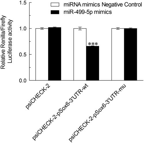
To examine whether pSox6 3'UTR can be directly targeted by miR-499-5p, the partial (276 bp) wild-type 3'UTR of pSox6 containing miR-499-5p binding site or the mutant 3'UTR with 7 bp mutation in seed region was cloned downstream of the luciferase gene and assayed in HEK 293 T cells. As shown in Figure 2, a statistical significant decrease (34%) was observed in luciferase activity when miR-499-5p mimics and the wild-type pSox6 3'UTR reporter were cotransfected into HEK 293 T cells. While cotransfection of miR-499-5p mimics with the mutant pSox6 3'UTR reporter did not result in reduction in luciferase activity (Figure 2), indicating that pSox6 is a target of miR-499-5p.
The result of luciferase reporter assay. Porcine Sox6 3'UTR reporter, its miR-499-5p site mutant, or empty vector was cotransfected with either miR-499-5p mimics or miRNA mimics Negative Control into HEK 293 T cells, followed by cell lysis and luciferase assays 48 h later. Data were presented as mean±SE (n=3). ***P<0.001.

miR-499-5p level inverse correlates with porcine Sox6 mRNA expression in extensor digitorum longus and Soleus muscles
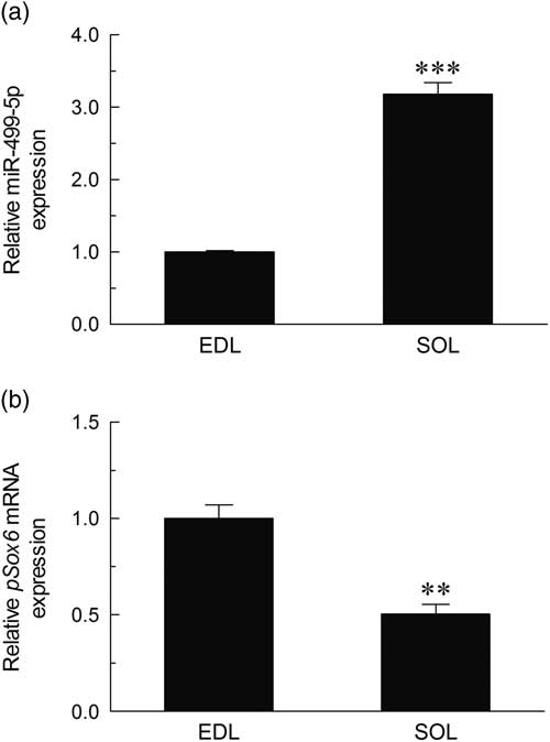
The expression profiles of miR-499-5p and pSox6 mRNA in EDL (fast) and SOL (slow) muscles of pigs were detected by real-time quantitative PCR. As shown in Figure 3a, the expression of miR-499-5p in SOL muscle is significant higher (more than three folds, P<0.001) than that in EDL muscle. On the contrary, the amount of pSox6 mRNA is more abundant in EDL, nearly two folds (P<0.01) than that in SOL muscle (Figure 3b). Obviously, miR-499-5p and pSox6 are inversely correlated.
Relative expressions of miR-499-5p and pSox6 mRNA in EDL and SOL muscles. Total RNA from EDL and SOL muscles of three healthy pigs was used to perform the real-time quantitative PCR. Samples were performed in duplicate. The amount of miR-499-5p (a) and pSox6 mRNA (b) were normalized to the amount of U6 snRNA or porcine β-actin mRNA, respectively. Data were the mean and standard errors (n=3). **P<0.01; ***P<0.001. EDL=extensor digitorum longus muscle; SOL=Soleus muscle.

Overexpression of miR-499-5p in porcine satellite cells promotes the formation of oxidative myofiber by downregulating porcine Sox6 expression
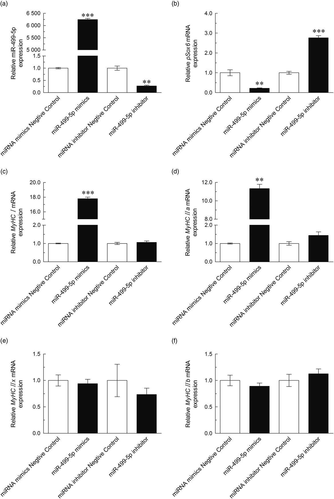
As shown in Figure 4a, compared to the cells transfected with miRNA mimics Negative Control, miR-499-5p mimics transfected cells had an extremely significant increase (more than 6200 folds, P<0.001) in miR-499-5p expression. While the miR-499-5p expression in cells transfected with miR-499-5p inhibitor was only a quarter of that in the miRNA inhibitor Negative Control transfected cells. This result proved that the transfection of miR-499-5p mimics and inhibitor in porcine satellite cells is really effective. As for the expression of pSox6 mRNA, it is just the other way around (Figure 4b), which exactly validated that the miR-499-5p and pSox6 mRNA are inversely correlated in myotubes derived from porcine satellite cells. As shown in Figure 4c and d, the expressions of MyHC I and MyHC IIa mRNA were markedly increased in miR-499-5p mimics transfected cells. However, neither miR-499-5p mimics nor miR-499-5p inhibitor had an effect (P>0.05) on MyHC IIx and MyHC IIb mRNA levels (Figure 4e and f). These data suggested that miR-499-5p might facilitate the formation of oxidative myofiber by decreasing the pSox6 expression.
Effects of miR-499-5p on the mRNA expressions of pSox6 and myosin heavy chain isoforms in porcine satellite cells. miR-499-5p mimics, miRNA mimics Negative Control, miR-499-5p inhibitor or miRNA inhibitor Negative Control was transfected, respectively, into porcine satellite cells. The miR-499-5p (a), pSox6 mRNA (b), MyHC I mRNA (c), MyHC IIa mRNA (d), MyHC IIx mRNA (e) and MyHC IIb mRNA (f) levels were detected by real-time quantitative PCR. The amount of miR-499-5p was normalized to the amount of U6 snRNA. The amount of porcine Sox6, MyHC I, MyHC IIa, MyHC IIx and MyHC IIb mRNA were normalized to the amount of porcine β-actin mRNA. Data were presented as mean±SE (n=3). **P<0.01; ***P<0.001.

Discussion
MyomiRs refer to those miRNAs that can express specifically in heart or skeletal muscle. MiR-208a, miR-208b and miR-499-5p belong to the myomiRs family, and are encoded by the corresponding intron of α-MHC (Myh6), β-MHC (Myh7) and Myh7b gene, respectively. These three miRNAs have a high homology, and van Rooij et al. (Reference van Rooij, Liu and Olson2008 and Reference van Rooij, Quiat, Johnson, Sutherland, Qi, Richardson, Kelm and Olson2009) have demonstrated that they all have the ability to regulate myosin switching and myofiber identity. In heart, genetic deletion of miR-208a resulted in the expression of fast skeletal muscle genes, such as fast troponins Tnnt3 and Tnni2 (van Rooij et al., Reference van Rooij, Sutherland, Qi, Richardson, Hill and Olson2007), and transgenic expression of miR-499-5p at the absence of miR-208a could replace the miR-208a to inhibit the Tnnt3 and Tnni2 expression (van Rooij et al., Reference van Rooij, Quiat, Johnson, Sutherland, Qi, Richardson, Kelm and Olson2009). In mice skeletal muscle, deletion of miR-208b and miR-499-5p caused a substantial loss of type I myofiber (van Rooij et al., Reference van Rooij, Quiat, Johnson, Sutherland, Qi, Richardson, Kelm and Olson2009). These results indicate that miR-208a, miR-208b and miR-499-5p are beneficial to the formation of slow-twitch fibers. In the present study, we also obtained a similar result that overexpression of miR-499-5p in porcine satellite cells led to a significant increase of MyHC I and MyHC IIa mRNA, implying that miR-499-5p plays a vital role in myofiber specification in pigs. However, when inhibiting the expression of miR-499-5p, the mRNA levels of four kinds of MyHC isoforms did not have significant changes, which is consistent with the result that deletion of either miR-208b or miR-499-5p in mice skeletal muscle did not affect the number of type I myofiber and verified the fact that the function of miR-208b and miR-499-5p in regulating myofiber specification is highly correlated (van Rooij et al., Reference van Rooij, Quiat, Johnson, Sutherland, Qi, Richardson, Kelm and Olson2009).
Sox6, first isolated from an adult mouse testis complementary DNA library (Connor et al., Reference Connor, Wright, Denny, Koopman and Ashworth1995), is an important transcription factor and has been proven to play an important role in embryonic muscle development and adult fast myofiber maintenance (Hagiwara et al., Reference Hagiwara, Ma and Ly2005 and Reference Hagiwara, Yeh and Liu2007; von Hofsten et al., Reference von Hofsten, Elworthy, Gilchrist, Smith, Wardle and Ingham2008; Quiat et al., Reference Quiat, Voelker, Pei, Grishin, Grange, Bassel-Duby and Olson2011; Wen et al., Reference Wen, Chen, Chen, Yu, Luo and Huang2016). In this study, we analyzed the levels of pSox6 mRNA in EDL and SOL muscles, and found that the expression of pSox6 mRNA in EDL muscle is nearly the double of that in SOL muscle, which is consistent with the previous studies that Sox6 is highly expressed in fast-twitch skeletal muscles (Cohen-Barak, et al., Reference Cohen-Barak, Hagiwara, Arlt, Horton and Brilliant2001; Hagiwara et al., Reference Hagiwara, Ma and Ly2005; Nachtigall et al., 2015). Up to now, Sox6 has been identified as a direct miR-499-5p target in rat (McCarthy et al., Reference McCarthy, Esser, Peterson and Dupont-Versteegden2009; Jia et al., Reference Jia, Wang, Shi, Liu, Wang, Tian, Lu, Chen, Ma and Zhou2016) mouse (Bell et al., Reference Bell, Buvoli and Leinwand2010; Li et al., Reference Li, Wang, Jia, Cui, Zhang, Wang, Chen, Ma and Zhou2013) and human (Hosoda et al., Reference Hosoda, Zheng, Cabral-da-Silva, Sanada, Ide-Iwata, Ogórek, Ferreira-Martins, Arranto, D’Amario, del Monte, Urbanek, D’Alessandro, Michler, Anversa, Rota, Kajstura and Leri2011). In the present study, we cloned a pSox6 3'UTR with the length of 1512-bp, and found it contains eight potential binding sites for miR-499-5p. At first we structured a vector that containing this 1512-bp pSox6 3'UTR and cotransfected it with miR-499-5p mimics into HEK 293 T cells, resulting in a 72% reduction of luciferase activity (P<0.01). But when cotransfecting miR-499-5p mimics with the mutant vector that contains mutations of all the eight potential miR-499-5p binding sites, the luciferase activity decreased 70% (P<0.01), which had no difference with the wild group and implied that it may contain undetected target sites of miR-499-5p in the 1512-bp pSox6 3'UTR. Then we inserted a 276-bp fragment which encompassing the first potential miR-499-5p binding site into the luciferase reporter vector. By analyzing the luciferase activity, we confirmed that pSox6 is indeed a target of miR-499-5p and the first potential miR-499-5p binding site (position 118 to 124 bp), which is highly conserved across different species, is a direct target of miR-499-5p. We also found that the expression of pSox6 was down regulated by miR-499-5p. Combined with the results above, we suggest a model that in porcine satellite cells miR-499-5p can lower the expression of pSox6 to facilitate oxidative myofiber formation. In summary, we elucidated for the first time that pSox6 is a target of miR-499-5p in pig and the data is consistent with a role for miR-499-5p in regulating porcine myofiber specification.
Acknowledgments
This work was supported by the National Natural Science Foundation of China (No. 31472110).