Introduction
Congenital ductus arteriosus aneurysm is defined as a focal saccular or diffuse tubular dilatation of the ductus arteriosus. Although the true incidence is unknown due to the high rate of asymptomatic patients and the possibility of spontaneous regression, it has been reported as between 1.5 and 8.8% in echocardiography series and in 0.8% of neonatal autopsies. Reference Falcone, Perloff and Roberts1–Reference Tseng and Jan4
The exact mechanism for aneurysm formation is unknown, but some theories have been proposed regarding the pathogenesis. Suggested hypotheses include: ductal wall weakening due to abnormal elastin expression or structure, reduced intimal cushions or extracellular matrix deposition, greater in utero curvature of the ductus, variation in natural hormonal processes (e.g. induction of labour with prostaglandin E2), and maternal risk factors (e.g. Diabetes mellitus). Reference Dyamenahalli, Smallhorn and Geva2,Reference Tseng and Jan4–Reference Dalldorf, Stern and Ezon9 Associated connective tissue diseases (Marfan syndrome, Ehlers-Danlos syndrome, etc) with ductus arteriosus aneurysm support the hypothesis of a ductal wall abnormality. Reference Dyamenahalli, Smallhorn and Geva2,Reference Morgan, Yim, Hayes, Martin, Hamilton and Stuart7–Reference Bannan, Aly, Yoo, Seed and Lam8
The most important diagnostic tool is echocardiography, and ductus arteriosus aneurysm is often detected incidentally during an examination performed for reasons such as a murmur or respiratory distress in the neonatal period. Computed tomography angiography or magnetic resonance imaging/angiography (MRI/MRA) may be helpful in detecting ductus arteriosus aneurysm and the presence of complications. Reference Dyamenahalli, Smallhorn and Geva2,Reference Lund, Jensen and Hjelms5,Reference Bannan, Aly, Yoo, Seed and Lam8
Most cases are asymptomatic and regress spontaneously, but complications have also been reported, Reference Jan, Hwang, Fu, Chai and Chi3,Reference Bannan, Aly, Yoo, Seed and Lam8,Reference Dalldorf, Stern and Ezon9–Reference Yukimoto, Okuma, Sohgawa, Nakano, Shimono and Miki11 such as thromboembolism, spontaneous rupture, erosion, infection, compression of airways, and even death. Reference Dyamenahalli, Smallhorn and Geva2,Reference Lund, Jensen and Hjelms5,Reference Hornung, Nicholson, Nunn and Hawker10,Reference Yukimoto, Okuma, Sohgawa, Nakano, Shimono and Miki11
In this study, we aimed to discuss the course and clinical features of patients with ductus arteriosus aneurysm in newborns.
Methods
A total of 22 newborns with echocardiographic images recorded in the hospital registry system were identified to have ductus arteriosus aneurysm between January 2019 and June 2024. The demographic, clinical, echocardiographic parameters and outcome data were extracted, including: sex, gestational age, delivery type, birth weight, APGAR score, maternal age, maternal diseases, family history (especially genetic and connective tissue diseases), indication for echocardiography, associated cardiac and extracardiac diseases, diameters of ductus arteriosus aneurysm, presence of complications, duration until closure/regression of aneurysm.
Echocardiographic studies were performed with commercially available standard ultrasound scanners (Philips iE 33, Philips Ultrasound LLC) using S8-3 transducer. The diagnosis of ductus arteriosus aneurysm was made based on the features previously described: saccular dilation of the ductus arteriosus with a maximal internal diameter equal to or larger than the adjacent transverse arch or descending aorta, an unusual ductal shunt jet, elongated ductal bump with terminal aneurysmal dilatation, and the configuration of the ascending aorta, main pulmonary artery and ductus arteriosus aneurysm in the high parasternal short-axis view (Figure 1). Reference Jan, Hwang, Fu, Chai and Chi3 Measurements from the echocardiography records included the maximal length (vertical) and width (horizontal) of the aneurysm, and dimensions at the aortic and pulmonary ends (Figure 1a). Data were analysed with a statistical programme and variables were given as mean±standard deviation, minimum and maximum and percentages. The study was approved by the local scientific committee. Since it was a retrospective study, informed family consent form could not be obtained and was judged not to be necessary.
Echocardiographic indicators of DAA: ( a ) Saccular dilation of DA; with a maximal internal diameter equal to or larger than the adjacent transverse arch or descending aorta, ( b ) Unusual ductal shunt jet: horizontal or left-low quadrant ductal shunt from the high parasternal short-axis view, ( c ) “Rabbit ear sign”: the right-side ear is the transverse arch of the aorta and descending aorta, and the left-side ear is the elongated ductal bump with terminal aneurysmal dilatation in the modified high parasternal short-axis view, ( d ) “Triple star sign”: the combination of the ascending aorta, main pulmonary artery and DAA in the high parasternal short-axis view. DAA = ductus arteriosus aneurysm, DAo = descending aorta, AAo = ascending aorta, Ao = aorta, MPA = main pulmonary artery.

Results
All patients were diagnosed in the postnatal period without an antenatal diagnosis of ductus arteriosus aneurysm. Of the 22 newborns, 13 were female. The mode of delivery was C/S in 11 patients and normal vaginal delivery in 11 patients, with a mean gestational age and weight at birth of 386/7 ± 15/7 (353/7 – 406/7) weeks and 3203 ± 469 (2445–4080) grams, respectively. Among the newborns, 15 were classified as appropriate for gestational age, five were large for gestational age, and two were small for gestational age. The mean 1st and 5th minute APGAR scores were 8.5 (6–9) and 9.8 (9–10).
Mean maternal age at the time of delivery was 27.9 ± 5.4 years (19–41). Maternal history was positive for gestational diabetes mellitus in five, hypertension in one, and early membrane rupture in one. Family history for connective tissue diseases was negative in all patients. The blood type distribution of both mothers and babies was the same as follows: 50% Type-A, 23% Type-0, 18% Type-B, and 9% Type-AB.
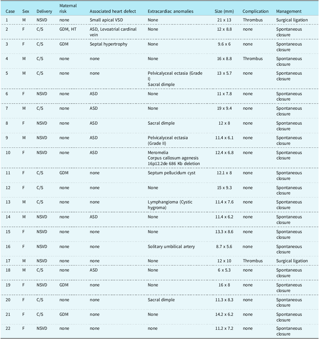
In eight of the newborns, extracardiac anomalies were present, as detailed in Table 1.
The demographic and clinical findings of the cases and measurements of DAA

*NSVD = Normal spontaneous vaginal delivery, C/S = Caesarean section, GDM = Gestational diabetes mellitus, HT = hypertension, EMR = early membrane rupture, ASD = atrial septal defect, VSD = ventricular septal defect.
Echocardiography was performed within 24 hours in 18 and 24–48 hours in 4 neonates. Indications for echocardiography were: murmur in 10, respiratory distress in three, family history or maternal disease in three, mild cyanosis in two, small or large for gestational age in two, solitary umbilical artery in one, and pericardial effusion in fetal ultrasound in one.
Ductus arteriosus aneurysm was an isolated finding in 12 of 22 newborns. Associated cardiac findings were present in 10: small atrial septal defect in eight, and apical muscular ventricular septal defect, levoatrial cardinal vein and septal hypertrophy in one case each. None of the associated cardiac diseases were haemodynamically important. According to measurements from the echocardiographic views: the mean maximal length, width, aortic end and pulmonary end diameters were: 12.78 ± 3.2 mm (6–21 mm), 7.8 ± 1.8 mm (5.3–13 mm), 5.13 ± 1.33 mm (2.3–7.2), and 2.18 ± 0.72 mm (1.3–3.6 mm), respectively.
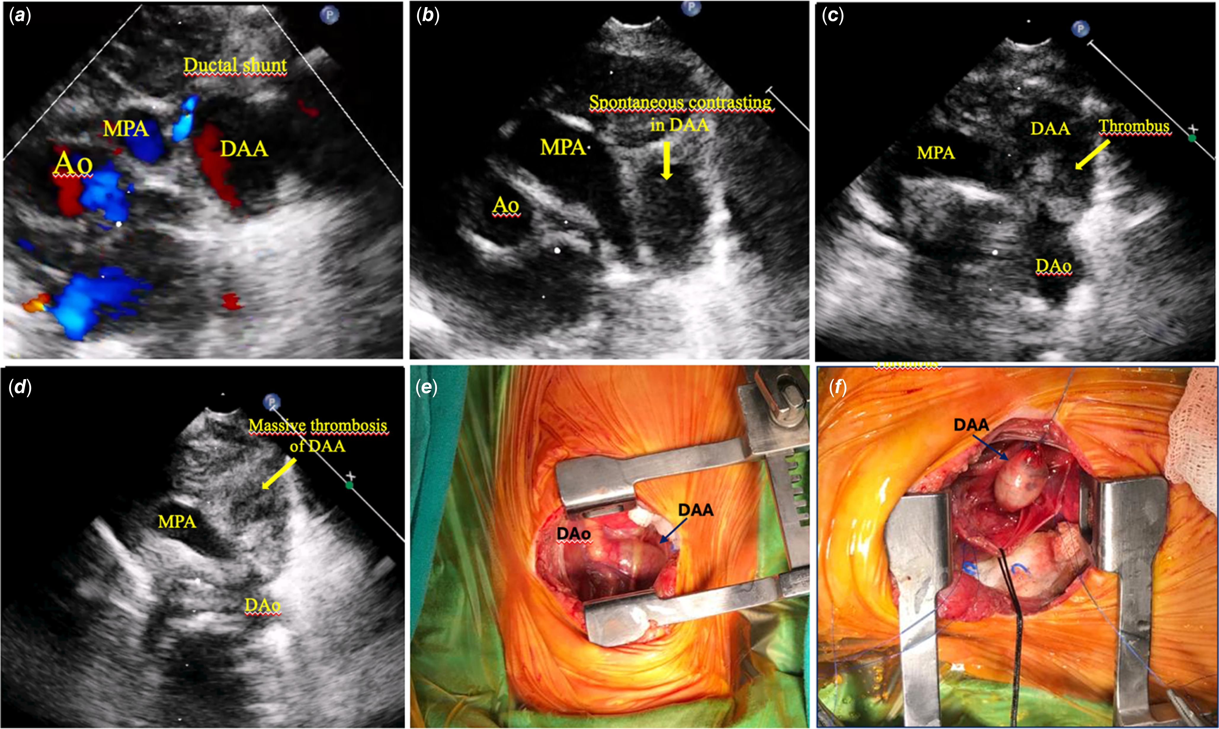
Serial follow-up echocardiograms were obtained until the ductus had closed. In the follow-up, regression of the aneurysm was seen at an average of 5.3 ± 2.7 days in 19 patients without any complication. Thrombus formation and spontaneous contrast enhancement were detected in three patients on the 3rd day of life, in one of whom there was regression and resolution by the 9th day. (Supplemental Mov.1). Two of these patients needed surgical resection of the ductus arteriosus aneurysm because of extension of thrombus to the aortic side without evidence of aneurysmal constriction on the 5th and 8th days. Resection of the aneurysm was performed after placing vascular clamps on proximal aortic and distal pulmonary ends through a left lateral thoracotomy. Post-operative unilateral diaphragm paralysis developed as a surgical complication in one patient. None of our patients received medication for ductus arteriosus or aneurysm closure. No death or additional complication was seen during a median follow-up of 3 years (1.7–4.5 years).
The demographic and clinical findings of the cases and measurements of the ductus arteriosus aneurysm are given in Table 1 and detailed summaries of each case in Table 2.
Summary of the characteristics and findings of cases with DAA

*C/S = Caesarean section, NSVD = Normal spontaneous vaginal delivery, GDM = Gestational Diabetes mellitus, HT = Hypertension, ASD = atrial septal defect, VSD = Ventricular septal defect.
Discussion
The true incidence of neonatal ductus arteriosus aneurysm is unknown because of the high rate of asymptomatic cases and spontaneous regression. In a prospective study by Jan et al., the incidence of ductus arteriosus aneurysm in 548 healthy newborns was reported to be 8.8% with a 100% spontaneous regression rate. In that study, echocardiographic screening was performed in the first 12 hours of life, and this may be the reason for relatively high rate. Reference Jan, Hwang, Fu, Chai and Chi3 In our study, all first echocardiography imaging, except four with different indications, was performed by age 24 hours and in 24–48 hours for the rest. We also observed a rapid and high rate of regression on subsequent days in most of the babies. Therefore, especially in babies who have their first echocardiography performed at an early stage, close follow-up and echocardiographic monitoring in the following days seem sufficient.
Although various theories have been proposed for the aetiology of ductus arteriosus aneurysm, the exact mechanism is unknown. Ductal wall abnormality, abnormal elastin expression or structure, reduced intimal cushions, greater in utero curvature of the ductus and prolonged exposure to systolic aortic pressure due to incomplete closure of aortic end are the most common theories. Reference Dyamenahalli, Smallhorn and Geva2,Reference Tseng and Jan4–Reference Dalldorf, Stern and Ezon9 In all of our cases, we observed that the pulmonary side of ductus arteriosus constricted to some extent; however, the aortic side remained wider. This may support continuing flow, systolic aortic pressure exposure and gradual enlargement of a ductus aneurysm. We also observed that in the case of ductus arteriosus aneurysm, the pulmonary side of the ductus coursed slightly downward and connected to the pulmonary artery at a higher point; this may lead to pressure exposure of the ductal wall rather than the pulmonary end (Figure 1b).
Maternal gestational diabetes mellitus and higher birth weight have been reported as risk factors for ductus arteriosus aneurysm. Reference Dyamenahalli, Smallhorn and Geva2,Reference Bannan, Aly, Yoo, Seed and Lam8,Reference Dalldorf, Stern and Ezon9 Gestational diabetes mellitus was present in five (22.7%) of our cases (three were large for gestational age), and all of them had complete regression of ductus arteriosus aneurysm without any delay in closure time (4–6 days). Dalldorf et al. [9] reported that 15% of fetal ductus arteriosus aneurysm had maternal diabetes. In the prospective study by Jan et al., Reference Jan, Hwang, Fu, Chai and Chi3 10.4% of the cases had maternal diabetes mellitus. They also reported that DAA developed in five (31%) of 16 newborns with maternal diabetes, 11 (24.4%) of 45 newborns who were large for gestational age and in four (80%) of five newborns with both conditions.
Male-to-female ratio in ductus arteriosus aneurysm cases was 0.69 (9/13) in our study and 2.42 (17/7) and 1.4 (28/20) in the series of Dyamenahalli et al. Reference Dyamenahalli, Smallhorn and Geva2 and Jan et al., Reference Jan, Hwang, Fu, Chai and Chi3 respectively. Although the mechanisms are different, our results seem to be consistent with the fact that patent ductus arteriosus is twice as common in girls.
In several studies, maternal blood group A was reported as a risk factor for ductus arteriosus aneurysm. In our study, Type-A was present in 50 % (11/22) in both mothers and babies. Similarly, Jan et al. Reference Jan, Hwang, Fu, Chai and Chi3 found that 47.9% had maternal type-A in their study.
Association with connective tissue diseases (e.g. Marfan syndrome, Ehlers-Danlos syndrome, Loey–Dietz syndrome etc.) has been reported, supporting the theory of ductal wall abnormality. Reference Dyamenahalli, Smallhorn and Geva2,Reference Morgan, Yim, Hayes, Martin, Hamilton and Stuart7,Reference Bannan, Aly, Yoo, Seed and Lam8 Dyamenahalli et al. Reference Dyamenahalli, Smallhorn and Geva2 reported that 6 of 24 patients had associated syndromes (Marfan, Smith–Lemli–Opitz, trisomies 21 and 13 and one possible Ehlers-Danlos). We didn’t investigate our patients for such connective tissue disorders in view of the absence of additional clinical findings during a mean 3-year follow-up. One of our patients had right-hand meromelia, corpus callosum agenesis, and a genetic mutation (16p12.2de 686 Kb deletion), which has been linked to neurodevelopmental delay, weak muscle tone, and heart malformations. In these patients, associated congenital heart disease includes hypoplastic left heart syndrome, ventricular septal defect, tetralogy of Fallot, patent ductus arteriosus, and absence of posterior pericardium. Reference Girirajan, Pizzo, Moeschler, Adam, Feldman and Mirzaa12 Our patient had an additional atrial septal defect, whilst the ductus arteriosus aneurysm regressed spontaneously on the 2nd day of life. Three of our patients had a sacral dimple without any abnormal findings on spinal ultrasonography.
Although the diagnostic criteria used for ductus arteriosus aneurysm are not clear, transthoracic echocardiography is the first-line imaging method for diagnosis and follow-up of neonatal ductus arteriosus aneurysm. Comparison of adjacent transverse arch or descending aorta, visualisation of ascending aorta, pulmonary artery and the aneurysm (triple sign) in high parasternal short-axis view, aorta and aneurysm in high parasternal view (rabbit ear sign) and unusual ductal shunting have been described for the diagnosis of ductus arteriosus aneurysm. Reference Jan, Hwang, Fu, Chai and Chi3,Reference Tseng and Jan4,Reference Bannan, Aly, Yoo, Seed and Lam8,Reference Dalldorf, Stern and Ezon9 The most important alerting echocardiographic findings in our cases were: saccular dilation and a narrowed ductal shunt from a higher-than-expected level, running downward before the pulmonary connection. In serial echocardiographic monitoring, attention should be paid to the possible development of thrombosis, especially if there is narrowing at the pulmonary end, no reduction in ductus arteriosus aneurysm size, slowing of the flow within the aneurysm, and spontaneous contrast enhancement (Figure 2 and Supplemental Mov.1). Computerised tomography angiography and MRA/MRI are helpful especially in adults and complicated cases. Reference Bannan, Aly, Yoo, Seed and Lam8,Reference Yukimoto, Okuma, Sohgawa, Nakano, Shimono and Miki11 In our study, we performed CT angiography in one patient to evaluate ductus arteriosus aneurysm thrombosis.
Serial echocardiography images for development of thrombosis in DAA from high parasternal views and intraoperative images; ( a ) narrowed pulmonary ending with slowed and swirling motion in aneurysm, ( b ) spontaneous contrast with decreased flow, ( c ) thrombus formation in aneurysm and ( d ) massive thrombosis of aneurysm extending to adjacent structures, ( e and f ) DAA seen during operation. DAA = ductus arteriosus aneurysm, Dao = descending aorta, Aao = ascending aorta, Ao = aorta, MPA = main pulmonary artery.

The different results in the series reported in the literature regarding the clinical features and outcomes of ductus arteriosus aneurysm raise the question of “whether we are over diagnosing or underestimating it.” Several previous fetal and neonatal series have demonstrated a 100% spontaneous regression rate without complications; Reference Jan, Hwang, Fu, Chai and Chi3,Reference Dalldorf, Stern and Ezon9 however, some serious complications have also been reported. Reference Dyamenahalli, Smallhorn and Geva2,Reference Lund, Jensen and Hjelms5 Previously reported complications of ductus arteriosus aneurysm include thromboembolism, rupture of the aneurysm, compression of adjacent structures (airway, nerve etc..), infection, erosion, and even death. These differing outcomes may be due to the criteria used for diagnosis of ductus arteriosus aneurysm or the age group of the patients. The incidence is high in the neonatal period, and complications are rare, but clinical consequences are more likely if ductus arteriosus aneurysm persists into adulthood. In our study, we detected extensive thrombosis of the ductus arteriosus aneurysm in three (13.6%) of the 22 newborns, two (9%) of whom required surgical excision. Dyamenahalli et al. Reference Dyamenahalli, Smallhorn and Geva2 reported that four of 24 patients had ductus arteriosus aneurysm-related symptoms, nine 9 had thrombus within the aneurysm, three had complications related to ductus arteriosus aneurysm thrombus extension into the pulmonary artery, spontaneous rupture, and an asymptomatic cerebral infarction. They reported a 4-month-old case who died of ductus arteriosus aneurysm rupture from the aortic side on autopsy. Reference Dyamenahalli, Smallhorn and Geva2
As in our cases, large thromboses developing without ductal constriction may create a mass effect and complications at an older age, and when a surgical or interventional procedure may be problematic due to the fragile nature of the aneurysm. Reference Yukimoto, Okuma, Sohgawa, Nakano, Shimono and Miki11,Reference Ise, Akasaka, Kamiya and Otani13 A thrombosed aneurysm without a ductal shunt from the aortic side may lead to a missed diagnosis of ductus arteriosus aneurysm with echocardiography. In adult cases, the diagnosis of ductus arteriosus aneurysm is often an incidental finding on CT or MRI rather than echocardiography.
Once detected in a neonate, clinical follow-up with serial echocardiography is often sufficient. Spontaneous regression within 6–12 weeks is the most common scenario reported in up to 100% in several series. Reference Jan, Hwang, Fu, Chai and Chi3,Reference Dalldorf, Stern and Ezon9 However, in the case of a ductus arteriosus aneurysm that fails to regress after 2 months, the presence of complications such as extending thrombosis, laryngeal nerve or airway obstruction, and rupture of aneurysm, endovascular (with emerging options for transcatheter stents and closure devices) or surgical intervention should be considered. Reference Dyamenahalli, Smallhorn and Geva2,Reference Lund, Jensen and Hjelms5,Reference Morgan, Yim, Hayes, Martin, Hamilton and Stuart7,Reference Bannan, Aly, Yoo, Seed and Lam8,Reference Doğan, Aksoy, Sayıcı and Çitli14
In our study, two patients were treated by surgical excision of the thrombus and aneurysm resection. Although thrombosis may be part of the normal process of spontaneous regression and aneurysm closure, the presence of a large aneurysm with thrombus extension into the descending aorta, led to surgical excision in our cases. In one patient, the thrombus and aneurysm resolved without an intervention. Post-operative unilateral diaphragm paralysis developed in 1 patient, requiring plication. Dyamenahalli et al. Reference Dyamenahalli, Smallhorn and Geva2 reported that six of 24 patients underwent surgical resection for ductal patency, ductus arteriosus aneurysm size or thrombus extension.
Interventional endovascular transcatheter closure using stent grafts or occlusion devices is potentially emerging new treatment option, especially for adults and high surgical risk patients. Reference Bannan, Aly, Yoo, Seed and Lam8,Reference Yukimoto, Okuma, Sohgawa, Nakano, Shimono and Miki11,Reference Doğan, Aksoy, Sayıcı and Çitli14 In a study involving eight adult ductus arteriosus aneurysm cases, four were treated with total arch replacement and 2 with thoracic endovascular repair. Reference Yukimoto, Okuma, Sohgawa, Nakano, Shimono and Miki11 Ise et al. Reference Doğan, Aksoy, Sayıcı and Çitli14 reported a 59-year-old case with an infected ductus arteriosus aneurysm treated by endovascular repair with a stent graft.
Conclusion
With increased awareness and earlier and more frequent application of echocardiography in newborns, the diagnosis of ductus arteriosus aneurysm is being reported more frequently. The most common scenario in these patients is spontaneous regression; however, it should not be forgotten that ductus arteriosus aneurysm can be associated with serious complications in both childhood and adulthood. In selected cases, invasive treatment may be an option to prevent more serious or late complications, although the indications for this remain unclear.
Supplementary material
The supplementary material for this article can be found at https://doi.org/10.1017/S1047951125101182.
Author contributions
VD: study concepts/study design, writing and data analysis, IUS: data acquisition or data analysis/interpretation, BK, ONA, KS: literature research, All authors: manuscript drafting or manuscript revision for important intellectual content, approval of final version of manuscript.
Financial support
The authors have no relevant financial or non-financial interests to disclose.
Competing interests
The authors have no conflicts of interest to declare that are relevant to the content of this article.