Introduction
A chronic discharging ear is still one of the commonest problems encountered by the otorhinolaryngologist in developing countries. Canal wall down mastoidectomy is the standard procedure for squamosal disease.
In canal wall down mastoidectomy, complete disease clearance is possible. However, this can lead to post-operative cavity problems and considerable hearing loss. In large mastoid cavities, epithelialisation and healing are particular problems and can be delayed. The most commonly encountered cavity problems include persistent or recurrent discharge and infection. This study aimed to reduce the healing time of the mastoid cavity after mastoidectomy with a simple technique of using fascia with a skin graft.
Materials and methods
This was a non-randomised controlled pilot trial with 2 groups of 30 patients in a tertiary referral teaching hospital. The study was completed in a span of 1.5 years after clearance was obtained from the institutional ethical review board (number: 447/KIMS/PGs/SYN/2017-18).
After meeting the eligibility criteria, the patients were allotted to either the case or the control group alternately. Full consent of the patients was obtained after an explanation of the procedures and their consequences was given.
Group A constituted the ‘cases’ that underwent placement of temporalis fascia with free skin graft in the mastoid cavity after canal wall down mastoidectomy. Group B constituted ‘controls’ who did not undergo placement of anything. Every eligible consecutive patient in the age group of 6–60 years with squamosal type chronic otitis media without complication confirmed by high-resolution computed tomography (CT) of temporal bone and who underwent canal wall down mastoidectomy was recruited into the study. Immunocompromised patients and those undergoing revision surgery were excluded from the study.
Surgical protocol and methodology
All patients underwent a standard canal wall down mastoidectomy through a postauricular approach. All visualised pathology, including cholesteatoma, granulations and unhealthy mucosa, were removed, and mastoidectomy with saucerisation of mastoid cavity, lowering of the facial ridge and adequate meatoplasty were performed.
In the cases group, temporalis fascia was harvested and then spread over the bare bone to cover the entire surface of the dissected mastoid cavity. Three pieces of free skin graft of size 2 × 0.25 cm and 0.02 to 0.03 cm thickness were harvested from the postauricular region at the site of the incision. These were spread and teased on a hard surface to denude the subcutaneous tissue. They were then placed carefully over the fascia along the margins of the cavity. This was further reinforced with pieces of gelfoam (absorbable gelatin sponge; Virchow Biotech Private, Andhra Pradesh, India) soaked in a nanocrystalline-silver gel (Meganano® containing 0.02 mg of silver nanoparticles per gram) to prevent displacement and migration of the fascia or skin pieces. Finally, a povidone-iodine tulle (Povi-10TM containing povidone-iodine IP 10 per cent w/w and Ornidazole IP 1 per cent w/w) piece was kept as a drain from the mastoid cavity to the external ear canal through the meatoplasty.
In the control group, the dissected bone was left bare, with no fascia or skin graft. However, the cavity was packed with the same gel-soaked gelfoam and a tulle drain was also kept. Based on the status of the ossicular chain, reconstruction of the middle ear was undertaken accordingly in both the groups. Postauricular wound sutures were removed on post-operative day 7 and the patient was discharged. Antibiotics were given for 14 days depending on the pre-operative ear swab report. The external auditory canal pack was removed on post-operative day 21. Follow up was six months, and data were recorded at the end of the first, third and sixth month.
At the end of the first, third and sixth month, cavities were assessed endoscopically and scored based on five criteria, as follows: dry (1) or wet (0) cavity; presence (0) or absence (1) of bacterial and fungal flora; presence (1) or absence (0) of wax; and major (1) or minor (0) epithelialisation. The maximum score was 5; cavities with a score of three or above were considered healed.
Epithelialisation was assessed by endoscopic examination in the following areas of the mastoid cavity: the tegmen plate, the sinus plate, the medial wall of epitympanum, the medial wall of the mastoid cavity and the floor of the mastoid cavity (each 20 per cent). If epithelialisation was found to be in four or more areas, it was considered to be major.
A swab was taken from all the cavities that were found wet during the follow-up period and sent for analysis of bacterial and fungal flora. If they were found to be positive, they were treated accordingly.
The data were analysed with a chi-square test and crude relative risk. The difference in mean values between cases and controls were compared using the independent t-test. A p-value less than 0.05 was considered statistically significant.
Results
The majority of the patients were under 30 years of age. Twenty (66 per cent) patients in the cases group belonged to the below 30 years age group whereas in the control group it was 22 patients (73.3 per cent). In the 31–50 years age group, 9 (30 per cent) and 6 (20 per cent) participants belonged to the case and control groups, respectively. In the 51–70 years age group, only 1 participant (3.3 per cent) from the cases group and 2 participants (6.6 per cent) from the control group belonged to this age group. In the cases group, 22 participants (73.3 per cent) were male and 8 (26.6 per cent) were female. Among the control group, 19 (63.3 per cent) patients were male and 11 (36.6 per cent) patients were female.
Analysis of healed cases and controls
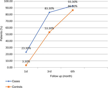
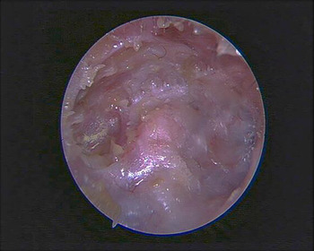
Table 1 and Figure 1 show the number of patients who had successfully healed mastoid cavities at the end of first month, third month and sixth month during post-operative follow up. Figure 2 shows the endoscopic view of a healed mastoid cavity in group A at three months after surgery. It was found that healing in the cases group was faster when compared with the control group.
Graph showing the percentage of healed cases and controls in the months of follow up.

Endoscopic view of a healed mastoid cavity in group A (cases group), three months after surgery.

Number of cases and controls that healed in the follow up

Analysis of unhealed cases and controls
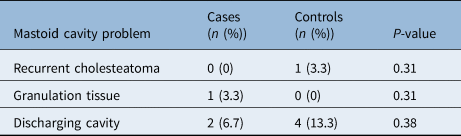
The cavity was considered to be unhealed if it did not dry up and heal in six months and showed the presence of discharge, cholesteatoma and or granulation tissue. It was found that there was no increase in the failure rate nor were there any new complications in the cases group where the application of skin graft with fascia was performed (Table 2).
Analysis of the unhealed mastoid cavities

Analysis of dry and wet cavity status
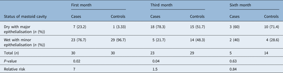
Participants were analysed based on a dry or wet cavity at the end of the first, third and sixth month follow-up period. It was found that the number of dry cavities in the cases group was far greater than in the control group, and it was found to be statistically significant at the end of the first and third month follow-up periods (Table 3). The number of patients in both cases and controls at the third month follow up excluded the dry cases of the first month.
Percentage of dry and wet cavities at each follow-up month

Analysis of bacterial and fungal swab
Fascia with epithelial grafting had almost no effect on bacterial or fungal growth. Even though the number of patients with either flora was higher among the controls, the difference was found to be insignificant (Table 4).
Percentage of bacterial and fungal growth in cases and controls

Analysis of wax formation
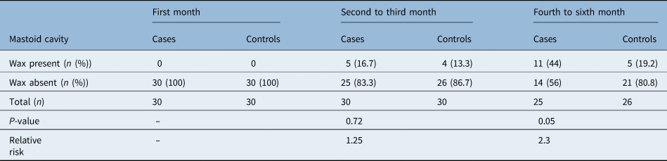
Wax formation was greater in cavities covered with fascia and skin graft. It was observed that the cavities with wax did not discharge in both cases and controls (Table 5). Therefore, wax could be considered as a reliable indicator of the healing process.
Analysis of wax formation at follow up for each month

Analysis of the epithelialisation
It was noted that there were a greater number of patients with a re-epithelialised cavity in group A, and this was also found to be statistically significant (Table 3). The follow up at the sixth month excluded the dry healed cases of both first and third month.
Discussion
In chronic otitis media squamous type, the mainstay of treatment is canal wall down mastoidectomy. However, creating a large cavity could result in problems such as a persistent discharging cavity, collection of debris and wax in the cavity, giddiness after exposure to cold water or air, and unsightly appearance because of a large meatoplasty.Reference Chhapola and Matta1
The main objective after a mastoid surgery is to create a discharge-free dry cavity. Sade et al.Reference Sade, Weinberg, Berco, Brown and Halevy2 stated that small and medium-sized cavities were much more likely to become dry because of a low facial ridge, large meatoplasty and the presence of air in the middle-ear cavity. Eustachian tube function was excluded from analysis.
The most morbid symptom after a canal wall down mastoidectomy is the persistent ear discharge. Several causes have been attributed to the discharging cavity including both mechanical and mucosal factors; they are inadequate meatoplasty, inadequate lowering of a facial ridge or incomplete removal of disease. Apart from these factors, a large mastoid cavity would take a lot more time to epithelialise and heal. These are the cases that would particularly benefit from grafting of temporalis fascia with a free skin graft. The other options are to obliterate such a cavity with a flap, bone chips, cartilage or some biomaterial like hydroxyapatite. Massive cavities with non-functional ears could also be closed with cul-de-sac procedures.
A study conducted by NadolReference Nadol3 found the presence of granulation tissue in the un-exenterated mastoid cells to be the cause of the discharging cavity. Other studies conducted by RamboReference Rambo4 and YoungsReference Youngs5 also found that retained infection and granulation tissue were responsible for continuous otorrhoea. This is in agreement with our study. Canal wall down procedures, despite having a low rate of recurrence, have more than 20 per cent of cases with intermittent or persistent otorrhoea that is resistant to antibiotic therapy.Reference Henatsch, Alsulami, Duijvestijn, Cleutjens, Peutz-Kootstra and Stokroos6 It seems that a major problem of unstable cavities is insufficient (re)epithelialisation of the mastoid cavity; (re)epithelialisation of the mastoid cavity occurs because of favourable local conditions and favourable cavity shape, size and host factors.Reference Henatsch, Alsulami, Duijvestijn, Cleutjens, Peutz-Kootstra and Stokroos6 Tansuker et al.Reference Tansuker, Öğreden, Özyılmaz, Cengiz, Beder and Oktay7 observed that the type of pre-operative pathology had no significant relationship with the epithelialisation time of the mastoid cavity.
Multiple techniques have been tried before to promote the epithelialisation of the mastoid cavity and reduce the complications and after-effects of surgery. A study conducted by Beales and HynesReference Beales and Hynes8 in 1958 used postauricular flap in the mastoid cavity to reduce the size of the mastoid cavity. Their study was based on the principle that raw surfaces of mastoid should not be allowed to heal spontaneously and should be covered with epithelium.
Beales and HynesReference Beales and Hynes8 also demonstrated that skin grafts placed in the mastoid cavity underwent atrophy and disintegration after an initial take and were replaced by granulation tissue which later on became infected leading to discharge. Therefore, skin grafts were not used in that period of time. In more recent years, investigators such as Guilford and Wright,Reference Guilford and Wright9 Cremers and Smeets,Reference Cremers and Smeets10 and many others have rediscovered the use of split-thickness and full thickness graft in the repair of the post-operative external auditory canal and mastoid cavities.
Pinch grafting was described by Takeda et al.,Reference Takeda, Saito, Takeuchi and Komada11 who showed that the secondary application of pinch grafts (multiple small grafts of the epidermal layer) was beneficial for healing of exposed mastoid cavities caused by the use of open technique tympanoplasty or radical mastoidectomy.
A study conducted by Walsh et al.Reference Walsh, Davies, McGlashan and Bowdler12 used the Davis skin graft technique, which uses the epidermis and small amounts of dermis harvested from the anterior abdominal wall for grafting mastoid cavities that underwent revision mastoid surgery and found it to be beneficial. In their study, 87 per cent of patients had a dry cavity at the end of the study period of 15 months, compared with our study where a dry cavity was obtained in 93 per cent of patients by the end of 6 months.
A study conducted by Renton and WetmoreReference Renton and Wetmore13 was similar to our study. In the study by Renton and Wetmore, split-thickness grafting was performed on post-mastoidectomy revision and lateral temporal bone resection cases. Epithelialisation of more than 85 per cent was taken as success. Average time taken for the graft to heal in this study was 2 months, with 5 cases out of 11 obtaining dry cavities within 1 month of revision surgery. When compared with our study, average time taken for healing was 2.8 months, with 7 out of 30 cases obtaining a dry cavity within 1 month of surgery.
A study published by Wetmore et al.Reference Wetmore, Bueller and Cost14 in 2014 used split-thickness graft harvested from the anterior thigh for the grafting of the mastoid cavity in patients undergoing canal wall down tympanomastoidectomy. In this study, graft success was taken as 80 per cent of epithelialisation.
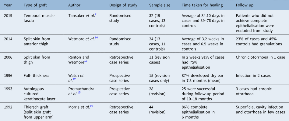
In Table 6, a few studies of the past have been compared. According to these studies, healing was much faster with split-thickness skin graft when compared with full-thickness grafts. However, most of these studies were case series and had a smaller sample size. Complications and after-effects noted in these studies were persistent otorrhoea, presence of granulation tissue, meatal stenosis, recurrence and tympanic membrane perforation. In our study, persistent otorrhoea and the presence of granulation tissue were the commonest after-effects noted.
Previous studies on the role of skin grafts in the healing of the mastoid cavity

It is a well-established fact that obliteration of the mastoid cavity by various flaps leads to a smaller cavity and hence faster healing. However, surgical flap techniques are elaborate, have greater morbidity and are more time-consuming than not using these techniques. Because our procedure did not involve three-dimensional obliteration of the cavity, it offered the advantage of easy endoscopic examination in the event of disease recurrence. Another advantage of free skin grafts when compared with all other studies mentioned in Table 6 is that the graft was obtained from the same postauricular incision. Therefore, the surgical time taken and post-operative morbidity was less.
Immediate application of fascia with skin graft was performed in our study, even though a few previous studies advised that grafts should be applied after 10–14 days.Reference Walsh, Davies, McGlashan and Bowdler12,Reference Premachandra, Woodward, Milton, Sergeant and Fabre15 This avoided an unnecessary second procedure after the discharge of the patients, thereby reducing the cost of the procedure.
Temporalis fascia was expected to serve as a scaffold over the irregular surface of the bone and also provide guidance for the epithelium to grow evenly over the walls of the mastoid cavity. Initially, the graft survived through serum imbibition and graft revascularisation took place later on.Reference Renton and Wetmore13
• Cavity healing after a canal wall down mastoidectomy is a challenge
• Incomplete epithelialisation is one of the causes of delayed healing
• Temporalis fascia superimposed with free skin grafts were used in this study
• This technique promoted healing mainly in the first three months after surgery
• This technique did not affect residual or recurrent disease or the flora of the post-operative discharge
• This technique can be easily adopted after every canal wall down mastoidectomy procedure
Conclusion
The technique of application of temporalis fascia superimposed with pieces of free skin graft in the mastoid cavity after canal wall down mastoidectomy is an easy and economical method to promote and facilitate the healing process. The difference in healing with this technique was obvious in the first three months after surgery. A dry cavity was achieved more commonly with graft than without it in the first three months after surgery. Therefore, this could be performed routinely after every canal wall down mastoidectomy in order to reduce the healing time. The formation of wax was a reliable indicator of the progress of the healing process. However, the grafting had no impact on residual or recurrent disease. Additionally, it did not affect the bacterial or fungal flora of the post-operative discharge. This study had a larger sample size and a control group that differentiates it from the previous studies.
Competing interests
None declared