Introduction
Kefir is a sour fermented milk whose consumption has been associated with an improvement in lactose digestion (Hertzler & Clancy Reference Hertzler and Clancy2003), an antioxidant activity (Liu et al. Reference Liu, Chen and Lin2005) and a modulation of the immune response (Vinderola et al. Reference Vinderola, Perdigón, Duarte, Thangavel, Farnworth and Matar2006; Hong et al. Reference Hong, Chen, Chen and Chen2009). Kefir has also been demonstrated to possess an antimicrobial activity against Escherichia coli, Salmonella typhi, Shigella sonnei, Candida albicans, and Staphylococcus aureus (Garrote et al. Reference Garrote, Abraham and De Antoni2000; Silva et al. Reference Silva, Rodrigues, Filho and Lima2009) and to inhibit Bacillus cereus spore germination (Kakisu et al. Reference Kakisu, Abraham, Pérez and De Antoni2007).
Several strains of Lactobacillus isolated from kefir grains were shown to have antagonistic properties against certain pathogens associated with foodborne diseases: Lb. kefir CIDCA 8321 and CIDCA 8348 adhered to Caco-2 cells and protected cultured epithelial cells against Salmonella invasion (Golowczyc et al. Reference Golowczyc, Mobili, Garrote, Abraham and De Antoni2007); while Lb. plantarum CIDCA 83114 exhibited in-vitro antimicrobial activity against Sal. enterica serovar Thypimurium and Esch. coli, adhered to Caco-2 cells (Golowczyc et al. Reference Golowczyc, Gugliada, Hollmann, Delfederico, Garrote, Abraham, Semorile and De Antoni2008) and decreased the adhesion of Esch. coli O157:H7 strain 69160 to Hep-2 cells so as to protect the cells from damage (Hugo et al. Reference Hugo, Kakisu, De Antoni and Perez2008).
Enterohaemorrhagic Esch. coli is an enteropathogenic bacterium that produces the Shiga toxin (Stx) and is involved in diarrhoea and food-transmitted illnesses. The serotype O157:H7 produces haemorrhagic colitis and the haemolytic-uremic syndrome and is the most prevalent in the epidemic cases of that disease (Banatvala et al. Reference Banatvala, Griffin, Greene, Barrett, Bibb, Green and Wells2001; Rivas et al. Reference Rivas, Miliwebsky, Chinen, Deza and Leotta2006). Shiga toxin is the major virulence factor of enterohaemorrhagic Esch. coli (O'Loughlin & Robins-Browne Reference O'Loughlin and Robins-Browne2001). The Shiga toxin family contains two major, immunologically non-cross-reactive groups referred to as Stx type I and II (Stx1 and Stx2). The Esch. coli strains producing Stx2 are commonly associated with an increased risk of developing the haemolytic-uremic syndrome (Boerlin et al. Reference Boerlin, Mc Ewen, Boerlin-Petzold, Wilson, Johnson and Gyles1999). The Shiga toxin contain two subunits, one A subunit of 32 kDa and a pentamer composed of five identical B subunit of 7·7 kDa each. The B subunits bind to the specific glycolipid globotriaosylceramide present on the plasma membrane of eukaryotic cells. The A subunit exhibits RNA N-glycohydrolase activity and cleaves a specific adenine residue on the 8S ribosomal RNA in the target-cell cytosol, thereby inhibiting protein synthesis by blocking aminoacyl-tRNA binding to the 60S ribosomal subunit (Paton & Paton Reference Paton and Paton1998) and subsequently inducing cell apoptosis (Smith et al. Reference Smith, Kane, Campbell, Acheson, Cochran and Thorpe2003).
The effectiveness of certain probiotic bacteria in combating diarrhoea is most likely based on their ability to protect the host against toxins (Oelschlaeger Reference Oelschlaeger2009). Some studies explain the protective effects of probiotic lactobacilli and bifidobacteria against the Shiga toxin of Esch. coli O157:H7 by a reduction in the bacterium's cytotoxic activity (Kim et al. Reference Kim, Han, Imm, Oh, You, Park and Kim2006), or by a down-regulation of Stx gene expression (Carey et al Reference Carey, Kostrzynska, Ojha and Thompson2008); the latter effect, in turn, being attributed to the elaboration of lactic and acetic acids by the lactobacilli. Alternatively, specific molecules on the bacterial surface, such as certain galactotrehalose copolymers or modified lipopolysaccharides, could bind to Stx and thus exert a neutralising activity (Pinyon et al. Reference Pinyon, Paton, Paton, Botten and Morona2004; Neri et al. Reference Neri, Nagano, Yokoyama, Dohi, Kabayashi, Miura, Inazu, Sugiyama, Nishida and Mori2007).
On the basis of these considerations, the aim of this study was to evaluate the effect of Lb. plantarum CIDCA 83114 in comparison with other lactobacilli isolated from kefir against the action on Vero cells of Stx2-containing supernatants of Esch. coli O157:H7 and to study the action of the Lactobacillus cell walls against the Shiga toxin.
Materials and Methods
Bacterial strains
Lb. plantarum strains CIDCA 83114 and CIDCA 8336 and Lb. kefir strains CIDCA 83113 and CIDCA 8348 were isolated from kefir grains (Garrote et al. Reference Garrote, Abraham and De Antoni2001) and grown in De Man, Rogosa and Sharpe broth (MRS Difco, Sparks, MD, USA) at 30 °C for 24 h. Lb. delbrueckii subsp. bulgaricus strain CIDCA 333 isolated from yoghurt (Abraham et al. Reference Abraham, De Antoni and Añon1993) was grown in MRS broth at 37 °C for 24 h.
Esch. coli O157:H7 strain 69160, a clinical isolate obtained from the Sor María Ludovica Interzonal Hospital (La Plata, Argentina), was grown in tryptic-soy broth (TSB, LW Lab, Córdoba, Argentina) under aerobic conditions at 37 °C for 18 h. The strain 69160 was characterised genetically as positive for Shiga toxin (stx 2+), intimin (eae+) and enterohaemolysin (ehly+).
All strains were stored frozen at −80 °C with 50% (w/v) milk as cryoprotectant and used for experiments in the second passage after thawing in the corresponding media.
Preparation of suspensions of lactobacilli
Ten millilitres of overnight cultures of lactobacilli in MRS were centrifuged at 10 000 g for 10 min and the pellet suspended in the same volume of phosphate buffered saline (PBS) and diluted appropriately in Dulbecco's modified Eagle's medium (DMEM, Gibco BRL Life Technologies, Rockville, MD, USA).
Preparation of spent culture supernatants of Esch. coli 69160
Cultures of Esch. coli, grown in tryptic-soy broth at 37 °C during 18 h, were centrifuged at 10 000 g for 10 min and filtered through 0·45 μm cellulose membranes (Millipore, Bedford, MA, USA) to obtain a spent culture supernatant (SCS).
Surface hydrophobicity of lactobacilli
Two millilitres of bacterial suspension in PBS (109 CFU/ml) were exposed to 0·5 ml n-hexadecane (Baker, Mallinckrodt Inc. NJ, USA) by vortexing for 2 min at 24 °C. The phases were allowed to separate by decantation. The aqueous phase was removed and the optical density (OD) at 550 nm measured. The decrease in the OD of the aqueous phase was considered a measure of the percentage of cell hydrophobicity (H%), which parameter was calculated by the formula H% = [(OD0 − ODt)/OD0] × 100, where OD0 and ODt are the optical densities before and after extraction with n-hexadecane.
Heat inactivation and proteolytic-enzyme treatment of lactobacilli
For heat inactivation, bacterial suspensions in PBS (107 CFU/ml) were incubated in a water bath at 100 °C for 10 min. The proteolytic enzymes were prepared in the appropriate buffer at a concentration of 2·5 mg/ml. Trypsin (Sigma, St. Louis, MO, USA) and α-chymotrypsin (Sigma) were prepared in 50 mm Tris–HCl, 100 mm NaCl; pH 8. These enzymes were inactivated by adding 1:10 (v/v) foetal-bovine serum (PAA Laboratories, GmbH, Pasching, Austria). Pepsin (Sigma) was prepared in 50 mm glycine–HCl, 100 mm NaCl; pH 2·2. This enzyme was inactivated at pH 7 with PBS. Proteinase k (Sigma) was prepared in 0·6 mm Tris/HCl buffer with 0·6 mm EDTA and 0·6% (w/v) sodium dodecylsulphate; pH 8. This enzyme was inactivated by adding phenylmethylsulphonyl fluoride.
For treatment with enzymes, 1 ml bacterial suspension (109 CFU/ml) was centrifuged at 10 000 g for 10 min and the pellet first washed with and then resuspended in 1 ml enzyme solution (2·5 mg/ml). Incubation was for 1 h at 37 °C. After inactivation of the enzyme, the pellet of lactobacilli was washed and suspended in PBS to an OD of 0·08, equivalent to 107 CFU/ml.
Preparation of cell walls
The lactobacilli in 1 L MRS culture were centrifuged at 10 000 g 10 min and washed with PBS. The pellet was lysed mechanically at −20 °C in a French Press XS-17523 (AB Biox, Järfälla, Sweden) by three consecutive disruptions at 100 kN. The suspension of disrupted cells was centrifuged at 10 000 g and 4 °C for 10 min and the supernatant ultra-centrifuged at 35 000 g (TL Optima, Beckmann Instruments Inc., Palo Alto, CA, USA). The resulting pellet was finally washed with PBS to a constant OD at 280 nm. The pellets were suspended in 1 ml PBS (equivalent to 1012 CFU) and stored at −20 °C.
Quantitative determination of haemolytic activity of Esch. coli 69160
Erythrocytes were obtained from rabbit blood after a 1:10 (v/v) dilution with 3·8% (w/v) sodium citrate and storage for 24 h at 4 °C. After 5 min of centrifugation at 250 g, the erythrocytes in the pellet were washed two times and suspended in PBS to obtain a 3% (v/v) erythrocyte suspension.
Haemolysis assays contained supernatants from Esch. Coli 69160 spent cultures serially diluted 1:2 to 1:256 along the above erythrocyte suspension and 0·1 m CaCl2 in a total volume of 200 μl. After incubation at 37 °C for 1 h and centrifugation at 250 g for 5 min, the release of haemoglobin was measured spectrophotometrically by absorbance at 600 nm. A value for total haemolysis was determined by mixing erythrocytes with 30% (v/v) Triton X-100 (Sigma, St. Louis, MO, USA). Haemolytic activity (HA) was expressed as HA% = [ODs − ODnc/ODpc − ODnc] × 100. Where s was the sample, nc was the negative control at no haemolysis and pc was the positive control at total haemolysis.
Cell cultures
Vero cell cultures were grown and maintained as previously reported (Kakisu et al. Reference Kakisu, Irigoyen, Torre, De Antoni and Abraham2011). Cells were inoculated in multiwell-culture plates (Greiner Bio One, Frickenhausen, Germany) with 1 × 10⁵ cells per well, to obtain a proliferation culture, and with 2·5 × 105 cells per well, to obtain a confluent culture.
Quantitative determination of Esch. coli– 69160-supernatant cytotoxicity on Vero cells
Crude supernatants from strain 69160 were prepared from filter-sterilised cultures and used at different dilutions in DMEM to investigate the biological effects of Stx2 on Vero cells.
To evaluate the Stx-inhibitory activity of different lactobacilli or their bacterial cell walls, the spent culture supernatant and a Lactobacillus suspension in PBS (107 CFU of whole bacteria or 50 μl bacterial cell-wall per well, equivalent to 107 CFU/ml), were added to confluent Vero cells in 24- to 48-well plates. The plates were incubated for 48 h. Gentamicin (100 μg/ml) was added in order to inhibit the growth of the lactobacilli during the experiment.
Cytotoxicity was evaluated by two different methods, one measuring the cell viability and the other the cell damage:
a Determination of succinate dehydrogenase mitochondrial activity as a measure of cell viability: The assay employing 3-(4,5-dimethylthiazol-2-yl) 2,5-diphenyl tetrazolium bromide (MTT) cleavage (Mossman, Reference Mosmann1983) was used for measuring cell viability. The MTT was used at a dilution of 5 mg/ml in DMEM without phenol red. This tetrazolium salt is metabolically reduced by viable cells to yield a blue formazan product that is measured spectrophotometrically as the optical density at 550 nm. Vero cells were seeded in the multiwell-culture plates and grown in DMEM with 5% (v/v) foetal-bovine serum. The cells were washed with PBS and exposed to a mixture of 69160 spent culture supernatant along with lactobacilli, or to each one separately. Untreated cells were used as a control in all the experiments. The plates were next kept at 37 °C in 5% (v/v) CO2 under growth-arresting conditions (i.e., in serum-free medium) for 48 h. The cultures were washed with PBS and then incubated in the above MTT solution for 3 h at 37 °C. The blue crystals formed within the cell layer were finally dissolved in 200 μl dimethyl sulphoxide and the optical density of the resulting solution measured spectrophotometrically at 550 nm. The assay was performed in duplicate for each sample. At least, three independent experiments were performed for each condition. The mean value for MTT reduction values was compared with the control to determine percent cell viability. The percentage of viable cells was calculated: ODs/ODc × 100, where ODs was the absorbance of the sample and ODc, the absorbance of the control cells without treatment.
b Determination of lactate dehydrogenase release (LDH) as a measure of cell damage
Extracellular lactate dehydrogenase (LDH) activity was evaluated as previously reported (Kakisu et al. Reference Kakisu, Irigoyen, Torre, De Antoni and Abraham2011).
Capture of Stx by lactobacilli
Pellets of Lb. plantarum CIDCA 83114 equivalents to 108 CFU were preincubated with 20 μl Esch. Coli 69160 supernatant for 1 h at 37 °C and then centrifuged at 10 000 g and 10 min. The cytotoxicity of the supernatants was tested on Vero cells by the LDH-release assay (Kakisu et al. Reference Kakisu, Irigoyen, Torre, De Antoni and Abraham2011).
The cell-free-supernatant proteins, including Stx2, were separated by Tricine 20% (v/v) sodium-dodecylsulphate-polyacrylamide-gel electrophoresis after the method of Schägger & von Jagow (Reference Schägger and von Jagow1987), through the use of a BioRad Mini Protean II (CA, USA) electrophoresis kit. The electrophoresis was performed in Tris–Tricine buffer at 30 V for 1 h and then at 90 V for 4 h. The gels were fixed in 50% (v/v) methanol and 10% acetic acid (v/v) for 30 min, coloured with Coomassie blue in 10% (v/v) acetic acid for 2 h, and finally decoloured with 10% acetic acid for 2 h.
Statistical analysis
All experiments were performed at least three times. The data shown are the means±se. The statistical significance between mean values obtained for two different experimental conditions was calculated by the Student's t-test. P-values <0·05 were considered significant.
Results and Discussion
Effects of lactobacillus against Esch. coli 69160-supernatant citotoxicity
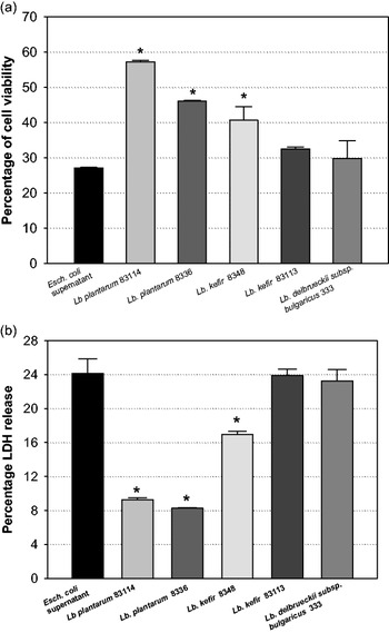
Spent culture supernatants of Esch. coli 69160 led to cytotoxic effects on cultured eukaryotic Vero cells. The damage exerted by the 69160 supernatant against Vero cells is evidenced by the MTT assay determining mitochondrial-dehydrogenase activity as a marker of cell viability (Fig. 1a, black bars) and by LDH release, as an indicator of membrane leakage as a result of damage (Fig. 1b, black bars). These figures indicate the cytotoxic effects of the 69160 supernatant in the presence of different lactobacilli. Lb. plantarum CIDCA 83114 and CIDCA 8336 and Lb. kefir CIDCA 8348 were effective in protecting Vero cells from the cytotoxic effects caused by the 69160 supernatant. The two Lb. plantarum strains were the ones with a greater degree of protection, whereas Lb. kefir CIDCA 83113 and Lb. delbrueckii subsp. bulgaricus CIDCA 333 exerted no significant inhibition of cytotoxicity (Fig. 1a). Essentially the same results were observed when damage was evaluated by the release of LDH (Fig. 1b), thus demonstrating that the ability of lactobacilli to antagonise the action of the Shiga toxin was strain-dependent (Fig. 1).
Effects of lactobacilli strains (107 CFU per well) on the cytotoxicity of Esch. coli 69160 supernatants to confluent Vero-cell cultures. (a) The percent cell viability was determined by the 3-(4,5-dimethylthiazol-2-yl)-2,5-diphenyltetrazolium–bromide assay (MTT). The 69160-supernatant dilution used was 1:100 (b). Cell damage was determined by the release of lactate dehydrogenase. The Esch. coli 69160 supernatant dilution used was 1:50. The percentage value of 100% of lactate-dehydrogenase release were refers to Vero cells treated with 3% (v/v) Triton X-100. Esch. coli 69160 supernatant (fx1); Lb. plantarum CIDCA 83114 plus 69160 supernatant (fx2); Lb. plantarum CIDCA 8336 plus 69160 supernatant (fx3); Lb. kefir CIDCA 8348 plus 69160 supernatant (fx4); Lb. kefir CIDCA 83113 plus 69160 supernatant (fx5); Lb. delbrueckii subsp. bulgaricus 333 plus 69160 supernatant (fx6). (*) Significant difference (P < 0·05) between Vero cells incubated with 69160 supernatant with or without lactobacilli.

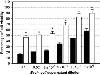
Therefore, on the basis of its superior ability to prevent the effect of Stx on Vero cells, Lb. plantarum CIDCA 83114 was selected to analyse the mechanism by which the cytotoxicity of the Esch. coli supernatant was being reduced. Different amounts of 69160 supernatant induced a dose-dependent decrease in cell viability as assessed by the MTT assay (Fig. 2, black bars). The presence of 107 CFU Lb. plantarum CIDCA 83114 per well, however, prevented the injury to Vero cells caused by the Stx2 (Fig. 2, white bars).
Antagonism by Lb. plantarum CIDCA 83114 (107 CFU per well) of the citotoxicity of different doses of Esch. coli 69160 supernatants to Vero cells. Cell viability was determined by the MTT assay. Esch. coli 69160 supernatant (fx1); Lb. plantarum CIDCA 83114 plus 69160 supernatant (fx2). (*) Significant difference (P < 0·05) between the viability of Vero cells incubated with 69160 supernatant in the presence and absence of Lb. plantarum CIDCA 83114.

Presence of haemolysin in Esch. coli 69160 supernatants
The cytotoxicity of Esch. coli 69160 supernatants to Vero cells could be attributed to the combined action of Stx2 and haemolysin, since the strain 69160 (stx–hly) has been characterised genetically as positive for both genes. In order to elucidate if both extracellular factors were responsible for the toxicity to Vero cells detected, the haemolysin expression of Esch. coli 69160 was measured by a haemolysis-activity assay. The percentage of haemolysis produced by the 69160 supernatant was only 7·1 ± 1·3% in comparison with the control values for total haemolysis. From the results, we assume that the haemolysin activity of the 69160 supernatant was only negligible.
Hydrophobicity test of the lactobacillus surface
To test the basis of the protective effect of these lactobacilli, we performed experiments focusing on their surface properties. The surfaces of Lb. kefir CIDCA 8348 and CIDCA 83113 exhibited hydrophobic characteristics (60·8% y 38·2% hydrophobicity, respectively), whereas the surface of Lb. plantarum CIDCA 8336, CIDCA 83114 and of Lb. delbrueckii subsp. bulgaricus CIDCA 333 had more hydrophilic values (at 2·4, 4·7 and 3·8% hydrophobicity, respectively). The protective effect of lactobacilli (Fig. 1) thus did not correlate with the degree of hydrophilicity of the lactobacilli surfaces, thus indicating that the interaction with Stx did not simply involve physical affinities.
The role of protein molecules in the protection against Shiga toxins by Lactobacillus cell walls
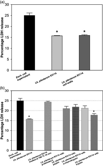
That the cytotoxic effect of 69160 supernatant was inhibited by the cell walls of Lb. plantarum CIDCA 83114 (Fig. 3a) with a protection proving as effective as that observed with Lactobacillus whole cells indicated that the cell surface was involved in the antagonism against Shiga toxin. The amount of Lactobacillus cell walls in this experiment was furthermore equivalent to that present on the intact bacteria.
(a) Effect of cell-walls on the cytotoxicity of Esch. coli 69160 supernatants to confluent Vero cells. Cell damage was determined by lactate-dehydrogenase release. The dilution of 69160 supernatant dilution used was 1:50. The amount of Lactobacillus cell walls used was equivalent to 107 CFU of intact bacteria per well. Esch. coli 69160 supernatant (fx1); Lb. plantarum CIDCA 83114 plus 69160 supernatant (fx2); Lb. plantarum CIDCA 83114 cell-wall plus 69160 supernatant (fx3). (*) Significant difference (i.e. P < 0·05) between Vero cells incubated with 69160 supernatant in the presence and absence of lactobacilli. (b) The effect of heat and proteolytic-enzyme treatment of Lb. plantarum CIDCA 83114 (107 CFU per well) on inhibition of the cytotoxicity of Esch. coli 69160 supernatant to confluent Vero cells. Cell damage was determined by lactate-dehydrogenase release. Esch. coli 69160 supernatant (fx4); Lb. plantarum CIDCA 83114 plus 69160 supernatant (fx5); Lb. plantarum CIDCA 83114 treated at 100 °C for 10 min plus 69160 supernatant (fx6); Lb. plantarum CIDCA 83114 treated with proteinase k plus 69160 supernatant (fx7); Lb. plantarum CIDCA 83114 treated with pepsin plus 69160 supernatant (fx8); Lb. plantarum CIDCA treated with α-chymotypsin plus 69160 supernatant (fx9); Lb. plantarum treated with trypsin plus 69160 supernatant (fx10). (*) Significant difference (i.e. P < 0·05) between LDH release by Vero cells incubated with 69160 supernatant in the presence and absence of lactobacilli.

Heat treatment or preincubation of strain CIDCA 83114 with proteolytic enzymes—proteinase K, pepsin and chymotrypsin— produced a significant decrease in the protection by this Lactobacillus strain (Fig. 3b), thus indicating that key thermosensitive peptides and/or proteins present on the surface of Lb. plantarum CIDCA 83114 are involved in the antagonism against Stx2. These peptides could be resistant to the proteolytic action of trypsin since the latter produced no significant decrease in the protective effect. These results demonstrate that a direct interaction between Stx and the protein structures of the Lb. plantarum cell walls produces the inhibition of the toxin.
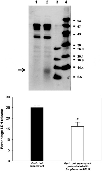
These findings prompted us to determine whether the diminution in the biological activity of Stx 2 by Lb. plantarum CIDCA 83114 resulted from an inhibition of toxin activity by a scavenging effect on the part of the bacterial cell surface. The supernatant of Esch. coli 69160 was treated with Lb. plantarum CIDCA 83114 and the Stx remaining therein after removal of the lactobacilli was revealed by SDS–PAGE (Fig. 4a, lanes 1 & 2). After exposure to strain 83114 a band of less than 14·6 kDa corresponding to the molecular weight of subunit B of Stx (lane 2) was no longer detected (lane 1). The absence of the band after incubation with the lactobacilli could be interpreted as either a binding or a proteolysis of the toxin by molecules present on the strain-83114 surface. In order to confirm the association between the absence of the band with a decrease in cytotoxicity, the same supernatant that had been present in Lane 1 was assayed on Vero cells. The cytotoxic activity measured as LDH release was accordingly found to be significantly lower than that observed with the 69160 supernatant in the absence of Lactobacillus treatment (Fig. 4b). The cytotoxic activity of the 69160 supernatant in the presence of either the whole lactobacilli or preparations of the cell walls from strain 83114 (Fig. 3a) was equivalent to the one achieved after pre-exposure of those bacteria-free supernatants to intact lactobacilli (Fig. 4b). These last observations would argue that the binding or proteolysis of the Shiga toxin by the lactobacilli could constitute al least one of the mechanisms involved in the bacteria's antagonism of the cytotoxicity of the 69160 supernatants to Vero cells.
(a) Protein profile of Esch. coli 69160 supernatants resolved by Tricine-sodium-dodecylsulfate-polyacrylamide-gel electrophoresis. Lane 1: 69160 supernatant preincubated (1 h at 37 °C with shaking) with Lb. plantarum CIDCA 83114 (108 CFU) and centrifuged to obtain a bacteria-free supernatant. Lane 2: Untreated Esch. coli 69160 supernatant. Lane 3: Very-low-molecular-weight markers. Lane 4: Low-molecular-weight markers (b) Antagonism by Lb. plantarum CIDCA 83114 (108 CFU) of the cytotoxicity of Esch. coli 69160 supernatants to confluent Vero cells. Bacteria-free supernatans of Esch. coli 69160 were incubated with 108 CFU of the lactobacilli for 1 h at 37 °C before exposure to the Vero cultures. Subsequent Vero-cell damage was determined by lactate-dehydrogenase release. Esch. coli 69160 supernatant (fx1); 69160 supernatant preincubated with Lb. plantarum CIDCA 83114 and centrifuged to obtain a bacteria-free supernatant (fx2).

Until now, few studies have been performed suggesting protective effects of bacterial surface proteins against Esch. coli O157:H7. A recent study, however, indicated that Lb. plantarum cell-surface adhesive proteins are critical for the protection of Caco-2 cells against the adhesion and tight-junction injury produced by enteropathogenic Esch. coli (Liu et al. Reference Liu, Shen, Zhang, Ma and Qin2011). Since Lb. plantarum CIDCA 83114 did not present S-layers proteins (Garrote et al. Reference Garrote, Delfederico, Bibiloni, Abraham, Pérez, Semorile and De Antoni2004) we cannot draw a similar conclusion; but the present results nevertheless demonstrate that the cell wall plays a crucial role in the inhibition observed. That receptors within the surface structure of certain bacteria similar to those on eukaryotic cells could be responsible of the inactivation of Shiga toxin had been suggested (Kitov et al. Reference Kitov, Sadowska, Mulvey, Armstrong, Ling, Pannu, Read and Bundle2000; Paton et al. Reference Paton, Morona and Paton2001a, Reference Paton, Rogers, Morona and Paton2001b). Furthermore, the Lb. plantarum genome encodes many cell-wall surface proteins involved in adhesion, enzyme action, phage functions, and other properties that are still unknown (Boekhorst et al. Reference Boekhorst, Wels, Keerebezem and Siezen2006). Since the Lactobacillus surface proteins could mimic the receptors on any specific target for pathogens and toxins, the expression of functional cell-wall proteins of that nature may be involved with the antagonistic effect against the Shiga toxin.
We previously formulated a promising two-strain starter culture containing Lb. plantarum CIDCA 83114 and observed a protection against Shiga toxin of Esch. coli 69160 supernatants (Kakisu et al. Reference Kakisu, Irigoyen, Torre, De Antoni and Abraham2011). The present work provides key evidence indicating the participation of the cell walls of this strain from kefir in the antagonism of Shiga-toxin cytotoxicity. The present findings concerning the nature of the blockade of this crucial host-pathogen toxin interaction constitutes an initial step in improving our understanding of possible inhibitory mechanisms against pathogenic-Esch. coli foodborne diseases.
E. Kakisu and C. Tironi Farinati are fellows of the National Research Council of Argentina (CONICET, Argentina). G. L. De Antoni is a researcher of CIC-PBA; A. G. Abraham and C. Ibarra are researchers of CONICET. G. L. De Antoni and A. G. Abraham are Professors of the School of Sciences of the National University of La Plata (UNLP). C. Ibarra is Professor of the School of Medicine of the University of Buenos Aires (UBA). This work was supported by the National Agency of Scientific and Technical Promotion (ANPCyT), CONICET, CIC-PBA, UNLP, UBA. The authors wish to thank Dr Donald F. Haggerty, a retired career investigator and native English speaker, for editing the final version of the manuscript.