Introduction
The weaning period in pigs represents a time of gastrointestinal and immunological instability, which results in a critical period of low voluntary feed intake and an increased susceptibility to infection. Therefore, various nutritional approaches for minimizing diseases or optimizing growth performance have been studied (Lalles et al., Reference Lalles, Bosi, Smidt and Stokes2007). Some feed additives, such as prebiotics, probiotics and some other substances, have been studied with regard to managing the health of weaning piglets (Lalles et al., Reference Lalles, Bosi, Smidt and Stokes2007). Among others, the use of probiotics such as lactobacilli and bifidobacteria has been proposed as the most promising approach for maintaining or improving animal health (Stein and Kil, Reference Stein and Kil2006; Lalles et al., Reference Lalles, Bosi, Smidt and Stokes2007). It has been reported that feeding certain probiotic strains to pigs could improve growth performance, prevent the growth of pathogenic bacteria and reduce the incidence of diarrhea (Shu et al., Reference Shu, Qu and Gill2001; Gardiner et al., Reference Gardiner, Casey, Casey, Lynch, Lawlor, Hill, Fitzgerald, Stanton and Ross2004; Taras et al., Reference Taras, Vahjen, Macha and Simon2006; Casey et al., Reference Casey, Gardiner, Casey, Bradshaw, Lawlor, Lynch, Leonard, Stanton, Ross, Fitzgerald and Hill2007). At the same time, feeding an FLD prepared with the use of bacteria, such as lactic acid bacteria, has also been shown to improve pig performance and to reduce the number of enteric pathogens (Canibe and Jensen, Reference Canibe and Jensen2003; Stein and Kil, Reference Stein and Kil2006). These positive influences may be due to the low pH, high short-chain fatty acid content and high lactic acid bacteria content of the FLD. However, the potential immunomodulatory effect of probiotic bacteria on weaning piglets has not been fully elucidated; nevertheless, the effect of feeding FLD on the systemic immune response remains unknown. In this paper, we assessed the immunomodulating potential of an FLD prepared with an inoculation of LQ80. The study analyzed its effect on the systemic and mucosal immune responses in weaning pigs.
Material and methods
Animals
Ten piglets (Landrace × Large white × Duroc) from one litter, which were obtained from our farm at the National Institute of Livestock and Grassland Science were used. The piglets were weaned at 28 days of age (BW, 9.8 ± 1.2 kg) and were randomly allocated to two treatment groups, which were balanced for sex and initial BW. They were conventionally housed individually in pens of 1.0 × 1.5 m with water and FLD or non-fermented liquid diet (NFLD) as the control diet ad libitum. No physical contact was allowed between the animals. The piglets were fed twice daily, at 0830 and at 1630 h, and the feed was renovated each time. BW and feed consumption were recorded to determine the average daily gain (ADG) and the average daily feed intake (ADFI). To determine the cytokine response by bacteria-treated whole blood cells, five piglets (Landrace × Large white × Duroc, aged 8 weeks) from another litter were used. All the animals received humane care as outlined in the Guide for the Care and Use of Experimental Animals (Animal Care Committee, National Institute of Livestock and Grassland Science).
Strain and diet
The strain LQ80 was originally isolated from fermented liquid feed for piglets and was provided by Dr Yimin Cai. The LQ80 strain used for the FLD was prepared by culturing it in the MRS broth (Difco Laboratories, Detroit, MI, USA) for 18 h at 30°C.
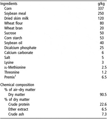
The basal feed formulation is shown in Table 1. NFLD and FLD were prepared by mixing dry basal feed and water in a 1 : 2 (wt/wt) ratio. In the preparation of NFLD, water was added just before the feeding. The FLD was inoculated with pre-cultured LQ80 (approximately 109 cfu/l) and stored in a tank at 30°C for 24 h before it was fed to the piglets. Finally, FLD was found to contain approximately 108 cfu lactobacilli/ml, and the pH was 3.6 to 4.0.
Ingredients and chemical composition of the basal diet

1Premix provided per kg of diet: vitamin A, 20 000 IU; vitamin D3, 400 IU; vitamin E, 20 mg; vitamin B1, 2.5 mg; vitamin B2, 17.5 mg; vitamin B6, 1.3 mg; calcium pantothenate, 27.3 mg; nicotinamide, 15 mg; choline chloride, 144 mg; Mn as manganese sulfate, 100 mg; Fe as iron sulfate, 100 mg; Cu as copper sulfate, 20 mg; Zn as zinc carbonate, 100 mg; I as calcium iodate, 2 mg.
Experimental procedure
Ten weaning piglets were divided into two groups to examine the effect of feeding FLD on the systemic and mucosal immune responses. Five piglets were fed FLD and the other five were fed NFLD for 28 days. The saliva and feces were collected from these piglets on the day of weaning (day 0) and on days 7, 14, 21 and 28 post weaning. At the end of the experiment (day 28), blood samples were collected from the jugular veins of the piglets.
Samples
The serum was obtained by centrifugation at 10 000 × g for 2 min after clotting and stored individually at −80°C until the assays were conducted. For the examination of cell-mediated immune response, i.e., the production of cytokines, lymphocyte proliferation and phagocytosis, blood was collected in a heparinized tube (Vacutainer; Becton Dickinson, San Jose, CA, USA). The saliva samples were obtained by determining the weight of the collected secretions, which was determined by weighing the swabs before and after sampling. Subsequently, saliva samples were rapidly diluted (0.1 g/ml) in phosphate buffered saline (PBS) (pH 7.2), supplemented with 20% fetal bovine serum (FBS), and stored at −80°C until the assays were conducted. Feces samples were collected and immersed in PBS (0.2 g/ml) supplemented with 10% FBS. The samples were mechanically disaggregated using pipette tips, vigorously mixed with a vortex mixer, and then centrifuged at 4°C for 10 min at 10 000 × g.
Detection of antibodies
Total porcine immunoglobulin M (IgM), immunoglobulin G (IgG) and immunoglobulin A (IgA) antibodies in the serum and total porcine IgA antibodies in the feces and saliva were measured by a sandwich ELISA. For the detection of IgG, IgM and IgA, microtiter plates (Maxisorp; Nunc, Roskilde, Denmark) were coated overnight at 4°C with goat anti-porcine IgG, IgM or IgA (Bethyl Laboratories, Montgomery, TX, USA). The wells were washed with PBS containing 0.05% Tween 20 (PBST), and blocked with 1% bovine serum albumin (BSA) in PBS for 30 min. After washing with PBST, the appropriately diluted samples were added and incubated for 2 h at room temperature. Thereafter, the wells were treated with horseradish peroxidase-conjugated goat anti-porcine IgG, IgM or IgA (Bethyl Laboratories) for 1 h at room temperature. The plates were washed, and 3,3′,5,5′-tetramethylbenzidine (TMB) solution (KPL, Gaithersburg, MD, USA) was added to each well as a substrate. After 30 min of incubation, the reaction was stopped by the addition of 1 M H2PO3. Absorbance was measured at 450 nm using a microplate reader (model 3550; BioRad, Hercules, CA, USA).
Production and detection of cytokines
Cytokine production was determined in whole blood cultures as previously described (Edfors-Lilja et al., Reference Edfors-Lilja, Wattrang, Marklund, Moller, Andersson-Eklund, Andersson and Fossum1998), with some modifications. Briefly, heparinized whole blood from the FLD-fed or NFLD-fed piglets was diluted to 1 : 10 for the detection of tumor necrosis factor-α (TNF-α), interleukin (IL)-8 and IL-6; 1 : 50 for the detection of interferon-γ (IFN-γ) with the culture medium (RPMI-1640 containing 50 μM 2-mercaptoethanol, 10 mM HEPES, 10 U/ml penicillin and 100 μg/ml streptomycin). Each diluted blood sample was seeded into 24-well plates (1 ml per well) and cultured in the presence of concanavalin A (ConA) (5 μg/ml) or Escherichia coli lipopolysaccharide (LPS; Sigma-Aldrich, St. Louis, MI, USA) (1 μg/ml). The plates were maintained at 37°C in a humid incubator with 5% CO2. The culture supernatant was collected at 6 h for TNF-α and IL-8 detection and at 72 h for the IL-6 and IFN-γ assays. The cytokine levels were determined with commercial ELISA reagents (IL-6, IL-8 and TNF-α; R&D Systems, Minneapolis, MN, USA and IFN-γ; PharMingen, San Diego, CA, USA), according to the manufacturer’s procedure.
Cytokine production by bacteria-treated whole blood cells
In order to determine the effect of bacteria on the cytokine production of porcine blood cells, we washed the bacteria two times with 0.85% NaCl and then resuspended them in the RPMI 1640 (Sigma-Aldrich) medium. Heat treatment of the bacteria was performed by heating at 100°C for 50 min.
Heparinized whole blood cells were obtained from five piglets (aged 8 weeks). Each blood sample was diluted to 1 : 10 for the detection of TNF-α and IL-6; 1 : 50 for the detection of IFN-γ with the culture medium. The blood cells were cultured with live or heat-treated LQ80 (final, 5 μg/ml). E. coli LPS at a concentration of 1 μg/ml was used as a control. The concentration of cytokines in the supernatants was determined by conducting a sandwich ELISA.
Blood phagocytosis assay
We measured blood phagocytosis by using whole blood as previously described (Shu et al., Reference Shu, Qu and Gill2001). Briefly, 100 μl of heparinized whole blood from each piglet was incubated for 30 min at 37°C with 10 μl heat-killed fluorescein isothiocyanate (FITC)-labeled E. coli (1 μg/ml). Phagocytosis was terminated by the addition of 100 μl of 8% formaldehyde, and the erythrocytes were lysed with 1 ml ice-cold water. Samples were centrifuged at 250 × g for 5 min and suspended in 0.5 ml PBS. After adding 50 μl of trypan blue, the samples were analyzed by flow cytometry using a FACSort flow cytometer (Becton Dickinson, Franklin Lakes, NJ, USA). The results were expressed as the percentage of cells showing phagocytic activity in the cell preparation.
Lymphocyte proliferation
Heparinized whole blood was diluted in the culture medium (1 : 4), and 50 μl of it was added in triplicate to the wells of a 96-well flat-bottom plate (Falcon; Becton Dickinson Labware, Franklin Lakes, NJ, USA). Then, the wells were stimulated with ConA (2.5 μg/ml) or the medium alone. The culture medium used in this experiment was RPMI 1640 supplemented with 5% FBS, 100 U/ml penicillin, 100 μg/ml streptomycin, 10 mM HEPES and 50 μM 2-mercaptoethanol. After 72 h of incubation at 37°C in a 5% CO2 atmosphere, the cells were pulsed with 0.5 μCi [3H]-thymidine per well. After 8 h, the cells were harvested, and radioactivity was counted using a liquid scintillation counter (Tri-Carb1600TR; Packard, Meriden, CT, USA). The stimulation index (SI) is defined as the mean counts per minute (cpm) of the response of the ConA-stimulated cells, divided by the mean of the response of cells cultured in medium alone.
Statistical analysis
Data were expressed as mean ± s.d. For the total IgA levels in the feces and saliva, data were analyzed by the GLM procedure of the SAS software version 9.1 (SAS Institute, Cary, NC, USA) with the following model:

where Yijk is the observation; μ the overall mean; αi the diet effect; βj (i ) the pig within diet effect; γk the day effect; (αγ)ik the interaction of diet and day; εijk the residual error. When day effect was significant (P < 0.05), differences between means were compared using the Tukey HSD test. For the antibody levels in the sera, cytokine levels, phagocytosis and lymphocyte proliferation assay, ANOVA using GLM procedure of the SAS software was used. The statistical significance of a given factor at different levels of the other factors (simple main effects), was obtained using the least-square means (LSMEANS) SLICE option. P-values less than 0.05 were considered significant.
Results
Growth performance
The averages of BW at 28 days of NFLD-fed and FLD-fed piglets were 24.0 ± 2.9 and 21.6 ± 1.9 kg, respectively. The ADFI during the 28 days experimental period of the NFLD- and FLD-fed piglets were 662.4 ± 112.9 and 561.5 ± 93.0 g/day, and the ADG were 510.7 ± 84.2 and 421.4 ± 48.2 g/day, respectively. There were no significant differences in ADFI or ADG between the NFLD- and FLD-fed piglets (P > 0.05). Mild diarrhea on day 14 to 21 was observed in both groups. All ill piglets recovered without any further health problems.
Cytokine production by bacteria-treated whole blood cells
In this study, we used the strain LQ80 for preparing the fermented liquid feed. It has been known that these organisms are capable of living in the gut when they are orally administered (Takahashi et al., Reference Takahashi, Egawa, Simojo, Tsukahara and Ushida2007). We tested the ability of this bacterium to stimulate IFN-γ and IL-6 production from the blood cells of piglets by co-culturing the blood cells with live and heat-treated bacteria. As shown in Table 2, IFN-γ secretion in the culture supernatant of the blood cells was significantly induced by the addition of LQ80. In contrast, the secretion of IL-6 was at the same level in the medium alone. The IFN-γ and IL-6 secretion ability of the live bacteria was almost the same as the heat-treated one.
Cytokine production by bacteria-treated whole blood cells

LPS = lipopolysaccharide; LQ80=Lactobacillus plantarum LQ80; IFN-γ = interferon-γ; IL-6 = interleukin-6.
The results are represented as mean ± s.d. (n = 5).
a,bMean values within a row with unlike superscript letters were significantly different (P < 0.05).
Effect of FLD feeding on antibody response
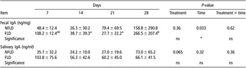
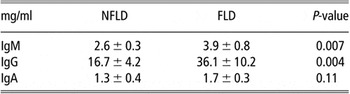
Table 3 shows the total IgA levels in the feces and saliva. The average fecal and salivary IgA levels at day 0 were 287.4 ± 483.2 and 21.2 ± 14.2 μg/g, respectively. Although the total salivary IgA levels of the FLD-fed piglets tended to be high (P < 0.1), no significant difference was observed between the two groups during the experimental period. With regard to the fecal IgA levels, no significant difference was observed between the two groups. In the only FLD-fed group, however, the mean fecal IgA levels at day 28 were higher than that at days 14 and 21 (P < 0.05). On the other hand, the total serum IgM and IgG antibody levels of the FLD-feeding piglets were significantly higher than those of the NFLD-fed piglets, although no differences in the serum IgA levels were observed (Table 4).
Total saliva and feces IgA levels in FLD-fed and NFLD-fed piglets

IgA = immunoglobulin A; NFLD = non-fermented liquid diet; FLD = fermented liquid diet.
The results are represented as mean ± s.d. (n = 5).
ns = P > 0.05.
a,bMean values with unlike superscript letters were significantly different (P < 0.05).
Total serum IgM, IgG and IgA levels in FLD-fed and NFLD-fed piglets

IgM = immunoglobulin M; IgG = immunoglobulin G; IgA = immunoglobulin A; NFLD = non-fermented liquid diet; FLD = fermented liquid diet.
The results of serum Ig are represented as mean ± s.d. (n = 5).
Effect of FLD feeding on cellular immune responses
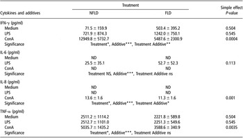
Cytokines are a part of an extracellular signaling network that controls every function of the innate and adaptive immune response such as inflammation, defense against virus infection and proliferation and differentiation of T and B cells. Therefore, we examined cytokine production by the blood cells (Table 5). On stimulation with ConA as a T-cell mitogen, IFN-γ production by the blood cells of the FLD-fed piglets was significantly lower than that of the NFLD-fed piglets. On stimulation with ConA, the levels of IL-8 and TNF-α, which are proinflammatory cytokines, in the blood cells of the FLD-fed piglets were also significantly lower than those of the NFLD-fed piglets (Table 5). Stimulation with LPS as a B-cell mitogen, revealed no significant differences, between the groups with regard to IL-6 production. On the other hand, the lymphocyte proliferative responses to ConA also tended to be less (P < 0.06) in the blood obtained from the FLD-fed piglets (SI, 93.3 ± 65.5), compared with those of the NFLD-fed ones (SI, 222.1 ± 55.3). These results indicate that the mitogen-induced T-cell response was suppressed in the FLD-fed piglets.
Effect of FLD-feeding on the cytokine production of blood cells

NFLD = non-fermented liquid diet; FLD = fermented liquid diet; IFN-γ = interferon-γ; LPS = lipopolysaccharide; ConA = concanavalin A; IL-6=interluekin-6; IL-8 = interleukin-8; TNF-α = tumor necrosis factor-α; ND = not detected.
The results are represented as mean ± s.d. (n = 5).
ns = P > 0.05.
Significance: *P < 0.05; **P < 0.01, ***P < 0.005.
Treatment by additive interaction effect was analyzed using a simple main effects analysis.
Effect of FLD-feeding on the phagocytic ability of macrophages
Macrophages are important for innate immune response and the cells initially respond to, and are rapidly activated by exposure to environmental agents such as pathogens. Therefore, we directly measured macrophage phagocytosis, which is a key macrophage function, by examining FITC-labeled E. coli by means of flow cytometry. The phagocytic activities of macrophages of the blood cells from NFLD- and FLD-fed piglets were 44.2 ± 8.6 (%) and 43.4 ± 28.6 (%), respectively. There was no significant difference in blood phagocytic activity.
Discussion
Various types of strain-specific immunomodulations can be brought about, as observed in numerous trials (Winkler et al., Reference Winkler, Ghadimi, Schrezenmeir and Kraehenbuhl2007). With regard to FLD, it has been reported that colostrum from FLD-fed sows showed greater mitogenic activity compared with that from dry feed-fed sows, and improved the health status of their piglets via the colostrum (Demeckova et al., Reference Demeckova, Kelly, Coutts, Brooks and Campbell2002); however, the direct immunomodulatory effects of fermented liquid feed have not been well studied. The present study investigated the influence of feeding FLD, prepared with LQ80, on the humoral and cell-mediated immune responses of weaning pigs. We observed the enhancing systemic antibody response of weaning piglets after feeding the FLD for 4 weeks.
T and B cells play an important role in the immune response, with T cells controlling most of the B cells producing antibodies. Mosmann et al. (Reference Mosmann, Cherwinski, Bond, Giedlin and Coffman1986) originally identified functionally distinct helper T-cell types (Th1 and Th2) on the basis of the patterns of cytokine production. IFN-γ secreted by Th1 cells stimulates cell-mediated immune response, and IL-4 or IL-6 secreted by Th2 cells stimulates humoral immune response. In the present study, LQ80 induced IFN-γ secretion from blood cells effectively, but it did not induce IL-6 secretion in vitro. Therefore, FLD containing LQ80 is expected to be able to stimulate a Th1-type immune response and have little effect on antibody response. However, systemic humoral immune response was stimulated in FLD-fed pigs. One of the reasons for the discrepancy is thought to be the difference of cell type to contact the bacteria. Antigen-presenting cells such as dendritic cells (DC) from different tissues, induce production of different T-cell cytokine profiles, while mucosal DC induce predominantly Th2 cytokines (Everson et al., Reference Everson, McDuffie, Lemak, Koopman, McGhee and Beagley1996). When the FLD is fed to piglets, LQ80 in the FLD might stimulate Th2 response via mucosal sites. Moreover, it has been reported that some types of B cells appear to be regulated in a T-cell-independent manner (Fagarasan and Honjo, Reference Fagarasan and Honjo2000; Macpherson et al., Reference Macpherson, Gatto, Sainsbury, Harriman, Hengartner and Zinkernagel2000). These antibodies are produced in the absence of any exogenous antigenic exposure without the help of T cells and play an important role in the first line of defense against bacterial and viral infections. In our study, T-cell-independent antibodies might have been produced in the FLD-fed piglets. Various factors could be considered to be involved in the stimulation of systemic immune responses. The FLD contains many factors that affect the host’s health, for example, short-chain fatty acids as the end-products of the fermentation of carbohydrates and proteins, antimicrobial compounds produced by some lactic acid bacteria and other immunomodulating agents (LeBlanc et al., Reference LeBlanc, Matar, Valdez, LeBlanc and Perdigon2002; Vinderola et al., Reference Vinderola, Matar, Palacios and Perdigon2007). Furthermore, the FLD might directly affect the composition of the gastrointestinal microbiota. In fact, the composition of fecal lactobacilli was markedly altered by an oral administration of LQ80 (Takahashi et al., Reference Takahashi, Egawa, Simojo, Tsukahara and Ushida2007). Therefore, the FLD containing LQ80 may affect the systemic immune response via a modulation of intestinal microbiota. Further studies are required to address the reasons for FLD-induced stimulation of systemic immune responses.
In the case of T-cell response, the blood cells from FLD-fed piglets showed a low level of IFN-γ secretion and a low level of mitogen-induced proliferation compared to the controls. The results suggest that T-cell reactivity was reduced by FLD feeding. This finding is similar to the results obtained after oral administration of probiotics in human subjects or rodents (Pessi et al., Reference Pessi, Sutas, Saxelin, Kallioinen and Isolauri1999; Winkler et al., Reference Winkler, Ghadimi, Schrezenmeir and Kraehenbuhl2007). Furthermore, we found that the production of inflammation cytokines such as TNF-α and IL-8, also significantly decreases in the blood cells of FLD-fed piglets. Weaning is probably the most stressful time in the life of a piglet and can lead to outbreaks of diarrhea as a result of inflammatory overreaction against non-pathogenic bacteria. Therefore, FLD-feeding may regulate immune response without unnecessary inflammatory reactions to harmless environmental agents such as normal bacterial flora, and prevent aberrant diseases.
Numerous studies have reported that oral administration of probiotics enhances mucosal immunity and increases the titer of IgA antibodies in the gut (Isolauri et al., Reference Isolauri, Sutas, Kankaanpaa, Arvilommi and Salminen2001; Corthesy et al., Reference Corthesy, Gaskins and Mercenier2007). However, FLD feeding had little effect on mucosal IgA response during the experimental period. The level of IgA antibodies in the sera of pigs, for approximately 2 to 4 weeks after weaning, is very low because of reducing maternal immunity and an immature mucosal immune system at that time. Therefore, we could not detect the differences of IgA levels in the sera and seromucos secretions. However, the average of fecal IgA levels of the FLD-fed pigs at day 28 was higher than that at days 14 and 21 (P < 0.05). The result suggests that mucosal immunity of FLD-fed piglets has matured enough for secretion of their own IgA antibodies at day 28.
In conclusion, the FLD used in this study could alter the immune responses, of weaning piglets by stimulating systemic immune responses without unnecessary inflammatory reactions. This indicates that the FLD feed prepared with the use of LQ80 may be a candidate feed, with regard to enhancing immune responses and preventing diseases in weaning piglets.
Acknowledgments
This work was financially supported by the Ministry of Agriculture, Forestry and Fisheries of Japan. We thank Dr Yimin Cai for providing strain LQ80. We also thank Dr Fuminori Terada for his help with the statistical analysis.