The Amplatzer septal occluder (AGA Medical Corp., Golden Valley, Minnesota, United States of America) is the most commonly used device to close secundum atrial septal defects in children, with high success and low complication rates.Reference Everett, Jennings and Sibinga 1 – Reference Diab, Cao, Bacha and Hijazi 5 Although accepted as a good alternative to surgery in most centres, rare life-threatening complications with damage to the aorta, the atrial roof, or the atrioventricular valves have been reported after the procedure.Reference Sarris, Kirvassilis and Zavaropoulos 2 , Reference Delaney, Li and Rhodes 6 – Reference Li, Han and Yu 13 Large rigid devices are particularly at risk for repeated micro injuries, and percutaneous closure of large atrial septal defects in children could be questionable.Reference Amin, Hijazi, Bass, Cheatham, Hellenbrand and Kleinman 7 On the contrary, growth of the heart is crucial to children and should modify the initial relationship between the device and cardiac structures, therefore decreasing the related late complications.
Methods
Study population
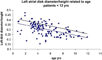
Between December, 1997 and February, 2002, 122 patients underwent atrial septal defect closure with an Amplatzer septal occluder at Sainte-Justine University Hospital Center. To define the relatively largest Amplatzer septal occluder for this population, we plotted the ratio of the left atrial disk diameter (mm) to the patient’s height (cm) against age (years). Of the patients, two were excluded because device placement was not possible and one patient was lost to follow-up. The 25 remaining patients with the highest ratios were selected as our study population (Fig 1).
Study population. The ratio of left atrial disk diameter (mm) to patient’s height (cm) is plotted against against the age of the patient. The 25 patients with the highest ration were selected as our study population.

Beyond the short hospitalisation period, all patients had routine echocardiographic evaluation within 3 weeks of closure (short term). All patients were asked to attend a mid-term follow-up in 2002 and a long-term follow-up in 2009. Until 2002, echocardiograms were carried out on Philips Sonos 2500 echographs (Philips Healthcare, Andover, Massachusetts, United States of America) using a 12, 8, or 4 MHz probe and recorded on videotapes. In 2009, they were carried out on GE (Fairfield, United States of America) or Philips (Amsterdam, Holland) echographs and recorded on DVD.
Echocardiographic evaluation
Atrioventricular valves
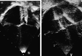
The minimal distance between the inferior extremity of each disk and the corresponding atrioventricular valve annulus was measured in the apical four-chamber view at the maximal size of the atria in diastole – last frame before opening of the atrioventricular valve (Fig 2). Contact between the atrioventricular valve and the disk was noted as was the existence of tricuspid or mitral regurgitation, which was graded on a 0–3 scale corresponding to mild, moderate, and severe regurgitation, respectively.Reference Zoghbi, Enriquez-Sarano and Foster 14 The distance between the left disk and the left atrial roof was measured in the same view and at the same time.
( a ) Measurement of the minimal distance between the lower extremities of the right and left disks and the respective atrioventricular valves. ( b ) Owing to the large size of the device, the left atrial disk is shifted towards the mitral annulus and the right disk towards the tricuspid annulus, with contact between the disks and respective atrioventricular valve, whereas the upper extremity of the disk is in contact with the atrial roof.

Aortic root
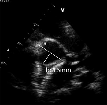
Position of both disks of the Amplatzer septal occluder in relation to the aortic annulus and eventual deformation of the aorta were studied in a parasternal short-axis view in diastole. A line between the margins of the two disks was drawn, and the perpendicular distance between this line and the posterior aspect of the aortic annulus was measured to quantify the straddling of the device over the aorta (Fig 3). A positive value was assigned when the extremities of the device straddled on the aorta anteriorly, and a negative value was assigned when there was a recess posterior to the aortic wall, that is no straddling. The aortic annulus was measured following the American Society of Echocardiography recommendations.Reference Lopez, Colan and Frommelt 15 Aortic insufficiency was noted and graded on a 0–3 scale corresponding, respectively, to mild, moderate, and severe.Reference Zoghbi, Enriquez-Sarano and Foster 14
Straddling on the aorta. A line is drawn between the extremities of the two disks ( a ). The perpendicular distance between this line and the posterior aspect of the aortic root is measured ( b ). A positive value is assigned when the extremities of the device straddled the aorta anteriorly. The straddling is then +16mm for this patient. Note the deformation of the non coronary sinus between the disks.

Venous returns
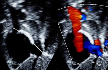
We measured the minimal distance between the upper extremities of the device and the superior vena cava in the sub costal view. We used the same view to measure the minimal distance between the left disk and the right upper pulmonary vein. Finally, we also measured the distance between the right disk and the inferior vena cava on this view (Fig 4). Evidence of dilatation of the coronary sinus was noted when present. Flow mapping of all venous returns was studied by colour and pulsed Doppler in search for turbulent flow. A flow velocity superior to 1.6 m/second and/or a monophasic flow were considered abnormal.Reference Smallhorn, Pauperio, Benson, Freedom and Rowe 16
Minimal distance between the extremities of the right disk and the vena cava in a subcostal bicaval view.

Chambers
A complete M-mode echocardiogram was obtained in the parasternal long-axis view where diastolic dimensions of the right ventricle and left ventricle were measured and converted into Z-scores.Reference Kampmann, Wiethoff and Wenzel 17
Device
The maximal width of the device was measured in a four-chamber view from the right to the left screw.
Statistical analysis
Descriptive statistics are presented as mean±standard deviation [range] for continuous variables, and categorical data presented as number of patients. Values were compared between the three sets of follow-up (early, mid, and long term). Paired Student t-test was used for continuous variables in case of normal distribution; otherwise a non-parametric test was conducted. Analysis of variance was also used for longitudinal assessment. The McNemar test was used to compare data distribution at the different stages of follow-up. A p-value <0.05 was considered significant. A correlation was calculated between echocardiographic measurements and variation of age, weight, height, and body surface area to establish the most reliable predictor of change in the relationship between the device and adjacent structures. Finally, to assess inter-observer correlation, five echocardiographic studies were reviewed independently (M.J.R., N.H.) and compared using paired t-test, Pearson’s correlation test for continuous data, and a Fisher exact test for nominal data.
Results
Population
At the time of intervention, patients were 4.6±2.9 years old [0.9–15.0], weighing 16.5±10.6 kg [6.8–62.5], with a height of 100.9±19.1 cm [69.9–163.0] and a body surface area of 0.66± 0.26 m2 [0.38–1.67]. The female to male distribution was 18/7. A specific feature of this group is the young age as four patients were <2 years of age, 12 were <5 years of age at the time of closure, and only one patient was over 9 years. Of the patients, seven (28%) had associated cardiac abnormalities: one had multiple regressive rhabdomyomas, three had mild pulmonary valvular stenosis, one had a moderate pulmonary valve stenosis requiring valvuloplasty, one had Ebstein’s anomaly, and one had mitral valve prolapse.
Echocardiographic evaluation
The intra-observer correlation analysis demonstrated an intraclass correlation coefficient of 0.998 (95th confidence interval 0.996–0.999; p<0.0001), with DFFIT analysis yielding no values changed>standard errors. Similarly, the inter-observer ICC was 0.981 (95th confidence interval 0.972–0.987; p<0.0001).
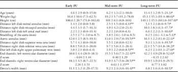
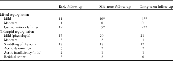
The results of the three echocardiographic evaluations are summarised in Tables 1 (numerical) and 2 (categorical variables). The time interval between the procedure and early, mid-term, and long-term follow-up was 2.1±3.4 days, [Reference Everett, Jennings and Sibinga1–Reference Smallhorn, Pauperio, Benson, Freedom and Rowe16] 17.8±10.5 months [5.7–38.3], and 8.8±0.9 years [7.5–10.5], respectively. The different measurements were feasible for all echocardiograms except for the distance between the left disk and the upper right pulmonary vein in two patients at early, seven at mid, and three at late follow-up.
Echocardiographic values at early, mid-, and long-term FU, numerical variables.

FU=follow-up.
*p<0.05 when the value is compared with mid-term follow-up
**p<0.05 when value compared with early follow-up
Comparison between echocardiograms at early, mid- and long-term follow-up, categorical variables.

*p<0.05 when value is compared with early follow-up
**p<0.05 when value is compared with mid-term follow-up
Mitral valve
There was a significant increase in the distance between the device and the mitral valve, leading to less contact between the left disk and the anterior leaflet and less mitral insufficiency. We observed 12 patients with mitral regurgitation at short-term follow-up, with a contact between the device and the anterior leaflet for 10 patients and a deformation of the annulus by the oversized device for two. Except for the latter two patients, the degree of mitral regurgitation was stable compared with preclosure trans-oesophageal echocardiography. The number of patients with mitral regurgitation decreased to 10 at mid-term and 4 at long-term follow-up.
Tricuspid valve
The distance between the device and the tricuspid annulus also increased significantly with time. Most patients had physiological tricuspid regurgitation except for three, who had moderate regurgitation. Tricuspid regurgitation was due to dilatation of the annulus for two patients and became mild at long-term follow-up. In one patient with Ebstein’s anomaly, the degree of tricuspid regurgitation did not improve after atrial septal defect closure.
Aorta
Straddling of the aortic valve by the device decreased significantly by a small distance of 2.2±2.1 mm. Of the 17 devices straddling the aorta at short-term follow-up, 12 were still in this position at long-term follow-up, and there was still aortic root deformation in 2/3, which consisted of pinching of the non-coronary sinus between the two disks (Fig 3).
Veins
In one patient, the right disk impinged on the opening of the superior vena cava into the right atrium without any flow acceleration, and the minimal distance evolved from 0 to 4 mm at long-term follow-up. In five patients, there was mild protrusion of the upper extremity of the left disk into the right upper pulmonary vein without flow acceleration at short-term follow-up. This was observed only in two patients at mid-term follow-up, and in none of the patients at long-term follow-up. There was protrusion of the right disk into the coronary sinus in one patient with mild dilatation of the latter, which resolved at mid-term follow-up.
Left ventricle
Owing to shunt occlusion, the left ventricular diameter increased both in diastole and systole at mid- and long-term follow-up, whereas the right ventricular diameter decreased from 18.3±4.5 to 14.9±4.7 mm at mid-term follow-up.
Device
The device flattened, its width decreasing from 11.1±3.2 to 6.8±1.6 mm at long-term follow-up.
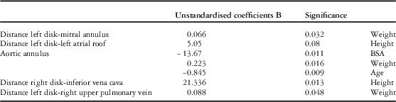
We tried to find a correlation between the different measurements and changes in weight, height, and body surface area. Only six variables showed a positive correlation with one of these factors. Results and level of significance of the analysis of variance test are given in Table 3. Changes in distance between the left disk and the mitral valve or the right upper pulmonary vein did show a correlation with weight gain – 0.066 and 0.088, respectively. In addition, the distance between the device and the inferior vena cava as well as with the left atrial roof did correlate with height (correlation 5.05 and 21.33, respectively).
Correlation between measurements and morphologic parameters.

Discussion
This is, to the best of our knowledge, the first echocardiographic study looking precisely at the relationship between large Amplatzer septal occluder and adjacent cardiac structures almost 10 years after atrial septal defect closure in a population of small children. Despite the proximity of the device to adjacent anatomic structures at the time of closure, there was no major interference with valvular function nor any obstruction, which could have been an indication for device removal.
Most of the complications reported in the literature after Amplatzer septal occluder implantation occur in adults and are related to damage to the aorta or to the atrial roof. Reports on atrioventricular valve regurgitation are less common.Reference Wilson, Smith, Prommete, O’Donnell, Gentles and Ruygrok 18 Li et alReference Li, Han and Yu 13 described a patient with late mitral regurgitation because of continuous traction on the mitral annulus by an oversized device. With 12 patients showing mitral regurgitation – two de novo mitral regurgitation because of annulus deformation – and two patients with moderate de novo tricuspid regurgitation, our series is the first one to show a high rate of atrioventricular valve insufficiency immediately after the procedure, without consequently removing the devices. Mitral regurgitation was most of the time secondary to contact between the anterior leaflet and the inferior extremity of the left disk, but we also observed stretching and deformation of the annulus by a large-size device in two patients. For the latter, the degree of mitral regurgitation increased compared with the preclosure echocardiogram but remained acceptable – one mild and one mild-to-moderate regurgitation. In the other patients, a mild degree of mitral regurgitation was present before closure and remained stable afterwards. At the time of the procedure, we chose to leave the device in place because the patients were asymptomatic, the wedge pressure was normal, and because we assumed that the atrial growth would decrease the contact between the device and the mitral valve. Indeed, this distance increased significantly and the number of patients with mitral regurgitation decreased. The same tendency was observed for the tricuspid valve, as the only patient with significant tricuspid regurgitation at long-term follow-up also had Ebstein’s anomaly. Although we considered acceptable a small progression in the severity of atrioventricular valve regurgitation during procedure for some patients, a significant progression of the degree of mitral regurgitation because of the device remains an indication for device removal.
Few series report cardiac perforation in children. Divekar et al reported 10 infants with a major cardiac event in a series of 29 patients.Reference Divekar, Gaamangwe, Shaikh, Raabe and Ducas 8 An absent aortic rim and a large atrial septal defect are two well-known risk factors for erosion of the atrial roof or of the aortic root or for device embolisation.Reference Amin, Hijazi, Bass, Cheatham, Hellenbrand and Kleinman 7 , Reference Divekar, Gaamangwe, Shaikh, Raabe and Ducas 8 , Reference Sauer, Ntalakoura, Haun, Le and Hraska 10 , Reference Huang, Hsieh, Lin and Lee 19 Large atrial septal defects in children are known to have a small or absent aortic rim. A large device will consequently fit behind the aorta in a straddling position. In this group, 17/25 devices straddled the aortic root. Our patients did not experience any perforation, but two patients had mild aortic regurgitation and three had aortic root deformation at short-term echocardiogram. However, the distance between the device and the atrial roof or aortic root increased at long-term follow-up, suggesting a shift away from those structures. For the same reason, the device itself tends to adopt a flatter shape. Interestingly, the amount of straddling was the measurement showing the least variation over time. The number of patients with significant aortic deformation or regurgitation – three and two, respectively, at initial echocardiogram – was probably too small to show statistically significant variation over time.
Depending on the cardiac structure considered, the distance between the device and the surrounding tissues did not vary to the same extent. Growth of the atrial septum is asymmetrical with maximal increase in the vertical diameter (right disk-inferior vena cava), the anteroposterior diameter (device–aortic root) showing smaller variations. Vertical measurements, as well as distance between the device and the inferior vena cava or the left atrial roof, are to some level proportional to the patient’s height. The fact that the distance between the device and the mitral annulus or the right upper pulmonary vein better correlated with weight is poorly understood.
Trans-thoracic echocardiography remains feasible and reliable after atrial septal defect closure in children, except for the right upper pulmonary vein, which may be difficult to see. Magnetic resonance imaging was used to evaluate the position of the Amplatzer septal occluder with regard to adjacent structures with similar conclusions on systemic venous return. With magnetic resonance imaging, evaluation of the pulmonary venous return was feasible in 100% of patients and could thus be a second choice if the echocardiogram is incomplete.Reference Lapierre, Raboisson, Miró, Dahdah and Guérin 20
Atrial septal defects seen in symptomatic young children are often large and known to be more challenging to close.Reference Vogel, Berger, Dähnert, Ewert and Lange 21 In addition, there are few reports of severe complications after surgery.Reference Sarris, Kirvassilis and Zavaropoulos 2 The risk of cardiac perforation owing to an oversized device makes the decision to close large atrial septal defects in the catheterisation suite debatable. Multiple attempts or mobilisation of the device should be avoided. Impingement on the venous structures without significant flow acceleration, as well as mild atrioventricular valve regurgitation owing to contact with the device, could reasonably be accepted in children. Contact with the left atrial roof and straddling of the aortic valve are also expected to decrease with time; however, those regions remain at risk for perforation and should be cautiously evaluated before releasing the device.
In this study, the initial side effects related to the large devices tended to decrease and there were no complications at long-term follow-up when the initial procedure was uncomplicated. Albeit frequent, the occurrence of atrioventricular valve regurgitation, aortic deformation, or various degrees of impingement on the venous structures remained without clinical significance 10 years after the procedure. However, our study has been conducted on a very small sample and careful long-term follow-up for these patients remains mandatory. Even though the follow-up in our series goes beyond 10 years, our conclusions about the safety of this practice need to be reinforced by larger series.
Conclusion
Closure of large atrial septal defects in children remains at risk for complications, especially atrioventricular valve regurgitation and aortic root deformation. Although frequently in close contact with the aortic root, mitral valve, or venous returns, those large devices did not lead to any acute complication, progressive distortion, or clinical side effect. With the patient’s growth, the device tends to centre, leading to a significant decrease in atrioventricular valve regurgitation and aortic straddling.
Acknowledgement
None.
Financial Support
This research received no specific grant from any funding agency, commercial, or not-for-profit sectors.
Conflicts of Interest
None.
Ethical Standards
This study received approval from the Institutional Review Board.