Implications
The basis for better understanding red deer growth relies on a fundamental knowledge of cervine gastrointestinal tract (GIT) development. In this deer calf study, the most rapid development of the rumen and reticulum was between the first and third months of age, continuing up to the last measurement at 351 days of age, with a seasonal hiatus during winter. This pattern of growth in farmed deer has possible implications for the timing of targeted nutrition to support both GIT development and maximum live weight gain. Computerised tomography (CT) scanning in the same animals over time has opened the way for studying less-invasive GIT development research.
Introduction
The ability of the young ruminant to sustain itself nutritionally is dependent on the growth and development of a functional rumen (Davis and Drackley, Reference Davis and Drackley1998). For a young pre-ruminant fed a milk-based diet to transition to mature ruminant status and consume predominately fresh forages, a number of changes within the gastrointestinal tract (GIT), particularly the rumen, must occur (Heinrichs, Reference Heinrichs2005). Associated with these changes is the ability of the rumen to host a variety of microorganisms to ferment plant forage structures, as well as develop associated absorptive capacity and the capability to metabolise and utilise end-products of fermentation to serve as the ruminant's primary energy source (Baldwin et al., Reference Baldwin, McLeod, Klotz and Heitmann2004).
The basis of deer farming in New Zealand is venison production, with an estimated 3000 farms with deer (Ministry of Agriculture and Forestry, 2012). In 2009, there were ∼1.2 million deer farmed in New Zealand, with 700 000 breeding hinds and 500 000 males (Ministry of Agriculture and Forestry, 2012). A successful venison production system in New Zealand requires early deer growth rates combined with the ability of animals to be biologically efficient so that venison carcass weights of 50 to 65 kg are achieved by 12 months of age or less (Hoskin, Reference Hoskin2005). To attain this, an understanding of cervine GIT development, from birth until slaughter, is essential so that weaning and rearing strategies are optimised to achieve rapid growth.
Most of the available literature on GIT development of ruminants is based on domestic sheep and cattle with limited information for deer. It has been assumed that by weaning (i.e. 3 to 4, 2.5 and 2 months of age for red deer, dairy calves and lambs, respectively), young ruminants are considered in nutritional terms as fully functioning adult ruminants (Leat, Reference Leat1969; Lyford, Reference Lyford1988; Greenwood et al., Reference Greenwood, Morrill, Titgemeyer and Kennedy1997). However, previous work has indicated that GIT development of various ruminant species can be associated with significant functional differences between species with regard to the efficient utilisation of available diets (Hervas et al., Reference Hervas, Ranilla, Mantecon, Tejido and Fructos2005). This is important for the production of venison because there could be potential to introduce strategies whereby deer calves are farmed to optimally utilise nutrients at certain stages of development and be weaned at the appropriate times successfully.
Previous research into GIT development has used serial slaughter for dissection that requires large numbers of animals, as well as causing disruption to the placement and relationships of organs during post mortem dissection. In addition, this technique does not account for biological variations between animals of the same age. CT scanning (Bajzik et al., Reference Bajzik, Berenyi, Biro, Bogner, Petrasi, Repa, Romvari, Sugar, Takacs and Tornyos1998) is a non-invasive diagnostic imaging procedure that provides the opportunity to examine the composition of the whole body at different time intervals using the same animal. However, this technique has been less used for GIT measurements, particularly with farmed ruminants. The objective of this study was to describe and provide quantitative data on the development of the red deer GIT from 1 to 12 months of age. Combinations of CT scanning and traditional slaughter plus dissection techniques were used, allowing CT scanning to be evaluated as a possible method for GIT development research.
Material and methods
Experimental design
An experiment to measure the development of the GIT in red deer from 1 to 12 months of age was conducted at AgResearch, Invermay campus, Mosgiel, New Zealand. The experiment was approved by the AgResearch Ltd, Invermay, Animal Ethics Committee, approval number AEC11046. Twenty-five female red deer calves (all a 25 : 75 composite of Scottish red deer (Cervus elaphus scoticus) and Eastern European red deer (C.e.hippelaphus)) were randomly assigned to two measurement groups. Group 1 (n = 5) animals were repeatedly CT scanned at 31 (January; summer), 63 (February; summer), 92 (March; autumn), 135 (May; autumn), 207 (June; winter), 275 (September; spring) and 351 (December; summer) days of age, whereas animals in Group 2 (n = 20) were used for scanning and slaughter at the same corresponding ages throughout the year, excluding 135 days of age. At each time point, four animals from Group 2 were randomly selected, CT-scanned, and then immediately euthanised with an intravenous dose of pentobarbitone (pentobarbitone 500 mg/ml; dose rate: 1 ml/10 kg, Provet New Zealand PTY Ltd, New Zealand) and dissected, allowing the CT-scanned results to be compared with actual anatomical measurements.
Measurements over the first 12 months of age included: birth date, birth weight, live weight (kg) and live weight gain (g/day); size (area, mm2; and tissue volume, l) and allometric growth of the rumen, reticulum, omasum, abomasum, caecum blind sac, left and right kidneys, spleen, liver and whole abdominal cavity as determined by CT scanning; empty and/or full weights (g) determined by traditional slaughtering plus dissection of the combined rumen and reticulum (RR), omasum, abomasum, small intestine (SI), caecum blind sac, kidneys, spleen, liver and colon; and volume (l) of the RR once removed from deer calf.
Animals and diets
Birth date and birth weight (mean ± s.d.) of the 25 calves were 11 December 2006 ± 7 days and 9.7 ± 1.27 kg, respectively. Calves grazed a permanent mixed sward of perennial ryegrass (Lolium perenne) and white clover (Trifolium repens) throughout the whole experiment. Before the first measurement, groups were grazed separately on a similar pasture at the same herbage allowance. At ∼92 days of age (7 March 2007) the calves were weaned.
Calves were weighed at monthly intervals to determine live weight gain, and were treated at 6-weekly intervals with Cydectin pour-on (Moxidectin 1 ml/10 kg, Fort Dodge Animal Health, Auckland, New Zealand) to control internal parasites. All calves were yarded at ∼0800 h on scanning day and weighed. Calves were then randomly selected for scanning and transported by trailer to the CT scanner. The first scan occurred at ∼0930 h, followed by subsequent scannings of the remaining animals at 20-min intervals.
CT scanning
Calves were scanned using an X-ray CT scanner (Somatom AR.C; Siemens Medical Systems, Erlangen, Germany), located at AgResearch Invermay, Mosgiel, New Zealand. Each calf selected for scanning was sedated with an intravenous injection of 1.8 ml/100 kg fentanyl citrate/azaperone/xylazine HCl (‘Fentazin 5′, Parnell Laboratories, New Zealand) under veterinary supervision. Sedation was reversed with an intravenous injection of 1 ml/40 kg naloxone HCl/yohimbine HCl (‘Contran H’, Parnell Laboratories, New Zealand) after scanning. Up until 135 days of age, calves were prepared for scanning by restraint in a prone/sitting position with hind legs under torso and forelegs extended cranially. From 207 days of age onwards, calves were restrained for CT scanning in a left-lateral position with hind legs and forelegs extended caudally and cranially, respectively, to accommodate their larger body size.
CT and the Cavalieri principle (Gunderson et al., Reference Gunderson, Bendtsen, Korbo, Marcussen, Moller, Nielsen, Nyengaard, Pakkenberg, Sorensen, Vesterby and West1988) were used to determine the relative positions and sizes (areas and volume) of the rumen, reticulum, omasum, abomasum, caecum blind sac, left and right kidneys, spleen, liver and whole abdominal cavity. The first cross-sectional image was located at sacral vertebra 1, with subsequent images taken until thoracic vertebra 7 was reached. The number of subsequent images taken varied from 14 to 25 depending on the size of the animal. Images were taken at 20-mm intervals throughout the GIT to form a scan sequence. Image slice thickness was 5 mm, the field of view was 450 mm, and the ‘Body 4’ algorithm was used to construct the CT image (Gunderson et al., Reference Gunderson, Bendtsen, Korbo, Marcussen, Moller, Nielsen, Nyengaard, Pakkenberg, Sorensen, Vesterby and West1988). The X-ray tube exposure setting was 140 kV, 70 mA, and the exposure time was set to 3 s.
Each organ was identified and located with help of previously published literature (Davis et al., Reference Davis, Garden, Young and Reid1987; Bajzik et al., Reference Bajzik, Berenyi, Biro, Bogner, Petrasi, Repa, Romvari, Sugar, Takacs and Tornyos1998). CT images were analysed by a specifically developed programme (‘CT-Tools’, Inner Vision, Mosgiel, New Zealand), which allowed the tracing of organ outlines to determine organ perimeter, area and average pixel density. The volume (l) of each organ was calculated by multiplying total organ area (mm2) by the distance (mm) between each image slice. The relationship between individual organ volumes (y) and live weight gain (x) were evaluated using Huxley's allometric equation of y = ax b. CT-scanned organs were classified as ‘early-maturing’ (b < 1.0), ‘average-maturing’ (b = 1.0) or ‘late-maturing’ (b > 1.0) in relation to live weight gain over the first 12 months of calf age.
Slaughter organ measurements
After CT scanning, Group 2 animals were immediately euthanised and the GIT dissected. Group 1 animals were slaughtered at the end of the experiment at 351 days of age. Crown rump, elbow–carpus and calcaneus–metatarsus lengths were recorded at each measurement date. For dissection, each calf was placed in a support in the dorsal recumbency position, and the GIT exposed by removal of a 50 to 100 mm strip of skin, sternum and abdominal wall tissue, which was taken along the ventral midline from the thoracic inlet to the pelvis. The RR, omasum, abomasum, SI, caecum blind sac, colon, kidneys, heart, lungs, spleen, liver and thymus were ligated with ties so that each compartment could be sealed off from each other. Total weights of all the organs, including those with digesta and their respective empty weight (digesta removed) were recorded.
The volume of the RR was determined using the water-displacement technique as described by Sibbald and Milne (Reference Sibbald and Milne1993). Briefly, this involved submerging an emptied RR (with any exit sites sealed off) in a water bath and filling it with water via the oesophagus until a pressure of 4 kg/cm2 was reached. The volume of water was displaced by the RR as it was filled with the represented RR volume.
Statistical analysis
A mixed procedure (Statistical Analysis System, Version 9.1 2006; SAS Institute Inc., Cary, NC, USA) was used to analyse live weight after data were normalised by arcsin square root transformation (Kuehl, Reference Kuehl2000). Measurement date and animal group were fitted as fixed effects for live weight, along with the first-order interaction. CT scanning involved repeated measurements in individual animals. This allowed changes with animal age to be examined on a within-animal basis, using a mixed model (SAS) with log live weight gain as a covariate and animal as a random effect. Growth of the internal organs (organ volume; y) over 12 months of age was analysed relative to increasing live weight gain (x) using the allometric equation of y = ax b (Huxley, Reference Huxley1972), where a was the scaling factor and b was the allometric coefficient. The data were log transformed for analysis to minimise the correlation between the means and variances, and linearise the allometric equation (log10 y = log10a + b log10x; Jopson et al., Reference Jopson, Thompson and Fennessy1997). A general linear model procedure (SAS) was used to compare organ volumes obtained from CT scanning with organ weights obtained from slaughter plus dissection. Prediction accuracy was determined using a general linear model procedure (SAS).
Results
Live weight
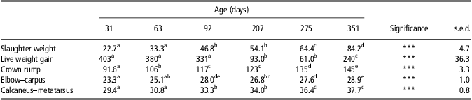
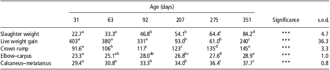
Calf live weight significantly increased (Table 1; P < 0.001) over the 12 months of the study, and there were no differences in live weight between Group 1 and Group 2 animals at any time. Live weight gain for each measurement period (Table 1) followed a seasonal pattern with lower growth rates during winter (207 to 275 days of age), compared with other seasons. Morphometric measurements (Table 1) of crown rump, elbow–carpus and calcaneus–metatarsus lengths increased with increasing live weight.
Slaughter weight (kg), live weight gain (g/day) and lengths (cm) of crown rump, elbow–carpus and calcaneus–metatarsus for red deer calves aged 31 to 351 daysFootnote 1
s.e.d. = standard error of the difference of the mean.
1 Total of four animals slaughtered at each age point, excluding 351 days of age when five animals were slaughtered.
* P < 0.05, **P < 0.01, ***P < 0.001, ns = non-significant.
a,b,c,d,e Means between columns within rows with different superscripts differ (P < 0.05).
Allometric growth of organs
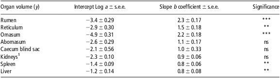
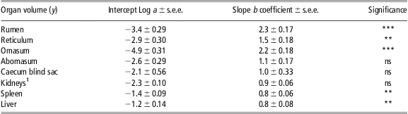
Individual organ allometric growth rates over 12 months of age, relative to deer live weight gain for both Group 1 and Group 2 animals combined are given in Table 2. Relative to live weight gain as a constant proportion, organ ‘maturity’ over the first 12 months of age ranked from greatest to least as: liver > spleen > kidneys > caecum blind sac > abomasum > reticulum > omasum > rumen.
Individual allometric organ growth rates of the rumen, reticulum, omasum, abomasum, caecum blind sac, kidneysFootnote 1, spleen and liver relative to deer live weight gain (g/day) over the first 12 months of age. Data were based on animal live weight gains and organ volumes (l) determined by repeat CT scanning in red deer calvesFootnote 2
CT = computer tomography; s.e.e. = standard error of the estimate.
1 Left and right kidney data combined.
2 Nine animals weighed and CT-scanned at each age point of 31, 63, 92, 135, 207 and 351 days, excluding 135 days of age when only five animals were used. Allometric organ growth rates were calculated using Huxley's equation of y = ax b, where y was organ size (i.e. volume); a was the scaling factor; x was live weight gain of the animals (g/day); and b was the coefficient value which compared y to x.
* P < 0.05, **P < 0.01, ***P < 0.001, ns = non-significant.
The b coefficient value in Table 2 indicates the relative growth rate of each organ in relation to live weight gain. The liver, spleen, kidneys and caecum blind sac had b coefficient values less than 1.0, and excluding the spleen and liver (P = 0.02) there were no significant relationships between organ growth and live weight gain. The forestomach organs (rumen, reticulum, omasum and abomasum) had b coefficient values greater than 1.0, and excluding the abomasum significant (P < 0.05) relationships between organ growth and live weight gain were found. These forestomach organs matured later and had faster growth rates, relative to live weight gain, compared with organs that had b coefficient values less than 1.0.
Changes in organ and digesta weights with increasing deer age
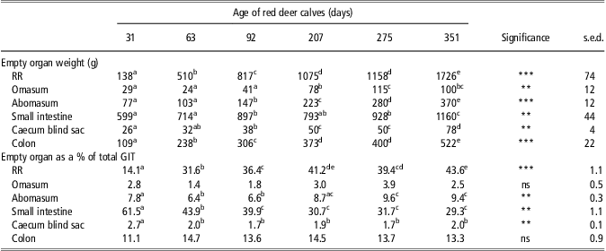
The purpose for measuring organ weights containing digesta (i.e. full organ weights) in this study was to compare organ weights with organ volumes determined by the CT scanner. Thus, only results considering empty organ weights (i.e. digesta removed) are presented here, as these represent changes in organ tissue that are independent of the amount of feed consumed before slaughter. The empty organ weights of the RR, omasum, abomasum, SI, caecum blind sac and colon in red deer calves had varying patterns of change over the first 12 months of age (Table 3). The empty weight of the RR had the greatest gain in tissue weight between 31 and 63 days of age (11.6 g/day) and between 63 and 92 days of age (10.6 g/day). Thereafter, the rate of tissue deposited by the RR was relatively small with next greatest gain of 7.5 g/day between the age of 275 and 351 days. By 351 days of age, the empty weight of the RR had increased (P < 0.001) almost 13-fold. The omasum empty weight was relatively static between 31 and 92 days of age, but thereafter there was a significant (P < 0.01) increase in weight up to 275 days, before decreasing by 15 g at 351 days. The abomasum increased (P < 0.001) in weight steadily over the 351-day measurement period, with the greatest incremental change between 275 and 351 days of age. Between 31 and 351 days, the SI almost doubled (P < 0.01) in empty weight. The caecum blind sac had small changes (P < 0.01) in empty weight over the total measurement period, but the greatest change occurred between 275 and 351 days. The empty weight of the colon increased (P < 0.001) about fivefold between 31 and 351 days of age.
Empty organ weights (g) (actual and expressed as a % of total GIT), as determined by removal of organs after slaughter from red deer calves aged from 31 to 351 daysFootnote 1

GIT = gastro-intestinal tract; s.e.d. = standard error of the difference of the mean; RR = rumen/reticulum.
1 Total of four animals slaughtered at each age point, excluding 351 days of age when five animals were slaughtered.
* P < 0.05, **P < 0.01, ***P < 0.001, ns = non-significant.
a,b,c,d,e Means between columns within rows with different superscripts differ (P < 0.05).
At the first measurement taken at 31 days of age, the SI occupied the greatest percentage of total GIT empty weight (60%; Table 3). However, by 351 days of age, the RR surpassed all other organs to occupy 44% of total GIT empty weight, and the contribution of the SI halved to 29%. The biggest change in RR occupancy of the total GIT empty weight was between 31 and 63 days of age (14% to 32%). Although changes were significant (P < 0.01), as a percentage of total GIT empty weight, the contributions of the omasum, abomasum and caecal blind sac all remained less than 10% over the 351 days. The colon maintained an average capacity of 13.5% of total empty GIT weight throughout the experiment.
Validation of CT scanning to determine organ volume
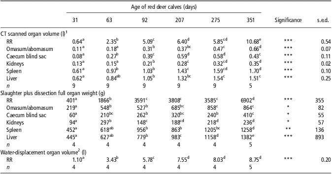
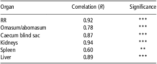
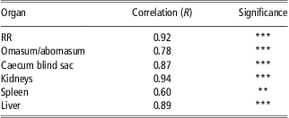
Three different methods were used for quantifying aspects of GIT development in red deer calves (Table 4). CT scanning was used to determine organ volume; full organ weights (including digesta) were determined by slaughter and dissection; and RR volume (once removed from the animal) was determined by the water-displacement method. According to the CT-scanned RR volume, the greatest change in volume was almost twofold, occurring between 275 and 351 days of age (5.85 to 10.68 l, respectively). This was supported by a similar change in RR full organ weight over the same time period (3585 to 6902 g, respectively). In contrast, the water-displacement method indicated little change in RR over the same time period (average RR volume of 8.39 l). It appeared that the water-displacement method and CT-scanned organ volumes were highly correlated with an R value of 0.92 (Table 5). CT-scanned organ volumes and organ weights obtained by slaughter plus dissection were also correlated (Table 5) with values varying from R = 0.94 for the kidneys (P = 0.001) to R = 0.60 for the spleen (P = 0.01).
Volumes (l; determined by CT scanning or the water-displacement technique) and full weights (g; determined using the traditional slaughter plus dissection technique) of the combined RR, combined omasum/abomasum, caecum blind sac, combined left and right kidneys, spleen, and liver from red deer calves aged 31 to 351 days
CT = computer tomography; RR = rumen/reticulum; s.e.d. = standard error of the difference of the mean.
1Organs contain digesta.
2Organs are without digesta and instead filled with water.
*P < 0.05, **P < 0.01, ***P < 0.001, ns = non-significant.
a,b,c,d,eMeans between columns within rows with different superscripts differ (P < 0.05).
The correlation (R) between volumes (l; determined by CT scanning or the water-displacement technique) and full weights (g; determined the traditional slaughter plus dissection technique) of the combined RR, combined omasum/abomasum, caecum blind sac, kidneys, spleen and liver from red deer calves over the first 12 months of age
CT = computer tomography; RR = rumen/reticulum.
RR correlation is between CT-scanned RR volume and RR volume determined by the water-displacement technique, not RR weight as used for the other organs.
*P < 0.05, **P < 0.01, ***P < 0.001, ns = non-significant.
Discussion
This study has provided a detailed description of the development of the farmed red deer GIT over the first 12 months of age during transition from pre-ruminant to ruminant status. Novel information on allometric growth of GIT organs in relation to live weight gain over the first 12 months in red deer calves has highlighted the slow maturity of the rumen compared with other GIT organs. CT scanning was also validated as a viable technique to follow GIT development in the same animals from 1 to 12 months of age, with CT-scanned data on organ volume significantly and positively correlated with actual organ weights, and with RR volume determined by the water-displacement technique.
Allometric growth of organs
Allometric organ growth indicated the relative maturity of individual GIT organs in relation to live weight gain during the first 12 months of age. In the early stages of life (i.e. 31 days), organs from most mature to least mature were liver > kidneys > spleen > abomasum > reticulum > caecum blind sac > omasum > rumen. With increasing live weight gain, those organs that were more mature at birth (e.g. liver) had a slower subsequent growth rate. This was because they had already reached a high proportion of their mature weight at birth. Of the forestomach organs, the rumen had the fastest growth rate, followed by the omasum, reticulum and abomasum. Although there are limited allometric organ growth data available in the literature, particularly with red deer, work by Abdallah et al. (Reference Abdallah, Shahin and Latif1982) (water buffalo) and Wardrop and Coombe (Reference Wardrop and Coombe1960) (sheep) found the rumen to have the fastest growth rate, followed by the reticulum, omasum and abomasum.
Earlier work, mainly with domestic sheep and cattle, indicated that internal organs can have markedly different growth rates between animal species, with their maximum growth rates occurring at different ages and live weights (Wardrop and Coombe, Reference Wardrop and Coombe1960; Oh et al., Reference Oh, Hume and Torell1972; Baldwin, Reference Baldwin2000). This information has been relatively unknown for red deer. In this study, deer calf GIT organs grew at different rates throughout the experiment, with the most rapid growth generally occurring in the earlier stages of development. The allometric growth of the RR in bovine calves and lambs has shown to be most rapid, relative to live weight, before 56 days of age (Wardrop and Coombe, Reference Wardrop and Coombe1960; Tamate et al., Reference Tamate, McGilliard, Jacobson and Getty1962). At 84 to 92 days of age, growth rates of the RR, as a proportion of total forestomach weight, was greater for red deer (87%, this study) and white-tailed deer (79%, Short, Reference Short1964), compared with cattle (66%, Godfrey, Reference Godfrey1961b), buffalo (71%, Singh et al., Reference Singh, Yadava and Rao1973) and sheep (71%, Wardrop and Coombe, Reference Wardrop and Coombe1960). This suggests that the absolute growth rate of the RR in red deer is greater than that observed in other ruminants at the same age. It was also found that, in both red deer (351 days old, from this study) and white-tailed deer (older than 351 days, Short, Reference Short1964), the RR, as a percentage of total forestomach weight, was greater (89% and 80%, respectively) compared with adult cattle (62%, Murray et al., Reference Murray, Tulloh and Winter1977), adult buffalo (70%, Abdallah et al., Reference Abdallah, Shahin and Latif1982) and adult sheep (73%, Wallace, Reference Wallace1948).
The RR was shown to be increasing in empty tissue weight up until the last measurement day taken at 351 days of age. Reports by Wardrop and Coombe (Reference Wardrop and Coombe1960), Godfrey (Reference Godfrey1961a) and Davis and Drackley (Reference Davis and Drackley1998) are indicative of species and/or experimental variation during the time that forestomach organs reach adult proportions, which is defined by no further changes in organ weight. This study found that individual forestomach organ weights were continuing to change at the last measurement taken at 351 days of age. However, as a proportion of total combined forestomach weight, individual organs did not make any considerable changes after 92 days of age, similar to white-tailed deer (Short, Reference Short1964). In contrast, individual organs of buffalo (Singh et al., Reference Singh, Yadava and Rao1973) and sheep (Wardrop and Coombe, Reference Wardrop and Coombe1960) had no further changes beyond 56 to 63 days of age. Information from domestic cattle is less defined, with forestomach maturity occurring between 56 and 119 days of age or later (Godfrey, Reference Godfrey1961a).
The differences in the age that the forestomachs mature in different ruminant species may be because of confounding factors, such as experimental methodology, body size, weaning time, type of diet consumed and seasonality. These factors could not be determined from the experiments cited here. In this study, it was likely that seasonality had a substantial influence on GIT development. Using CT scanning, it was observed that the RR volume increased to a maximum capacity at 135 days of age (8.73 l; autumn), before decreasing between 135 and 207 days (7.55 l) during winter, and once again increasing up to 351 days of age by spring (8.75 l). In red deer, seasonality affects rumen pool sizes, rumen digesta load, voluntary feed intake and body growth (Domingue et al., Reference Domingue, Dellow, Wilson and Barry1991a and Reference Domingue, Dellow, Wilson and Barry1991b; Sibbald and Milne, Reference Sibbald and Milne1993). It cannot be ignored that seasonality may have had a significant effect on the rumen, GIT capacity and development in red deer in this study, in addition to the effects of age per se. When the rumen volume was compared with the rumen digesta pool weight, the proportion of rumen volume occupied by digesta was 0.50 at day 207 and 0.79 at day 351. These values are very similar to those calculated by Freudenberger et al. (Reference Freudenberger, Toyakawa, Barry, Ball and Suttie1994), who supported the view that some of the variation between sampling times were influenced by seasonality of intake rather than age.
CT scanning
This study indicates that CT scanning for diagnostic imaging has the potential to be useful for following GIT development in the same animal, rather than slaughtering a series of animals. CT scanning has the ability to provide information on the effect of different weaning methods and demonstrate the effect this can have on GIT development in young ruminants. This would be a useful tool and an indicator for better weaning management practices and hence more efficient animals. The timing of weaning and its relative success depends on the development of an RR that is able to digest forages effectively to supply nutrients for body growth. This study has indicated that the most critical time for RR development of farmed red deer grazing pasture is from 30 to 60 days of age, and hence weaning earlier than 60 days of age may not be advisable for optimum rumen development. However, there is no indication of the ideal time for weaning to occur with regard to GIT development. Thus, the impact of weaning and management, in terms of diet and optimal growth performance, needs evaluation in further studies.
Conclusions
The growth and development of the red deer GIT continues until at least 351 days of age. Red deer calves appear to have a similar or greater rate of RR development up until ∼60 to 92 days of age compared with sheep and cattle. However, the final increments of GIT maturity in red deer may take longer to complete than in sheep or cattle, with a hiatus being brought about by the seasonal nature of voluntary feed intake during winter. The CT-scanning technique was highly accurate in determining organ volume, particularly the RR. Information on allometric GIT organ growth of red deer is now available, highlighting the slow rate of RR maturity compared with other GIT organs in relation to live weight gain. Further research on GIT development is needed to confirm these results and investigate other variables such as the effects of diet type and weaning management practices.
Acknowledgements
The authors thank Wendy Bain for CT-scanning expertise, Dr Jason Archer and Jamie Ward for help with most aspects of the study, as well as a number of staff from AgResearch Invermay involved with animal management, handling and dissection. Financial support for this study was given by AgResearch Ltd, DEEResearch, the Foundation of Research Science and Technology and the Institute of Veterinary, Animal and Biomedical Sciences (IVABs), Massey University. Dr Kirsty Hammond acknowledges financial assistance from the Dave McGrath Memorial Scholarship, Kathleen Stewart Scholarship, Hurley Fraser Postgraduate Scholarship and the Purehuroa Summer Research Award.