The dry period is a critical part of the lactation cycle in dairy cows, and is essential for achieving optimal milk yield in the subsequent lactation (Capuco et al. Reference Capuco, Akers and Smith1997; Bachman & Schairer, Reference Bachman and Schairer2003). The non-lactating period is required for mammary gland remodelling processes, including regression (apoptosis), proliferation and differentiation of mammary cells to prepare for the ensuing lactation (Capuco et al. Reference Capuco, Akers and Smith1997; Bachman & Schairer, Reference Bachman and Schairer2003). The importance of mammary remodelling in dairy cows has been demonstrated by early experiments using split-udder designs, which established that the need for a dry period is related to local changes in the mammary gland (Smith et al. Reference Smith, Wheelock and Dodd1966). In the absence of a dry period, milk yield in the subsequent lactation may be reduced as much as 20% (Capuco & Akers, Reference Capuco and Akers1999; Bachman & Schairer, Reference Bachman and Schairer2003). Several studies have been carried out to redefine the optimal dry period length to maximize subsequent milk (Bachman & Schairer, Reference Bachman and Schairer2003). In addition, any influence on mammary remodelling that accelerates mammary involution during the dry period may contribute to an enhanced milk yield during the subsequent lactation.
Recent studies have quantified mammary cell turnover in dairy cows (Capuco et al. Reference Capuco, Wood, Baldwin, Mcleod and Paape2001; Sorensen et al. Reference Sorensen, Norgaard, Theil, Vestergaard and Sejrsen2006; Norgard et al. Reference Norgaard, Theil, Sorensen and Sejrsen2008); however, the mechanisms regulating apoptosis and cell proliferation during the lactation and dry period cycle are still unclear. Controlled cell death in the bovine mammary gland is mediated by apoptosis and, to a lesser extent, autophagy (Zarzynska et al. Reference Zarzynska, Gajkowska, Wojewódzka, Dymnicki and Motyl2007). There are two major pathways leading to apoptosis: an extrinsic and an intrinsic pathway. The extrinsic pathway is induced by activation of death receptors located on the cell surface. The intrinsic pathway of apoptosis involves the mitochondria and can be triggered by intracellular stressors such as oxidants. A variety of proteins that are involved in maintaining a stable intramitochondrial environment and membrane integrity have been identified (Fernandez-Checa, Reference Fernández-Checa2003).
Panax ginseng C. A. Meyer as a traditional medicine has been utilized in China for at least 2000 years. Ginseng saponins, or ginsenosides, are believed to be the active substances in total ginseng extracts. They are chemically triterpenoid glycosides of the dammarane series. At present, more than 30 ginsenosides have been identified in P. ginseng (Song et al. Reference Song, Chen, Li and Hu2010). The therapeutic effect of ginseng root (GS) may be related to its stimulation of the natural resistance against infections (Owen, Reference Owen1981). Ginseng extracts consisting mainly of saponins have been found to possess various effects on the immune system, such as enhancement of lymphocyte proliferation, stimulation of macrophages for cytokine production and improvement of macrophages and polymorphonuclear leucocytes phagocytic activity (Jie et al. Reference Jie, Cammisuli and Baggiolini1984; Yun et al. Reference Yun, Moon, Oh, Jo, Kim and Yun1987; Scaglione et al. Reference Scaglione, Ferrara, Dugnani, Falchi, Santoro and Fraschini1990; Kim et al. Reference Kim, Germolec and Luster1990; Scaglione et al.Reference Scaglione, Cattaneo, Alessandria and Cogo1996; Larsen et al. Reference Larsen, Moser, Hoiby, Song and Kharazmi2004). Adjuvant activities are ascribed to GS saponins’ chemical structure, suggesting that may act by triggering innate immunity stimulating secretion of a broad range of cytokines (Song & Hu, Reference Song and Hu2009). In addition, GS has potential as a chemopreventive agent through mechanisms that include inhibition of DNA damage (Park et al. Reference Park, Yeo, Jin, Lee, Jung, Choue, Cho and Hahm2005), induction of apoptosis by oxidative stress (Volate et al. Reference Volate, Davenport, Muga and Wargovich2005) and inhibition of cell proliferation (Kang et al. Reference Kang, Kim, Kim, Chae, Koh, Kim, Choo, Kim and Hyun2005). It is also becoming increasingly clear that ginseng has potent effects on the inflammatory cascade and may inhibit the inflammation-cancer sequence (Hofseth & Wargovich, Reference Hofseth and Wargovich2007).
Previous investigations in vivo and in vitro with GS have shown that the dry root extract has immunomodulatory and adjuvant effects in bovine udder (Hu et al. Reference Hu, Concha, Johannisson, Meglia and Waller2001, Reference Hu, Concha, Lin and Persson Waller2003). However, information about the in-vivo direct effects of ginseng in bovine mammary gland is not clear. In addition, to our knowledge, there is no available literature published about the intramammary effects of ginseng at drying off. Therefore, the objective of this study was to describe the effects of a single intramammary infusion of Panax ginseng extract on cell proliferation and death mechanism in bovine mammary gland during early involution.
Materials and Methods
Ginseng extract
Ginseng dry extract containing saponins equivalent to 27% ginsenoside Rg1, was kindly provided by Indena Company (Indena® SPA, Milan, Italy). The inoculation solution was prepared by dissolving the extract in 0·89% saline to a final concentration of 3 mg ginseng extract per ml. The solution was sterilized by filtering it through a 0·22-μm membrane filter and then sealed in sterilized 250-ml glass bottles. The solution was prepared 1 d before the start of the experiment and stored at 4°C. Sterility was checked by seeding 100 μl in agar Columbia added with 5% calf blood and incubating overnight at 37°C.
Prior to the present study, a dose-response trial was carried out to select the ginseng extract dose. Briefly, four Holstein non-pregnant cows in late lactation from the Rafaela Experiment Station of INTA were used for these tests. One quarter of each cow was intramammarily infused with ginseng extract concentrations of 3, 5 and 10 mg/ml while the remaining quarter was infused with placebo (vehicle alone). The dose yielding the highest somatic cell count (SCC) response without gross mammary swelling or systemic adverse effects (i.e. elevated rectal temperature and increase in respiratory frequency) was selected.
Animals and experimental design
Six Holstein non-pregnant cows from parity 3–5 in late lactation (weeks 31–36) from the Rafaela Experiment Station of INTA herd were used. Cows were milked twice daily and produced an average of 10 kg milk/d before interruption of lactation. All procedures were carried out according to the Guide for the Care and Use of Agricultural Animals in Agricultural Research and Teaching (FASS, 1999). The animals were selected based on previous bacteriological studies and SCC. All the quarters used in this work were free of infection. The unit of study was the mammary quarter. Eight quarters were infused with 10 ml of ginseng solution (3 mg/ml), eight quarters were treated with 10 ml of placebo (vehicle alone) and eight quarters were maintained as uninoculated controls. Two quarters of placebo-inoculated and of non-inoculated cows were not considered for the experiment owing to high SCC at the time of inoculation. Milking was stopped after infusion. Cows were given only alfalfa hay and had free access to water for the remainder of the experiment.
Mammary secretion samples were aseptically collected for bacteriological analysis using standard procedures (Hogan et al. Reference Hogan, Harmon, González, Nickerson, Oliver, Pankey and Smith1999) 3 d before ginseng administration, immediately before inoculation and every 48 h after infusion. The first two streams of milk from each teat were discarded and the next 5 ml were collected in sterile plastic vials for bacteriological analysis. Teats were dipped in a 0·5% iodophor solution after samples were taken. Animals included in the three groups were slaughtered at 7 d after inoculation at a local abattoir and samples for histological analysis were taken.
Bacteriological examination
Mammary secretion samples (10 μl) were streaked onto blood agar plates supplemented with 5% bovine blood and incubated for 48 h aerobically at 37°C. Plates were examined for bacterial growth at 24 h and 48 h. Isolated colonies were identified according to standard procedures (Hogan et al. Reference Hogan, Harmon, González, Nickerson, Oliver, Pankey and Smith1999). Intramammary infection was defined as isolation of the same organism from two consecutive samples.
Tissue sample preparation
Immediately after cows were slaughtered tissue samples were obtained from three zones of mammary quarters following previous descriptions (Capuco et al. Reference Capuco, Akers and Smith1997). Mammary tissue was obtained from the base of the gland adjacent to the gland cistern (zone 1), midway between the gland cistern and dorsal boundary of the gland (zone 2) and near the dorsal mammary border (zone 3). All zones were approximately oriented along an axis through the centre of the gland in line with the teat. Tissue samples of approximately 1 cm3 were fixed in 4% neutral buffered formalin, for 8 h at 4°C and then washed in phosphate-buffered saline (PBS; pH 7·4 0·01 m). For light microscopy, fixed tissues were dehydrated and embedded in paraffin wax. Sections (5 μm) were mounted on slides previously treated with 3-aminopropyltriethoxysilane (Sigma-Aldrich, St. Louis MO, USA) and assigned for staining with haematoxylin and eosin (HE) for mammary gland structures preliminary observation and morphometric studies or for use in immunohistochemistry (IHC) procedures (Dallard et al. Reference Dallard, Ortega, Lorente and Romano2005). Additional sections of mammary tissues were transferred into a freezing vial, weighed and placed in liquid nitrogen for further use in gene expression assays.
Immunohistochemistry
A streptavidin-biotin immunoperoxidase method was performed as described by Dallard et al. (Reference Dallard, Baravalle, Ortega, Ruffino, Heffel and Calvinho2008, Reference Dallard, Baravalle, Ortega, Tumini, Canavesio, Neder and Calvinho2009). Briefly, sections were dewaxed, hydrated and subjected to microwave pretreatment in citrate buffer (pH 6·0) for antigen retrieval. Endogenous peroxidase activity was blocked with H2O2 (1% in methanol) and non-specific binding was blocked with 10% normal goat serum (Sigma-Aldrich). Sections were incubated overnight at 4°C with rabbit polyclonal antiserum against bcl-2 protein (Abcam, Cambridge, UK) diluted 1:100, bax polyclonal antibody diluted 1:30 (BioGenex, San Ramon CA, USA), active caspase-3 polyclonal antibody diluted 1:400 (R&D Systems, Minneapolis MN, USA) and Ki-67 monoclonal antibody diluted 1:40 (BioGenex). Slides were washed with PBS and incubated at room temperature for 30 min with rat pre-absorbed biotinylated secondary antibodies specifically selected against one of each of the two types of primary antibodies used (monoclonal or polyclonal). Visualization of antigen-antibody reaction was by the streptavidin peroxidase method (BioGenex) and 3,3-diaminobenzidine (Liquid DAB-Plus Substrate Kit-Zymed, San Francisco CA, USA) was used as chromogen. Finally, the slides were washed in distilled water and counterstained with Mayer's haematoxylin, dehydrated and mounted. Adjacent control sections were performed as described by Dallard et al. (Reference Dallard, Baravalle, Ortega, Ruffino, Heffel and Calvinho2008, Reference Dallard, Baravalle, Ortega, Tumini, Canavesio, Neder and Calvinho2009).
Terminal deoxynucleotidyl transferase dUTP nick end labelling (TUNEL)
In-situ detection of apoptotic cells utilized terminal deoxynucleotidyl transferase dUTP nick end labelling (TUNEL) to visualize cells that exhibited endonucleolytic degradation of DNA, a key feature of apoptotic cells. A commercial kit (ApopTag Plus Peroxidase, Chemicon International, Temecula CA, USA) was used following previous descriptions (Dallard et al. Reference Dallard, Baravalle, Ortega, Ruffino, Heffel and Calvinho2008). Briefly, after deparaffinization and hydration, slides were incubated with proteinase K (Dako North America Inc., Carpinteria CA, USA) for 15 min at 25°C. Tissue sections were quenched in 3% H2O2 in PBS, incubated in equilibration buffer for 10 min, and then with terminal deoxynucleotidyl transferase (TdT) for 60 min at 37°C. Sections were washed with stop wash buffer for 10 min and then incubated with antidigoxigenin-peroxidase for 30 min at 25°C. Tissue sections were washed in PBS and then incubated with diaminobenzidine for approximately 6 min at room temperature for colour development. Sections were washed in distilled water, counterstained with Mayer's haematoxylin, dehydrated and mounted.
RNA extraction
Total RNA was extracted from frozen mammary tissue samples using Trizol LS Reagent (Invitrogen, Carlsbad CA, USA) following the manufacturer's instructions with slight modifications. Briefly, 50–100 mg of tissue was homogenized with 1 ml of Trizol reagent (Invitrogen) and incubated for 10 min at 25°C. RNA was purified by vigorously homogenizing with chloroform and incubating for 15 min at 4°C. After centrifugation at 12 000 g, the aqueous phase was incubated with an equal volume of isopropanol for 30 min at −20°C and centrifuged at 12 000 g to obtain the pellet of mRNA that was then washed with 75% ethanol for 10 min at 4°C. Alcohol was replaced by diethylpyrocarbonate (DEPC)-water (Sigma-Aldrich) pre-warmed to 55–60°C. Total RNA content was determined spectrophotometrically at 260 nm; then samples were aliquoted and stored at −80°C until further use.
Reverse transcriptase polymerase chain reaction (RT-PCR)
Apoptotic and antiapoptotic gene expression was assessed using a semiquantitative multiplex RT-PCR measuring bcl-2 and bax. To avoid putative genomic DNA contamination, RNA samples were treated with DNase (Invitrogen) according to the manufacturer's instructions. Single-stranded cDNA was reverse transcribed from total RNA that had been treated with DNase using a master mix (MMLV buffer, DTT, RNA out, M-MMLV reverse transcriptase, dNTP) and random primers (Invitrogen). The reverse transcription conditions consisted of 10 min of annealing at 25°C, 50 min of cDNA synthesis at 37°C and 15 min of inactivation at 70°C. Conditions for the enzymic amplification were optimized. Ten nanograms of cDNA previously quantified by the Qubit method (Invitrogen) were used for the PCR.
Details of procedures for primer design used in the present study to measure mRNA levels of bcl-2 and bax have been described previously (Colitti et al. Reference Colitti, Wilde and Stefanon2004). Primer sequences used were: bcl-2 forward 5′-ATGTGTGTGGAGAGCGTCAA-3′, reverse 5′-CAGACTGAGCAGTGCCTTCA-3′ and bax forward 5′-TTTGCTTCAGGGTTTCATCC-3′, reverse 5′-TGGGTGTCCCAAAGTAGGAG-3′. The β-actin gene sequence was included as housekeeping: forward primer 5′-CGGAACCGCTCATTG CC-3′, reverse 5′-ACCCACACTGTGCCCATCTA-3′ and the primer sequence was designed from the bovine sequence (GenBank accession No BT030480). Conditions used for multiplex PCR amplification of bcl-2+bax mRNA have been previously described (Colitti et al. Reference Colitti, Wilde and Stefanon2004). PCR products were analysed by agarose gel electrophoresis and the relative signal intensities of all examined factors were assessed after normalization based on the β-actin PCR signal intensities.
Image analysis
Image analysis was performed using Image Pro-Plus 3.0.1 system (Media Cybernetics, Silver Spring MA, USA). Images were digitized by a CCD colour video camera (Motic 2.000, Motic China Group, China) mounted on top of a conventional light microscope (Olympus BH-2, Olympus Co., Japan) using an objective magnification of 40×. Resolution of the images was set to 1200×1600 pixels. Each pixel of the image corresponded to 0·13 μm at the respective magnification and each field represented a tissue area of 0·031 mm2. Details of image analysis as a valid method for quantifying expression levels and the methodological details were previously described (Dallard et al. Reference Dallard, Ortega, Lorente and Romano2005; Reference Dallard, Ruffino, Heffel and Calvinho2007). Briefly, the immunohistochemical stained area (IHCSA) for antibody reaction was calculated as a percentage of total area evaluated through the colour segmentation analysis that extracts objects by locating all objects of the specific colour (brown stain). The brown stain was selected with a sensitivity of 4 (maximum 5) and a mask was next applied to make separation of colours permanent. The IHCSA (% of black area) was calculated from at least 50 images in each one of the following structures: alveoli, ducts and interstitial tissue. Bcl-2, bax and active caspase-3 expressions were evaluated by positive IHCSA.
The percentage of tissue area occupied by stroma was determined by a quantitative morphometric analysis described previously (Dallard et al. Reference Dallard, Ortega, Lorente and Romano2005, Reference Dallard, Ortega, Iguzquiza, Salvetti, Quaino and Calvinho2010). Two independent tissue blocks for each zone within a quarter and twenty random fields of the sections from each block were evaluated. Image resolution was set to 640×480 pixels. At the magnifications used, each pixel of the image corresponded to 2·68, 1·04 and 0·26 μm2, respectively, and each field in the monitor represented a tissue area of 2·2, 0·33 and 0·02 mm2, respectively.
Quantification of TUNEL-positive cells in the epithelium and stroma of mammary gland was measured as described previously (Dallard et al. Reference Dallard, Baravalle, Ortega, Ruffino, Heffel and Calvinho2008). Cells were classified as epithelial, stromal, labelled epithelial or labelled stromal cells. Labelled brown nuclei were readily visible and a cell was classified as labelled when the nuclear staining was at least twice as intense as the background.
Quantification of epithelial and stromal cells labelled with anti Ki-67 protein was performed in a similar manner as for TUNEL. All immunostained mammary cells showing a characteristic nuclear staining pattern were considered positive. A minimum of 1000 cells in at least 20 microscopic fields at 40× magnification were counted in each slide. Epithelial and stromal cells showing an intense nuclear staining were quantified and results were expressed as a percentage of immunopositive cells.
Balance between dying and proliferating cells was estimated using the ratio of apoptosis to cell proliferation (A/P). The mean ratio of the number of TUNEL or Ki-67 positive cells to total cell number in each set of tissue sections was calculated following previous descriptions (Colitti et al. Reference Colitti, Wilde and Stefanon2004; Dallard et al. Reference Dallard, Baravalle, Ortega, Ruffino, Heffel and Calvinho2008).
For bax and bcl-2 mRNA expressions, images of gels were captured by a digital camera (Olympus). Bax and bcl-2 mRNA levels were expressed as a ratio between the integrated optical density (IOD) band of both mRNA and the corresponding band of β-actin. Balance between pro- and anti-apoptotic genes was estimated using the ratio of IOD band of bax and bcl-2 mRNA.
Statistical analysis
A statistical software package (SAS, 1999) was used to perform statistical analysis. Differences between treatments were analysed by Kruskal-Wallis non-parametric test, followed by Bonferroni as a multiple comparison test. A value of P<0·05 was considered significant. Results are expressed as mean±sem.
Results
Bacteriological examination and side effects
Macroscopic changes in mammary secretions and moderate swelling were observed in mammary quarters of GS-treated group at 24 h pt. All samples from mammary gland secretions were negative for pathogenic bacteria. With the dose selected, no side effects, other than those mentioned, were observed during the experimental period.
Morphometric analysis
Analysis of mammary tissue area occupied by stroma, at 7 d after infusion, showed no differences between zones within the gland (P>0·15). Percentages of connective tissue stroma were significantly affected (P<0·05) by treatment, averaging 69·40±2·47, 57·10±5·14 and 54·18±7·53% in quarters treated with GS, placebo and in untreated controls, respectively. In every experimental group, at 1 week after cessation of milking, small and large alveoli showing cuboidal epithelial cells were observed. Secretory epithelial cells contained large fat droplets at the apical area and a small number of cells presented irregular nuclei with chromatin condensation and a highly vacuolated cytoplasm. There was no evidence of extensive sloughing of alveolar epithelial cells. Mammary tissue was composed mainly of alveolar lumen and little interalveolar tissue was present.
Evaluation of apoptosis
Immunohistochemistry for bcl-2, bax and active caspase-3
The positive immunohistochemical reaction with each protein (bcl-2, bax and active caspase-3) was detected by brown cytoplasmic staining and evaluated by the IHCSA. Differences in the pattern of localization and IHCSA for each protein were observed within the mammary gland. Effects of GS treatment on IHCSA for bax, bcl-2 and active caspase-3 at drying off are summarized in Table 1. No differences in percentages of IHCSA for the three evaluated proteins in control and treated groups between zones were detected.
Effects of intramammary infusion of Panax ginseng (GS) extract on the relative expression of bcl-2, bax and active caspase-3 at drying off. Values are means of percent immunohistochemical stained area across zones within the gland±sem

† Means within a row with different superscripts differ (P<0·05)
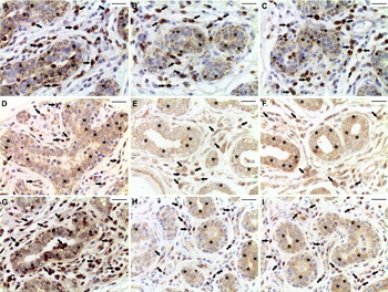
Immunostaining for bcl-2 protein was observed in all tissue sections, ranging from weak to intense, and was mainly associated with the mammary parenchyma structures. Positive reaction was observed in nuclei, particularly around the inner nuclear membrane and in the cytoplasm of epithelial and stromal cells (Fig. 1). No differences in percentages of stained area for bcl-2 protein between control and treated groups at either zone were detected.
Immunohistochemical localization of bcl-2, bax and active caspase-3. A) Bcl-2 immunostaining in mammary quarters treated with Panax ginseng (GS), B) placebo and C) uninoculated controls at 7 d of drying off. D) Bax immunodetection in mammary quarters treated with GS, E) placebo and F) uninoculated controls at 7 d of drying off. G) Active caspase-3 immunostaining in mammary quarters treated with GS, H) placebo and I) uninoculated controls at 7 d of drying off. Positive staining is shown as a brown colouring of the cytoplasm (asterisks) and nucleus (heads) of the cells. Magnification is the same for all panels and is indicated by 25-μm bars. [A colour version of this Figure is available online as Supplementary Material on Cambridge Journals Online (http://journals.cambridge.org)].

Expression of bax protein was primarily associated with the epithelium of the alveoli and ducts of the mammary parenchyma. The pattern of localization for bax was similar to bcl-2 expression. A significant increase of immunostained area for bax protein was observed in GS-treated quarters compared with placebo-treated and untreated controls (P<0·05) (Fig. 1).
Immunostaining for active caspase-3 was observed in the cellular nucleus and cytoplasm of epithelial and stromal cells. In GS-treated quarters the percentages of IHQSA for this proteinase showed a significant increase compared with controls (P<0·05) (Fig. 1).
RT-PCR analysis for bcl-2 family
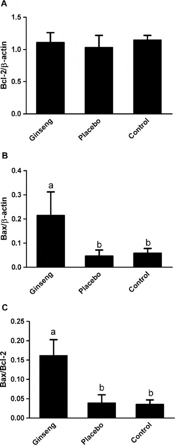
Multiplex PCR analysis was used to compare the relative levels of transcriptional activity for bcl-2 and bax in mammary tissue. Analysis of PCR products was expressed as the ratio of densitometric readings for bcl-2 and bax to β-actin. Expression of bcl-2 mRNA (201 bp) and bax mRNA (431 bp) was detected in all samples analysed. A high expression of bcl-2 mRNA was evident in all samples compared with bax mRNA expression; however the levels of bcl-2 mRNA showed non-significant differences between treatments. Expression of bax mRNA was significantly higher in GS-treated quarters than in controls (P<0·05). The bax/bcl-2 ratio, which is considered as an index of cell susceptibility to apoptosis, indicated a significant predominance of bax expression over bcl-2 expression in GS-treated quarters compared with controls (P<0·05) (Fig. 2).
Semi-quantification of bcl-2/β-actin mRNA (A), bax-2/β-actin mRNA (B) and bax/bcl-2 mRNA ratio (C) in mammary quarters treated with Pananx ginseng (n=8), placebo (n=6) and uninoculated controls (n=6). Values represent the mean of integrated optical density (IOD)±sem. Means without a common letter (a–b) differ (P<0·05).

Detection of apoptosis in situ (TUNEL)
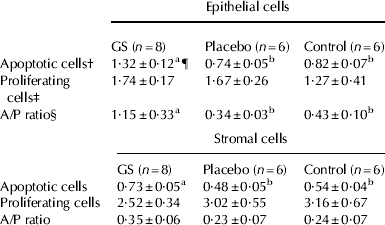
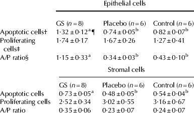
Apoptotic epithelial and stromal cells were distributed apparently at random through the alveolar epithelium and interalveolar stroma, identified as single cells rather than clusters (Table 2). Apoptotic events were evident as TUNEL-positive cells juxtaposed with healthy cells. Percentages of apoptotic cells in parenchymal and stromal tissue were affected by GS treatment. A significant increase of TUNEL-positive epithelial cells was observed in quarters treated with GS compared with placebo-treated quarters and untreated controls (P<0·05). Regarding stromal cells immunostaining, significant differences in percent of apoptotic cells between GS-treated quarters and controls (P<0·05) were observed. The percentages of labelled epithelial and stromal cells for the different treatment groups were not influenced by zone within the mammary gland (P>0·15). Percentages of TUNEL-positive epithelial cells were higher than percentages of stromal cells (P<0·05) both in GS-treated quarters and in controls.
Effect of treatment with Panax ginseng (GS) extract on percentage of apoptotic cells and proliferating cells. Values represent means of percent epithelial and stromal stained cells across zones within the gland ±sem
† Assessed by TUNEL
‡ Assessed by Ki-67 staining
§ A/P ratio: ratio of apoptotic to proliferating cells
¶ Means within a row with different superscripts differ (P<0·05)
Cellular proliferation
The proliferation index was evaluated by Ki-67 immunohistochemistry. No differences were detected in epithelial and stromal cells proliferation index among control and treated groups at either zone. Epithelial cells showed greater percentages of proliferation than stromal cells in every group evaluated, but the differences were not significant. Percentages of proliferation of mammary epithelial and stromal cells are summarized in Table 2.
Ratio of apoptotic to proliferating cells
Ratio of apoptotic to proliferating cells (A/P) is shown in Table 2. A significant increase of A/P ratio was observed in quarters treated with GS compared with controls at 7 d of involution (P<0·05) for epithelial cells. Regarding stromal cells, no differences in A/P ratio between treatments were detected.
Discussion
In dairy cows, a non-lactating period is required for mammary gland remodelling processes, including regression (apoptosis), proliferation and differentiation of mammary cells to prepare for the ensuing lactation (Capuco et al. Reference Capuco, Akers and Smith1997; Bachman & Schairer, Reference Bachman and Schairer2003). Whereas the need for an adequate dry period has been established, physiological and biochemical events during involution and influence of biological response modifiers on these events are poorly understood (Bachman & Schairer, Reference Bachman and Schairer2003).
In the present study, the effect of P. ginseng extract on cell proliferation and death mechanism administered in a single dose at the end of lactation in Holstein cows was determined. The ginseng dose used in this study proved to be safe for cows, causing only mild inflammation. The percentage of bovine mammary tissue area composed of epithelium and lumen decreases during gland involution while the percentage of stromal area increases, reaching a peak between 14 and 25 d after milking is interrupted (Sordillo & Nickerson Reference Sordillo and Nickerson1988; Capuco et al. Reference Capuco, Akers and Smith1997). Changes in mammary epithelial cells observed in this study in untreated quarters were in accord with those described in naturally involuting mammary glands 7 d after drying off (Holst et al. Reference Holst, Hurley and Nelson1987; Wilde et al. Reference Wilde, Addey, Li and Fernig1997; Dallard et al. Reference Dallard, Baravalle, Ortega, Ruffino, Heffel and Calvinho2008). Percentages of area occupied by stroma showed a significant increase in GS-treated quarters, indicating an intense tissue remodelling in all zones evaluated, which was mainly due to the presence of abundant interlobular connective tissue. In addition, following GS inoculation mammary morphology was not altered since alveolar and stromal structures were mantained.
Mammary gland remodelling depends on a dynamic equilibrium between two opposite processes: mitosis and apoptosis. The latter is responsible for the loss of cells during mammary involution after natural weaning or litter removal in rodents (Quarrie et al. Reference Quarrie, Addey and Wilde1996) and during drying off in ruminants (Quarrie et al. Reference Quarrie, Addey and Wilde1994; Wilde et al. Reference Wilde, Addey, Li and Fernig1997). Biochemically, apoptosis is characterized by the activation of caspases, highly specific proteinases that cleave a wide array of intracellular substrates (Thornberry & Lazebnik, Reference Thornberry and Lazebnik1998). The variations in the expression of pro- and anti-apoptotic proteins network in the bovine mammary gland have not yet been fully explored, and the exact role of apoptosis-regulating genes at drying off is not entirely clear (Zarzynska & Motyl Reference Zarzynska and Motyl2008). In order to elucidate the mechanism by which GS induce apoptosis, regulatory proteins (bax and bcl-2) and an executor of apoptosis (active caspase-3) were analysed by immunohistochemistry. The staining for these proteins was located in the nuclei, particularly around the inner nuclear membrane and in the cytoplasm of epithelial and stromal cells, this coincided with bax, bcl-2 and caspase-3 distribution in sow involuting mammary gland (Motyl et al. Reference Motyl, Gajkowska, Wojewódzka, Wareski, Rekiel and Płoszaj2001). The results from this experiment revealed that GS inoculation increased bax expression with a concomitant maintenance in bcl-2 protein levels. It has been shown that bax accumulation on organellar membranes precedes the activation of caspase-3, an apoptosis executor (Zhang et al. Reference Zhang, Heim and Meyhack1998). Immunohistochemical labelling of CPP-32 (caspase-3) in goat mammary glands revealed a significant increase of this enzyme content, concurrent with a degree of mammary tissue involution, suggesting the involvement of caspase-dependent pathways in the execution of apoptosis in this tissue (Wareski et al. Reference Wareski, Motyl, Ryniewicz, Orzechowski, Gajkowska, Wojewodzka and Ploszaj2001). In this study, labelling of active caspase-3 showed a significant increase in this enzyme content in GS-treated quarters compared with controls, suggesting that this compound induces apoptosis via the mitochondrial pathway. These results are in accord with a recent study using a lipopolysaccharide (LPS)-based biological response modifier (BRM) infused in bovine mammary glands at drying off which caused a pronounced increase in active caspase-3 expression at 7 d post inoculation (Dallard et al. Reference Dallard, Baravalle, Ortega, Ruffino, Heffel and Calvinho2008).
An indicator of sensitivity of cells for undergoing apoptosis is the ratio between bax and bcl-2 (Schultz & Harrington Reference Schultz and Harrington2003). The relatively greater expression of bcl-2 than bax mRNA in every experimental group, indicated by multiplex PCR analysis, is consistent with a modest apoptotic index. While this event could be associated with maintenance of constitutive levels of anti-apoptotic factors, a high expression of bax/bcl-2 ratio in GS-treated quarters compared with controls suggests that GS treatment increased pro-apoptotic protein synthesis. Therefore, GS treatment might contribute to the shift in the balance of death inducer to death repressor gene expression.
To our knowledge there are no data available showing that GS enhances apoptosis rates in bovine mammary glands. However, recent studies indicate that ginseng has potential as a chemopreventive agent. Volate et al. (Reference Volate, Davenport, Muga and Wargovich2005), in an azoxymethane (AOM)-induced rat colon cancer model demonstrated that ginseng induced apoptosis in a specific region of colon mucosa, suggesting a potentially beneficial effect on decreasing precancerous lesions in the large bowel. Interestingly, the rise of epithelial and stromal cell apoptosis in situ by TUNEL was more marked in quarters treated with GS than in control quarters.
A recent study revealed a transient increase in epithelial and stromal cell proliferation, measured by Ki-67 expression, at 7 d of involution following intramammary inoculation of a LPS-based BRM (Dallard et al. Reference Dallard, Baravalle, Ortega, Ruffino, Heffel and Calvinho2008). In the current study, intramammary inoculation of GS had no effect on the number of epithelial and stromal cells committed to the cell cycle, as indicated by immunohistochemical labelling of the proliferating cell nuclear antigen Ki-67. Percentages of Ki-67 labelled cells were in agreement with those reported in previous studies for mammary epithelial and stromal cells at 7 d of involution (Capuco et al. Reference Capuco, Ellis, Hale, Long, Erdman, Zhao and Paape2003, Dallard et al. Reference Dallard, Baravalle, Ortega, Ruffino, Heffel and Calvinho2008, Norgaard et al. Reference Norgaard, Theil, Sorensen and Sejrsen2008). Maintenance of alveolar structure and lack of differences in expression of Ki-67 in mammary epithelial and stromal cells in GS-treated and control quarters suggested absence of adverse effects on mammary gland function and structure.
The ratio of apoptotic to proliferating cells in epithelial and interstitial tissue suggested that, on average, the balance between generation and loss of cells tended towards net cell loss of epithelial cells in quarters treated with GS, rather than of interstitial components. Conversely, infusion of a LPS-based BRM caused a decrease of A/P ratio in mammary epithelial cells and an increase of A/P ratio in stromal cells at 7 d post infusion (Dallard et al. Reference Dallard, Baravalle, Ortega, Ruffino, Heffel and Calvinho2008), indicating increased mammary epithelial cell proliferation as a result of this BRM infusion.
Our data show for the first time that intramammary inoculation of P. ginseng extract at drying off increased the rate of mammary cell apoptosis without inhibiting cell proliferation leading to enhancement of mammary regression rate during early involution. Moreover, the present results provide new insights into the molecular mechanisms of mammary cell apoptosis in cows and may have practical implications for control and regulation of mammary gland remodelling, which is a prerequisite for subsequent successful lactation. Use of compounds such as GS may be useful to accelerate early mammary involution in high producing cows; however, further studies will be needed to validate this hypothesis.
The authors express their appreciation to O Warnke and M Marín for field technical assistance. This work was supported by CAI + D (Universidad Nacional del Litoral), Argentine National Agency for the Promotion of Science and Technology (ANPCyT) (PICT 604/2006) and INTA AESA 52:203992.