Introduction
Pain and stress have a complex web of interactions, with stress having potent effects on pain in terms of processing, perception and modulation. Stress can be described as the biological response of an animal to maintain homeostasis in response to change, whilst a stressor is a stimulus that has the possibility to result or does result in an alteration of an animal's homeostasis and elicits a stress response (Yamada and Nabeshima, Reference YAMADA and NABESHIMA1995; Muir, Reference MUIR2009; Blas, Reference BLAS2015). Pain, both the noxious stimuli and the perceived pain, is a stressor and can alter homeostasis (Amit and Galina, Reference AMIT and GALINA1986; Butler and Finn, Reference BUTLER and FINN2009). The complexity of the relationship between pain and stress goes beyond this, with the exposure to a stressful stimulus having the ability to alter an animal's experience of pain (Amit and Galina, Reference AMIT and GALINA1986; Butler and Finn, Reference BUTLER and FINN2009). Intense stressors have been shown to alter the pain perceived by animals and can result in an increase or decrease in pain sensitivity. These two phenomena, known as stress induced analgesia (SIA) and stress induced hyperalgesia (SIH) are well documented in both humans and other mammals (Amit and Galina, Reference AMIT and GALINA1986). There is some evidence that they occur in avian species. The objective of this review is to elucidate the underlying mechanisms of both SIA and SIH, and provide evidence that these phenomena could occur in avian species.
Pain and stress in birds
pain
Pain is defined as being the negative sensory and emotional experience resulting from actual or potentially dangerous tissue damage (IASP, 1994). It is intended as an early warning signal that helps an animal respond to a noxious stimulus and take actions to minimise the potential damage and dangerous alteration of homeostasis, which aids in survival (Aguggia, Reference AGUGGIA2003; Butler and Finn, Reference BUTLER and FINN2009). Pain in birds, as with mammalian species, is associated with peripheral detection and transmission of noxious stimuli, and the central processing of noxious information into perceived pain (Machin, Reference MACHIN2005; Trebilcock, Reference TREBILCOCK2015; Douglas et al., Reference DOUGLAS, GUZMAN and PAUL-MURPHY2018). Nociceptors detect and encode noxious stimuli or stimuli indicative of actual or potential tissue damage. Activation of a nociceptor in response to intense temperature or mechanical shearing force stimulation leads to depolarisation of the neuron and initiation of an action potential that transmits the pain signal via either primary afferent Aδ or C fibres to the dorsal root ganglion of the spinal cord (Machin, Reference MACHIN2005; Douglas et al., Reference DOUGLAS, GUZMAN and PAUL-MURPHY2018). The dorsal horn neurons integrate nociceptive stimuli and have the ability to modulate the nociceptive information. Dorsal horn projection neurons then transmit the nociceptive signal to the supraspinal structures of the avian brain (Trebilcock, Reference TREBILCOCK2015; Douglas et al., Reference DOUGLAS, GUZMAN and PAUL-MURPHY2018), which is responsible for subcortical neural processing (Douglas et al., Reference DOUGLAS, GUZMAN and PAUL-MURPHY2018) and relays the somatosensory and nociceptive information to the pallium for cognitive processing of the noxious stimuli into perceived pain (Aguggia, Reference AGUGGIA2003; Trebilcock, Reference TREBILCOCK2015; Douglas et al., Reference DOUGLAS, GUZMAN and PAUL-MURPHY2018). If the avian central nervous system (CNS) perceives a noxious stimulus as painful it will elicit protective physiological and behavioural responses that allow the bird to evade the source of the noxious stimuli and results in learned avoidance to minimise future exposure to such stimuli (Gentle, Reference GENTLE2011).
Two important neuromodulator systems of pain in birds are the endogenous opioid and endocannabinoid systems (Gentle and Corr, Reference GENTLE and CORR1995; Fowler et al., Reference FOWLER, NILSSON, ANDERSSON, DISNEY, JACOBSSON and TIGER2001; Machin, Reference MACHIN2005). The endogenous opioid system consists of neuropeptides named endogenous opioids (EO), such as endorphins, enkephalin and dynorphins, which bind to specific opioid receptors (Douglas et al., Reference DOUGLAS, GUZMAN and PAUL-MURPHY2018). EO modulate pain as the binding of EO to the receptor decreases the neuron excitability, thus decreasing neuron firing and inhibiting neurotransmitter release and preventing further transmission of the pain signal (Kapitzke et al., Reference KAPITZKE, VETTER and CABOT2005; Dickenson and Kieffer, Reference DICKENSON and KIEFFER2013). The location and density of receptors determine the effect the EO has on the pain pathway (Dickenson and Kieffer, Reference DICKENSON and KIEFFER2013). Similarly, the endocannabinoid system has two receptor types, CB1 and CB2, to which endocannabinoid neurotransmitters bind and inhibit neurotransmitter release (Fowler et al., Reference FOWLER, NILSSON, ANDERSSON, DISNEY, JACOBSSON and TIGER2001; Woodhams et al., Reference WOODHAMS, SAGAR, BURSTON and CHAPMAN2015; Starowicz and Finn, Reference STAROWICZ and FINN2017; Douglas et al., Reference DOUGLAS, GUZMAN and PAUL-MURPHY2018). Endocannabinoids are retrograde neurotransmitters, meaning that they are released from the postsynaptic neuron into the synapse. They diffuse across the synapse to bind to cannabinoid receptors on a presynaptic neuron, resulting in hyperpolarisation of the presynaptic neuron. This hyperpolarisation prevents the neuron from firing, thus inhibiting neurotransmitter release and altering the pain pathway (Hohmann and Rice, Reference HOHMANN and RICE2013; Woodhams et al., Reference WOODHAMS, SAGAR, BURSTON and CHAPMAN2015; Starowicz and Finn, Reference STAROWICZ and FINN2017).
stress
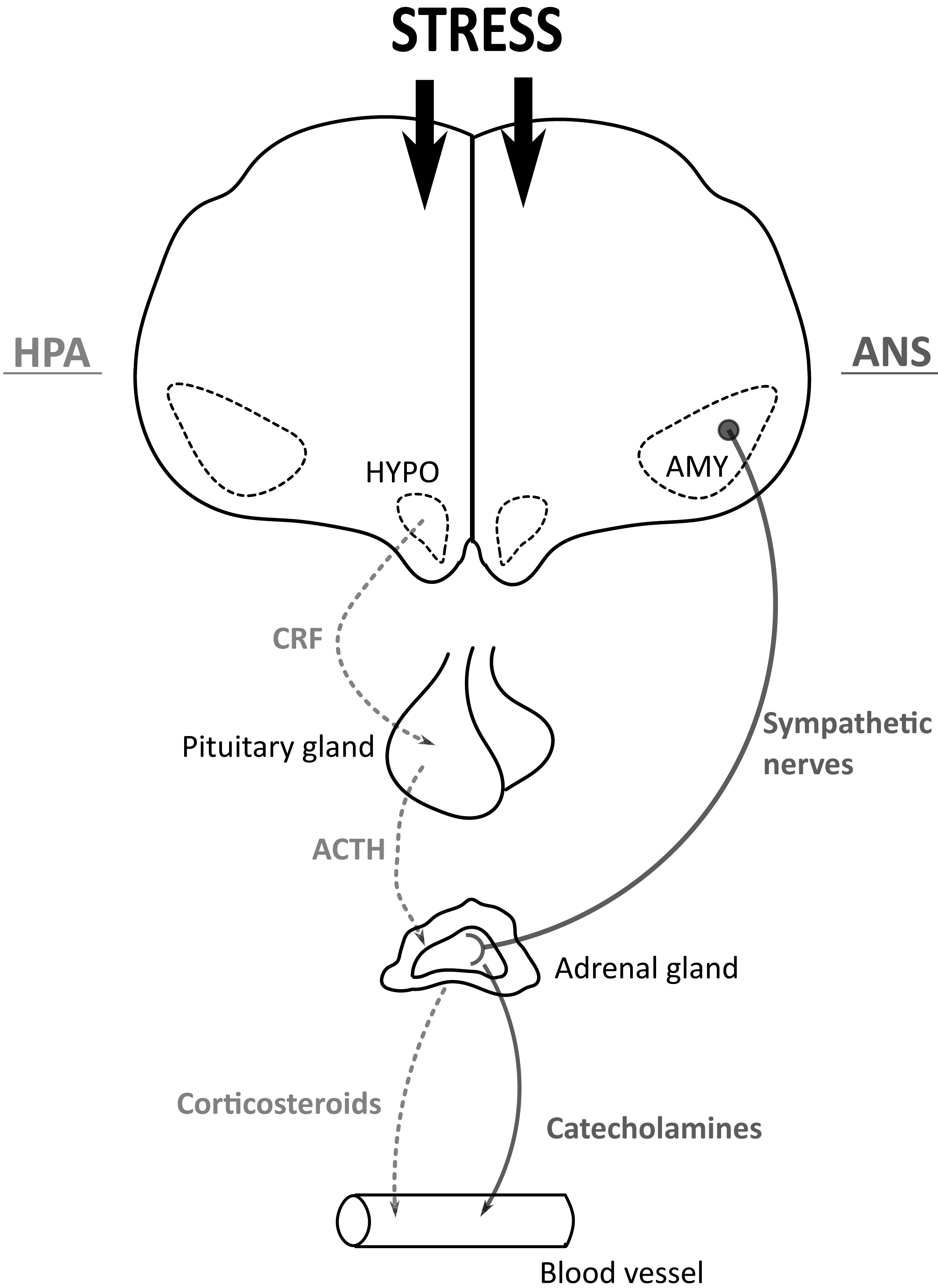
Just as pain is a defence response to potential damage, stress is a defensive response to a stressor or aversive stimulus (Siegel, Reference SIEGEL1980; Yamada and Nabeshima, Reference YAMADA and NABESHIMA1995). A stressor is the aversive stimulus, either endogenous or exogenous, that challenges the maintenance of homeostasis, whilst the stress response is the behavioural and physiological changes that occur in response to a stressor (Siegel, Reference SIEGEL1980; Yamada and Nabeshima, Reference YAMADA and NABESHIMA1995; Muir, Reference MUIR2009). The principal response to a stressor by birds is the activation of the hypothalamic-pituitary adrenal cortical axis (HPA) and autonomic nervous system (ANS; Siegel, Reference SIEGEL1980) which results in the activation of neural pathways and the release of neurogenic substrates that are involved in the aforementioned process of noxious stimulus transmission and pain perception (Figure 1; Jørum, Reference JØRUM1988; Butler and Finn, Reference BUTLER and FINN2009; Yilmaz et al., Reference YILMAZ, DIERS, DIENER, RANCE, WESSA and FLOR2010; Parikh et al., Reference PARIKH, HAMID, FRIEDMAN, NGUYEN, TSENG, MARQUEZ and LUTFY2011). The ANS responds to a stressor by activation of the sympathetic nervous system, known as the ‘Flight or Fight’ response, in order to mobilise energy to actively cope with a stressor and reinstate homeostasis. This response results in various physiological changes throughout the body; such as increased heart rate, blood pressure and breathing rate, peripheral vasoconstriction and altered glucose metabolism, all of which aid with immediate action to cope with the stressor (Muir, Reference MUIR2009; Ulrich-Lai and Herman, Reference ULRICH-LAI and HERMAN2009). The hypothalamic-pituitary adrenal cortical axis (HPA) is the neuroendocrine response, and involves a cascade of hormones which mobilise stored energy and potentiate the effects of ANS activation (Mormède et al., Reference MORMÈDE, ANDANSON, AUPÉRIN, BEERDA, GUÉMENÉ, MALMKVIST, MANTECA, MANTEUFFEL, PRUNET, VAN REENEN, RICHARD and VEISSIER2007; Ulrich-Lai and Herman, Reference ULRICH-LAI and HERMAN2009).
One of the neurobiological mechanisms involved in the stress response, which has been shown to be a mediator of pain modulation and SIA, is the release of corticotropin-releasing factor (CRF) (Siegel, Reference SIEGEL1980; Yamada and Nabeshima, Reference YAMADA and NABESHIMA1995; Lariviere and Melzack, Reference LARIVIERE and MELZACK2000). CRF is a neurohumoural factor involved in the activation of the HPA axis and is released from the hypothalamus in response to a stressor. The increase in CRF will stimulate the release of adrenocorticotropic hormone (ACTH) and β-endorphins from the pituitary gland. Together, ACTH and β-endorphins stimulate the release of corticosteroids from the adrenal cortex (Siegel, Reference SIEGEL1980; Lariviere and Melzack, Reference LARIVIERE and MELZACK2000). Released CRF will be distributed throughout the brain, where it has both excitatory and inhibitory actions in multiple regions of the brain that are involved in pain processing and SIA (Lariviere and Melzack, Reference LARIVIERE and MELZACK2000; Muir, Reference MUIR2009). The release of CRF is just one component of the stress response that interacts with pain processing and perception, with many other neural pathways and neural substrates involved in pain are being modulated by the stress response and are involved in SIA and SIH.
Diagram of stress response.
The perception of stress activates the hypothalamic-pituitary adrenal cortical axis (HPA) and the autonomic nervous system (ANS). With HPA, stress stimulates corticotrophin-releasing factor (CRF) from the hypothalamus (HYPO), stimulating the anterior pituitary gland to secrete adrenocorticotropic hormone (ACTH). Increased ACTH will stimulate the release of corticosteroids, such as corticosterone, from the adrenal cortex into the bloodstream. With ANS, stress activates sympathetic nerves originating in the amygdala (AMY), to stimulate the adrenal medulla to release catecholamines, such as norepinephrine and epinephrine, into the bloodstream.

Stress and its effect on the modulation and perception of pain
The potent effects of stress on nociception and pain perception, and the phenomena of SIA and SIH have been demonstrated in numerous studies in both humans and other mammals. The biphasic effects of stress on pain are dependent on both the type of noxious stimuli and stressor (Imbe et al., Reference IMBE, IWAI-LIAO and SENBA2006; Butler and Finn, Reference BUTLER and FINN2009; Jennings et al., Reference JENNINGS, OKINE, ROCHE and FINN2014). The duration, intensity and type of stimulus, as well as whether it is processed centrally or peripherally, all have modulatory influences on whether pain transmission is inhibited or facilitated (Imbe et al., Reference IMBE, IWAI-LIAO and SENBA2006; Butler and Finn, Reference BUTLER and FINN2009 Jennings et al., Reference JENNINGS, OKINE, ROCHE and FINN2014). More severe stressors are proposed to evoke somatic analgesia, whilst less severe stressors evoke somatic hyperalgesia (Tramullas et al., Reference TRAMULLAS, DINAN and CRYAN2012). Often the same stressor will have biphasic effects, as illustrated by research using restraint stress on rats, with some seeing an increase in pain sensitivity and others seeing a decrease (Jørum, Reference JØRUM1988). Furthermore, the type of emotional response, a ‘fear’ versus ‘anxiety’ response, has been shown to lead to a different response in nociceptive processing and the pain experience (Yilmaz et al., Reference YILMAZ, DIERS, DIENER, RANCE, WESSA and FLOR2010).
Stress induced analgesia (SIA)
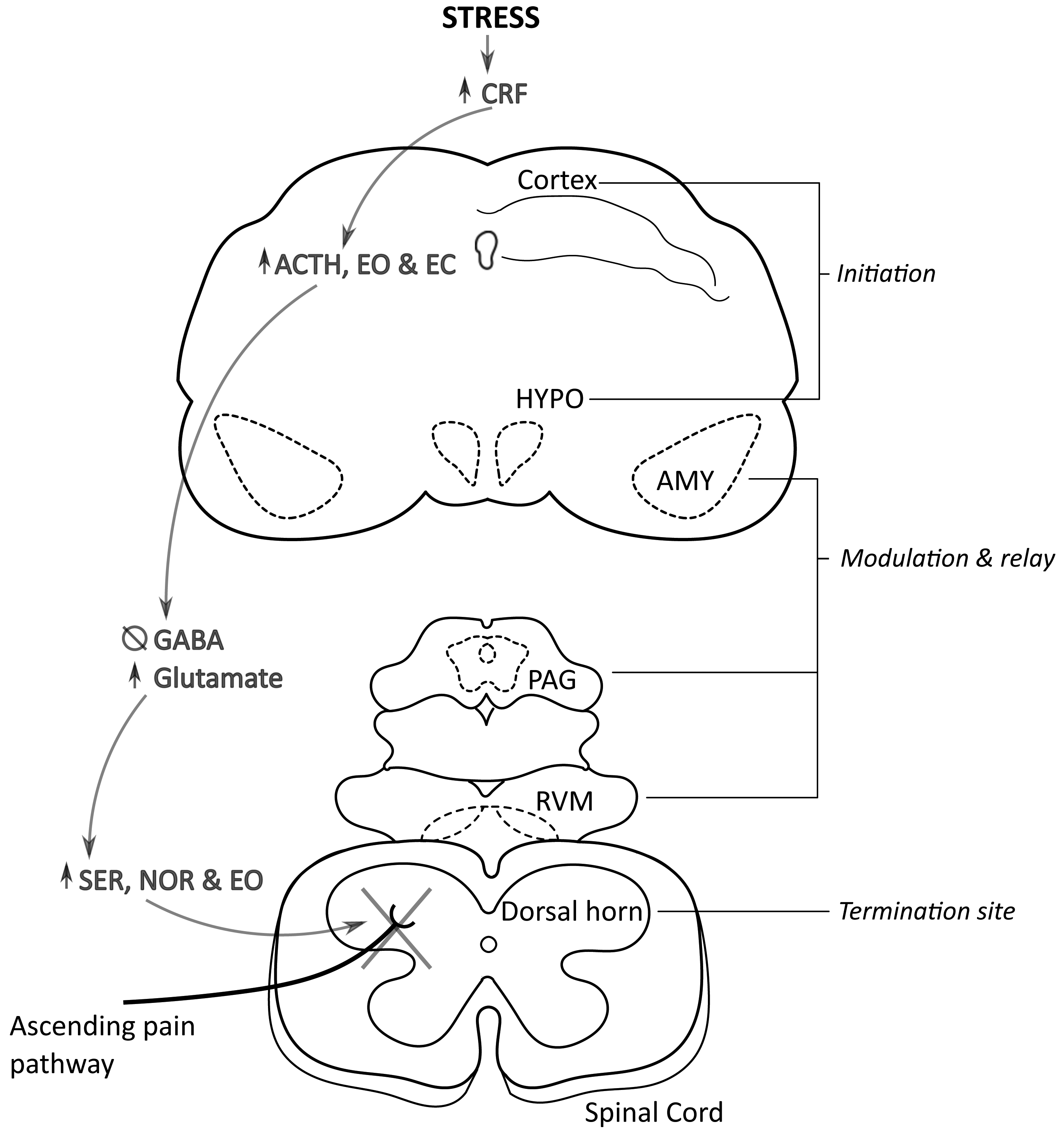
The modulation of pain and alteration of pain perception, as occurs with SIA, occurs through a complex system of interactions between neurons, neurotransmitters and neuropeptides and their receptors on both a spinal and supraspinal level (Carsten, Reference CARSTEN1987; Butler and Finn, Reference BUTLER and FINN2009). Modulation of pain means either increasing or decreasing the perceived pain level by the inhibition or facilitation of nociceptive information transmission to the higher centres of the brain. The modulation of dorsal horn activity occurs via the descending pain pathway. The key components in the system are initiation by the cortex and hypothalamus, further modulation and relaying of information by the amygdala, periaqueductal grey (PAG) and rostral ventral medulla (RVM), with the dorsal horn as the termination site and site of ascending nociceptive transmission inhibition as well as the neurochemicals involved in signalling (Figure 2) (Bodnar et al., Reference BODNAR, KELLY, BRUTUS and GLUSMAN1979; Bodnar, Reference BODNAR1986; Butler and Finn, Reference BUTLER and FINN2009; Jennings et al., Reference JENNINGS, OKINE, ROCHE and FINN2014).
Stress induced analgesia pathway.
The descending pain pathway (DPP) initiates in the cortex and hypothalamus (HYPO), as corticotrophin-releasing factor (CRF) is released in response to stress, stimulating adrenocorticotropic hormone (ACTH), endogenous opioid (EO) and endocannabinoid (EC) release. Increased circulating EO and EC will bind to GABAergic interneurons in the amygdala (AMY), periaqueductal grey (PAG) and rostral ventral medulla (RVM), inhibiting GABA release. With no GABA to inhibit glutamate release, the glutamate release increases. This activates DPP neurons, allowing modulating and relaying of DPP signal. The signal terminates in the dorsal horn, stimulating the release of serotonin (SER), norepinephrine (NOR) and EO, inhibiting the ascending pain pathway and nociceptive information transmission.

The primary neurosubstrates (and their various receptor subtypes) responsible for both inhibitory or facilitatory pain modulation are monoamines, endocannabinoids, EO as well as both gamma-aminobutyric acid (GABA) and glutamate. The descending pain pathway is initiated by the cortex or hypothalamus in response to a stressor, as the increase in circulating CRF drives the release of ACTH, EO and/or endocannabinoids. Within the amygdala, PAG and RVM, the circulating EO and/or endocannabinoids will bind to receptors on a GABAergic interneuron and inhibit the release of GABA from the interneuron. The inhibition of GABA means it is no longer able to block glutamate release and the increased glutamate will activate descending inhibitory pathway neurons. The pain inhibition signal will be transmitted via an interneuron to the spinal cord, where the activation of the descending pathway neuron drives serotonin, norepinephrine and EO release. These will bind to their respective receptor subtypes on the primary afferent nerve terminal and inhibit the release of neurotransmitters that mediate ascending nociceptive transmission (Bodnar et al., Reference BODNAR, KELLY, BRUTUS and GLUSMAN1979; Bodnar, Reference BODNAR1986; Butler and Finn, Reference BUTLER and FINN2009; Jennings et al., Reference JENNINGS, OKINE, ROCHE and FINN2014).
Pain modulation and SIA can be classified as either opioid or non-opioid mediated (Butler and Finn, Reference BUTLER and FINN2009; Yilmaz et al., Reference YILMAZ, DIERS, DIENER, RANCE, WESSA and FLOR2010; Parikh et al., Reference PARIKH, HAMID, FRIEDMAN, NGUYEN, TSENG, MARQUEZ and LUTFY2011). Mediation of SIA by opioid or non-opioid mechanisms has been shown to be dependent on the severity of the stressor, with a severe stressor resulting in non-opioid mediated SIA, whilst less severe stressors are mediated by EO (Parikh et al., Reference PARIKH, HAMID, FRIEDMAN, NGUYEN, TSENG, MARQUEZ and LUTFY2011). The duration of the stressor and whether it is applied intermittently or continuously impacts whether SIA is opioid or non-opioid mediated, with short continuous stressors resulting in non-opioid SIA, whilst opioid-mediated SIA is seen with stressor that are applied intermittently or for a long duration of time (Amit and Galina, Reference AMIT and GALINA1986; Parikh et al., Reference PARIKH, HAMID, FRIEDMAN, NGUYEN, TSENG, MARQUEZ and LUTFY2011). Overall, it seems that the mechanism of SIA pain modulation consists of a complex pathway of neurotransmitters that can work via either EO or non-opioid mechanisms to inhibit ascending nociceptive transmission and result in an analgesia effect.
SIA plays an adaptive role in protecting an animal from a dangerous circumstance by disrupting the pain experienced and related behaviours to allow the animal to escape from a potentially harmful situation (Amit and Galina, Reference AMIT and GALINA1986; Kavaliers and Colwell, Reference KAVALIERS and COLWELL1991). A few examples of stressors that have shown to result in SIA with rodents under research conditions are stress tests such as the cold water swim (Parikh et al., Reference PARIKH, HAMID, FRIEDMAN, NGUYEN, TSENG, MARQUEZ and LUTFY2011) elevated maze test (Butler and Finn, Reference BUTLER and FINN2009), application of electric foot shocks (Amit and Galina, Reference AMIT and GALINA1986) as well as immobilisation and restraint (Amit and Galina, Reference AMIT and GALINA1986; Butler and Finn, Reference BUTLER and FINN2009). Food restriction (Butler and Finn, Reference BUTLER and FINN2009), negative experiences with conspecifics such as social conflict, experiencing defeat and social isolation (Sufka and Hughes, Reference SUFKA and HUGHES1991; Butler and Finn, Reference BUTLER and FINN2009; Jennings et al., Reference JENNINGS, OKINE, ROCHE and FINN2014), as well as exposure to predators or the scent of predators have been shown to induce SIA (Kavaliers and Colwell, Reference KAVALIERS and COLWELL1991; Butler and Finn, Reference BUTLER and FINN2009). Overall, this indicates that a wide range of both physical and non-physical non-noxious stimuli can induce SIA.
Stress induced hyperalgesia
Stress induced hyperalgesia (SIH), or an exacerbation of pain perception resulting from an exposure to a stressor, is mediated via similar mechanisms to those involved in SIA. The descending pain pathway is biphasic and enables descending facilitation alongside descending inhibition. Descending facilitation transmits signals from the supraspinal region to the dorsal horn, but amplifies ascending nociceptive transmission, instead of inhibiting it. This is further supported by the biphasic pain modulation effects of the RVM, which can inhibit or facilitate pain depending on the cell subtype activated (Imbe et al., Reference IMBE, IWAI-LIAO and SENBA2006; Martenson et al., Reference MARTENSON, CETAS and HEINRICHER2009; Jennings et al., Reference JENNINGS, OKINE, ROCHE and FINN2014), with activation of ON cells in the RVM facilitating the descending pain pathway, whilst the activation of the OFF cells is inhibitory (Imbe et al., Reference IMBE, IWAI-LIAO and SENBA2006; Martenson et al., Reference MARTENSON, CETAS and HEINRICHER2009). Increased CRF and corticosterone due to stress alter activity in PAG and activate the ON cells in the RVM, thus facilitating nociceptive information transmission to the brain (Jennings et al., Reference JENNINGS, OKINE, ROCHE and FINN2014). This change from inhibition to facilitation of nociceptive information has been attributed to maladaptive changes or dysfunction in the HPA axis and pain pathway, or to the repeated exposure to a stressor having anxiogeneic effects or leading to the development of a tolerance to a stressor (Jørum, Reference JØRUM1988; Butler and Finn, Reference BUTLER and FINN2009; Martenson et al., Reference MARTENSON, CETAS and HEINRICHER2009; Jennings et al., Reference JENNINGS, OKINE, ROCHE and FINN2014). Chronic stress has been shown to induce hyperalgesia, with increased duration and repeated exposure to a stressor resulting in alterations throughout the pain pathways and impairing endogenous pain modulation (Jørum, Reference JØRUM1988; Butler and Finn, Reference BUTLER and FINN2009; Martenson et al., Reference MARTENSON, CETAS and HEINRICHER2009; Jennings et al., Reference JENNINGS, OKINE, ROCHE and FINN2014). Research performed on rodents has shown hyperalgesia resultant from neurochemical changes in the prefrontal cortex and amygdala, such as a reduced binding EO and lower densities of opioid receptors in the amygdala (Jennings et al., Reference JENNINGS, OKINE, ROCHE and FINN2014), increase in pro-inflammatory mediators (Jennings et al., Reference JENNINGS, OKINE, ROCHE and FINN2014) and over excitation of the dorsal root ganglion (Jennings et al., Reference JENNINGS, OKINE, ROCHE and FINN2014). Research involving mice without β-endorphin has shown that the hyperalgesic response could be due to an imbalance between the nociceptive inhibitory and excitatory mechanisms (Parikh et al., Reference PARIKH, HAMID, FRIEDMAN, NGUYEN, TSENG, MARQUEZ and LUTFY2011).
Stressors shown to induce SIH in rodents are often identical to those in SIA, but SIH occurs when the animal's exposure to stress is either long term or repeated (Imbe et al., Reference IMBE, IWAI-LIAO and SENBA2006; Butler and Finn, Reference BUTLER and FINN2009; Martenson et al., Reference MARTENSON, CETAS and HEINRICHER2009; Jennings et al., Reference JENNINGS, OKINE, ROCHE and FINN2014). A few experimental stressors shown to induce SIH in rodents include repeated cold environment exposure (Imbe et al., Reference IMBE, IWAI-LIAO and SENBA2006; Jennings et al., Reference JENNINGS, OKINE, ROCHE and FINN2014), repeated forced swim and water avoidance tests (Imbe et al., Reference IMBE, IWAI-LIAO and SENBA2006; Parikh et al., Reference PARIKH, HAMID, FRIEDMAN, NGUYEN, TSENG, MARQUEZ and LUTFY2011; Jennings et al., Reference JENNINGS, OKINE, ROCHE and FINN2014), repeated restraint or immobilisation (Imbe et al., Reference IMBE, IWAI-LIAO and SENBA2006; Jennings et al., Reference JENNINGS, OKINE, ROCHE and FINN2014) and social defeat stress (Jennings et al., Reference JENNINGS, OKINE, ROCHE and FINN2014). Hyperalgesia has been shown to occur with chronic exposure to mild stress or environmental stressors, such as noise, vibration or daily cage rotation (Jennings et al., Reference JENNINGS, OKINE, ROCHE and FINN2014), and occurs in adult rodents as a result of maternal separation or deprivation as well as general early life stress (Imbe et al., Reference IMBE, IWAI-LIAO and SENBA2006; Jennings et al., Reference JENNINGS, OKINE, ROCHE and FINN2014).
As aforementioned, the animal's emotional state can affect their reaction to pain, with an anxious emotional state resulting in hyperalgesia and a fear emotional response to stressor resulting in analgesia (Kavaliers and Colwell, Reference KAVALIERS and COLWELL1991; Yilmaz et al., Reference YILMAZ, DIERS, DIENER, RANCE, WESSA and FLOR2010; Jennings et al., Reference JENNINGS, OKINE, ROCHE and FINN2014). Research into these phenomena studied the effect of using the same stressor on animals with different emotional behavioural responses and found that exposure to vibration produced analgesia with quiet rats and hyperalgesia in agitated or hyperemotional rats (Jørum, Reference JØRUM1988). The research into SIH suggested that a default response of pain after exposure to a stressor would be analgesia, but repeated or chronic exposure to a stressor or an anxious emotional state will result in hyperalgesia.
Stress induced analgesia and hyperalgesia in birds
SIA and SIH have been extensively researched in humans and mammals, but there is limited available research on either SIA or SIH occurrence in birds. Many of the key components of the mechanism of pain modulation or their homologues have been found in birds. The regions in the CNS that have been elucidated to play a major role in modulating pain perception in mammals are the cortex, amygdala, and the PAG and RVM (Butler and Finn, Reference BUTLER and FINN2009). Within the mammalian cortex there are two key regions involved in pain processing; the primary somatosensory which is integral in discriminating between nociceptive signals, and the prefrontal cortex which is vital in establishing the stimulus intensity (Butler and Finn, Reference BUTLER and FINN2009). Although the avian forebrain is not structurally homologous to the mammalian cortex, it does contain regions that are functionally and hodologically comparable to the primary sensory cortex and prefrontal cortex (Douglas et al., Reference DOUGLAS, GUZMAN and PAUL-MURPHY2018). Located within the avian hyperpallium are the hyperpallium apicale (HA) and caudomedial nideopallium (NCM), which have both been shown to be comparable to mammalian pain centres (Kuenzel, Reference KUENZEL2007; Blas, Reference BLAS2015; Trebilcock, Reference TREBILCOCK2015). Both HA and NCM, have been shown to be integral in the processing of somatosensory and nociceptive information from the thalamus (Reiner et al., Reference REINER, YAMAMOTO and KARTEN2005; Blas, Reference BLAS2015; Trebilcock, Reference TREBILCOCK2015) as well as in pain integration (Trebilcock, Reference TREBILCOCK2015). The posterior amygdala, and nucleus taenia of the avian amygdala have been shown to be homologous in function, development, hodology and neurochemistry to the key regions of the amygdala within mammals (Reiner et al., Reference REINER, YAMAMOTO and KARTEN2005; Blas, Reference BLAS2015). The avian midbrain contains two corresponding sub-regions, namely the central mesencephalic grey (GCt) and adjacent nucleus intercollicularis (ICo) which together are key in relaying pain information to the higher brain centres and pain modulation, and have been shown to be a homologue to the PAG and RVM in the mammalian brain (Melleu et al., Reference MELLEU, LINO-DE-OLIVERIA and MARINO-NETO2017). The dorsal horn is both anatomically and functionally comparable in birds and mammals (Douglas et al., Reference DOUGLAS, GUZMAN and PAUL-MURPHY2018). The neurotransmitters and neuropeptides and their respective receptors that are integral to mediating SIA and SIH in mammals have been found in avian species (Douglas et al., Reference DOUGLAS, GUZMAN and PAUL-MURPHY2018). The various EO, dynorhpins, enkephalins, endorphins and endomorphins as well as the β-, δ- and κ-opioid receptors are present across species of birds (Douglas et al., Reference DOUGLAS, GUZMAN and PAUL-MURPHY2018). Similarly, endogenous cannabinoids and their respective receptors are present in birds (Fowler et al., Reference FOWLER, NILSSON, ANDERSSON, DISNEY, JACOBSSON and TIGER2001; Douglas et al., Reference DOUGLAS, GUZMAN and PAUL-MURPHY2018). GABA, glutamate and monoamines, such as serotonin, dopamine and norepinephrine, have been established as playing a key role in pain and be are abundant in the avian CNS and pain pathway (Wallnau and Gallup, Reference WALLNAU and GALLUP1977; Douglas et al., Reference DOUGLAS, GUZMAN and PAUL-MURPHY2018). Scientific evidence has shown that birds contain the CNS components and neural substrates needed for conscious pain perception as well as for stress induced alter pain perception. This suggests that the phenomena of both SIA and SIH could occur in birds.
Past research on avian species demonstrated conflicting evidence in terms of the presence of SIA in birds. Experiments by Sufka and Hughes (Reference SUFKA and HUGHES1991), and Feltenstein and colleagues (2002) have shown that, in young domestic chickens, a reduction in pain response is seen when the chicks are exposed to a stressor. Both research groups found that social isolation and handling increased the chick's pain threshold, which was indicative of SIA in young birds. These experiments showed that, if the chicks were habituated to handling, it reduced the aversiveness of handling and both attenuated the stress response and increased the pain response (Sufka and Hughes, Reference SUFKA and HUGHES1991; Feltenstein et al., Reference FELTENSTEIN, FORDA, FREEMAN and SUFKAA2002). This may indicate that as the stressor became non-aversive, its analgesic effect decreased and SIA no longer occurred. Both the evidence of handling and isolation having analgesic effect and habituation to the stressor attenuating the analgesic response, are further evidence to suggest that the SIA effect does occur in birds. Multiple studies by Gentle and colleagues into endogenous analgesia led to the conclusion that, although birds are capable of an endogenous mechanism of pain suppression, this is not due to stress; instead it is due to attentional and motivational shifts (Gentle and Corr, Reference GENTLE and CORR1995; Wylie and Gentle, Reference WYLIE and GENTLE1998; Gentle and Tilston, Reference GENTLE and TILSTON1999). An investigation into the effects of novel environments and pre-lay behaviour on pain suppression saw marked hypoalgesia or complete analgesia in birds, but without the performance of classic fear or distress behaviours. From this it was concluded that the analgesia was not induced by stress but attentional shift (Gentle and Corr, Reference GENTLE and CORR1995). A second study investigated the effect of access to feed after food deprivation on pain suppression in broilers, and, as access to feed had an analgesic effect, it was concluded that it caused motivational analgesia and not SIA (Gentle and Tilston, Reference GENTLE and TILSTON1999). Although it is a well-established fact that endogenous analgesia can be induced by stimuli that are not aversive (Amit and Galina, Reference AMIT and GALINA1986), the evidence presented in the two aforementioned studies was not sufficient to disprove SIA in birds. This was particularly noted as the stimuli used in the experiment were classified as non-stressors, but with closer examination both stimuli involved the deprivation of a highly motivated behaviours which has been shown to be a significant stressor in birds (Bokkers et al., Reference BOKKERS, KOENE, RODENBURG, ZIMMERMAN and SPRUIJT2004; Weeks and Nicol, Reference WEEKS and NICOL2006). As feeding is a highly motivated behaviour in broilers (Bokkers et al., Reference BOKKERS, KOENE, RODENBURG, ZIMMERMAN and SPRUIJT2004), 16 h feed deprivation prior to re-access to food used in this study could be a significant stressor, thus the analgesic effect shown could be classified as stress induced. Similarly, in the initial experiment in which pre-laying behaviour was shown to be analgesic, the inability of birds to perform highly motivated pre-laying behaviours would be a significant stressor (Weeks and Nicol, Reference WEEKS and NICOL2006). The fact that the birds were kept in a pen with wood shavings, a feeder and a drinker and demonstrated stereotypical pacing as well as escape attempts, suggest the environment may not have been sufficient to satisfy the highly motivated pre-lay behaviours, such as nest site finding and nesting, thus the environment and deprivation of behaviour may be argued to be a stressor, and the resultant analgesia as SIA. This suggested that, although distraction/attentional analgesia is a form of endogenous analgesia, there is evidence that indicates that SIA occurs in birds. To the best of the author's knowledge, evidence for the occurrence of SIH is not available, however, birds have been shown to experience SIA and possess the anatomical and physiological components required to SIH, suggesting SIH could occur in birds.
Tonic immobility and its relationship to stress altered pain perception
Tonic immobility (TI) is another fear or stress response present in animals, including birds, and has been linked to SIA and SIH. TI is a defensive fear response characterised by a catatonic state and reduced responsiveness to external stimuli and results from a short-lived restraint stress (Jones et al., Reference JONES, BEUVING and BLOKHUIS1987; Porro and Carli, Reference PORRO and CARLI1988; Gentle et al., Reference GENTLE, JONES and WOOLLEY1989; Melleu et al., Reference MELLEU, LINO-DE-OLIVERIA and MARINO-NETO2017). Research in both chickens (Gentle et al., Reference GENTLE, JONES and WOOLLEY1989) and rabbits (Porro and Carli, Reference PORRO and CARLI1988) using an electroencephalogram (EEG) showed that, during a state of tonic immobility, electrical activity within the brain was reduced, but there was still a response to stimuli. This indicated that the animal was still capable of processing sensory information but with limited ability to sustain arousal (Gentle et al., Reference GENTLE, JONES and WOOLLEY1989). Research performed on rabbits has shown that when TI is induced during acute pain it has a hypoalgesic effect and abolishes motor and EEG reactions to pain (Porro and Carli, Reference PORRO and CARLI1988). However, in lizards, TI has been shown to cause hyperalgesia (Mauk et al., Reference MAUK, OLSON, LAHOSTE and OLSON1981). This suggested that the analgesic effects of TI were not uniform across species, and there is no scientific evidence whether TI has analgesic or hyperalgesic effects in birds. Studies of the anti-nociceptive effect of TI with rats, rabbits and guinea pigs, have shown TI to be opioid and serotonin mediated, and that its effects can be potentiated by morphine as well as attenuated by opioid antagonists (Porro and Carli, Reference PORRO and CARLI1988; da Silva and Menescal-de-Oliveira, Reference DA SILVA and MENESCAL-DE-OLIVEIRA2007). Similar research performed with chickens showed that although it can be morphine potentiated, TI may not be opioid mediated as it cannot be attenuated by naloxone (Wallnau and Gallup, Reference WALLNAU and GALLUP1977; Peters and Hughes, Reference PETERS and HUGHES1978). This indicated that TI in chickens is mediated primarily by serotonin and not opioids, which may influence whether TI is analgesic or hyperalgesic in birds.
In mammals, the PAG and RVM have prominent roles in mediating TI (da Silva and Menescal-de-Oliveira, Reference DA SILVA and MENESCAL-DE-OLIVEIRA2007; Melleu et al., Reference MELLEU, LINO-DE-OLIVERIA and MARINO-NETO2017) and are attributed to analgesic effects in rabbits (Porro and Carli, Reference PORRO and CARLI1988) and guinea pigs (da Silva and Menescal-de-Oliveira, Reference DA SILVA and MENESCAL-DE-OLIVEIRA2007). Avian species contain a homologue to the PAG, known as the ICo-GCt complex in the avian midbrain (Melleu et al., Reference MELLEU, LINO-DE-OLIVERIA and MARINO-NETO2017). The presence of structures involved in the analgesic effect of TI suggests, but does not confirm, that TI is analgesic in birds. Further research is needed to gain conclusive evidence.
Another reason that TI analgesic properties may be related to SIA is that it results from fear. Stressors that induce a fear response have been shown to have an analgesic effect, which mirrors the analgesic effect of the TI fear response and this is why TI has been suggested as a form of SIA in mammals (Butler and Finn, Reference BUTLER and FINN2009; Martenson et al., Reference MARTENSON, CETAS and HEINRICHER2009; Yilmaz et al., Reference YILMAZ, DIERS, DIENER, RANCE, WESSA and FLOR2010; Jennings et al., Reference JENNINGS, OKINE, ROCHE and FINN2014; Melleu et al., Reference MELLEU, LINO-DE-OLIVERIA and MARINO-NETO2017). Although there is no direct evidence about an SIA-like effect occurring with TI in birds, the resemblance of the avian brain regions and neural substrates to those that mediate TI-induced analgesia in mammals suggest there is a possibility of an analgesic effect with TI. However, this should be thoroughly researched to be confirmed, as TI has been shown to have a hyperalgesic effect in lizards. Although the anatomy and physiology are comparable this is not conclusive evidence that TI is analgesic in birds nor that it is another form of SIA.
Conclusions
Both pain and stress pathways are highly interactive and both SIA and SIH have been shown to occur in humans and various mammalian species. The same underlying mechanisms are responsible for both SIA and SIH, but the two result from different stressors and a different response of the CNS and pain pathways. There is evidence to suggest that SIA occurs in birds, as all the vital components are either present or homologues have been illustrated by research. Furthermore, there is some scientific evidence based on avian research that stress can induce analgesia and cause pain suppression in birds. The presence of the necessary components suggests that SIH could occur in birds, although scientific evidence looking at specific stressors inducing SIH in birds is limited. TI, which is seen as a specialised form of SIA in mammals, occurs in birds, although its analgesic effects are not fully understood and there is a possibility it results in hyperalgesia instead of analgesia. Further research on SIA in birds is needed to clarify the analgesic effects of specifics stressors, as well as into the presence of SIH in birds and whether TI has a possible analgesic or hyperalgesic effect in birds.