Case
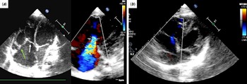
A full-term female was diagnosed with atrial septal defect with left to right shunt and congenital tricuspid valve regurgitation due to tricuspid valve dysplasia (Fig 1a). Her trivuspid valve regurgitation was severe, but no symptoms of heart failure were observed while growing up; hence, she was discharged and followed up at an outpatient clinic. Her somatic growth deteriorated over time, and the right atrium and ventricle dilated. As a result, at the age of 1 year and 1 month, with a body weight of 8.5 kg, tricuspid valve repair was performed.
Echocardiography. ( a ) Before the operation. Restrictive mobility and tethering of the anterior leaflet (green arrow) and septal leaflet (white arrow) with severe tricuspid valve regurgitation were seen. ( b ) One year after surgery. Tricuspid valve regurgitation improved to be trivial.

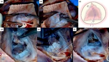
At the operation, the tricuspid valve, especially the anterior and the septal leaflets were dysplastic. The characteristics of each leaflets were normal; however, the anterior and the septal leaflets were fixed to the anterior papillary muscle and the interventricular septum, respectively. The leaflets were also tethered due to the right ventricular dilatation (Fig 2a and b). At first, annuloplasty using De Vega technique was performed. However, saline test revealed that regurgitation could not be controlled at all and sutures were removed. Papillary muscle approximation was performed in the light of these findings. Sutures were applied at the base of the anterior papillary muscle and the interventricular septum with autologous pericardial pledgets using 5–0 Prolene® (Johnson & Johnson Services, Inc., New Brunswick, NJ, USA). TR improved after a saline test. Atrial septal defect was directly closed. Transesophageal echocardiography after unclamping, demonstrated moderate tricuspid valve regurgitation between the anterior and the posterior leaflets, deviated to the interatrial septum. Free edge of the anterior leafet was longer than that of the posterior leaflet. Commissural edge-to-edge suture between the anteroposterior commissure was revealed to be ineffective by a saline test. Therefore, first suture of the approximation was removed, and the new approximation suture was applied so that the anterior papillary muscle was positioned slightly toward the anteroseptal commissure (Fig 2c, d and e). A saline test revealed that regurgitation could be controlled better than the first repair, and residual regurgitation remained between the anterior and the septal leaflets. This residual regurgitation was treated successfully with a commissural edge-to-edge suture (Fig 2f). The tricuspid annular diameter after the repair was 16 mm measured by Hegar dilator. Regurgitation had improved to be mild without stenosis by transesophageal echocardiography following unclamping. Cardiopulmonary bypass time and aortic cross-clamp time were 148 and 96 minutes, respectively.
Intraoperative image of tricuspid valve repair. ( a ) Dysplastic septal leaflet was fixed to the interventricular septum. ( b ) Dysplastic anterior leaflet was fixed to the anterior papillary muscle. ( c ) Schema of papillary muscle approximation. A mattress suture is placed from the base of the anterior papillary muscle to the interventricular septum with pledgets. ( d ) Papillary muscle approximation. Suture of the anterior papillary muscle. ( e ) Papillary muscle approximation. Suture of the interventricular septum. ( f ) After repair.

She was discharged 8 days after surgery with an excellent postoperative course. One year after surgery, she is doing well with trivial tricuspid valve regurgitation (Fig 1b).
Discussion
Various surgical techniques for congenital tricuspid valve regurgitation, including Ebstein’s anomaly, have been reported. Reference Said, Burkhart and Dearani1,Reference Hetzer, Hacke and Javier2 However, isolated congenital tricuspid regurgitation with tricuspid valve dysplasia, especially in the anterior and the septal leaflets, were rare. In our case, among various surgical techniques, leaflet augmentation and artificial chordae might be viable options. In our case, because leaflet properties were normal, leaflet augmentation may not be applicable. Also, artificial chordae may be impractical, because most chordae of the anterior and the septal leaflets must be resected, and a lot of artificial chordae were required.
In adults, papillary muscle approximation for mitral or tricuspid valve regurgitation caused by tethering of the leaflets is used. Reference Nappi, Lusini and Spadaccio3,Reference Matsumiya, Kohono and Matsuura4 We referred to it and considered that papillary muscle approximation was effective in our case with tethering of the leaflets. Yamauchi et al. reported the efficacy of papillary muscle approximation of the tricuspid valve in an ex vivo study. Reference Yamauchi, Vasilyev and Marx5 In this report, the direction of approximation had no statistical difference. However, in our case, the direction of approximation was closely linked to the management of tricuspid valve regurgitation. In our case, only the posterior leaflet was not dysplastic and functioned normally. We presumed that the direction of approximation affected whether the posterior leaflet functioned sufficiently. In tricuspid valve dysplasia, different from functional tricuspid valve regurgitation, the direction of approximation may need to be adjusted.
Sebening stitch Reference Komoda, Stamm, Fleck and Hetzer6 is the other reparative technique at the subvalvular apparatus. In this technique, sutures of the anterior papillary muscle are placed at the head. In our technique, different from Sebening stitch, sutures are placed at the base of the anterior papillary muscle. Therefore, tearing of the tissues and prevention of inflow from the right atrium to the right ventricle may be less likely.
Finally, tricuspid valve repair with papillary muscle approximation for congenital tricuspid valve regurgitation due to tricuspid valve dysplasia could be one of the viable repair options.
Financial support
This research received no specific grant from any funding agency, commercial, or not-for-profit sectors.
Conflicts of interest
None.
Ethical standards
The authors assert that all procedures contributing to this work comply with the ethical standards of the relevant national guidelines on human experimentation and with the Helsinki Declaration of 1975, as revised in 2008, and has been approved by the institutional review board (Hyogo Prefectural Amagasaki General Medical Center, ID: 3–178).