

In large psychiatric hospitals, entire wards used to be designated for managing ‘catatonic patients'; however, this is now a footnote in history. In modern in-patient psychiatric practice, patients presenting with catatonic features invariably necessitates the uncovering of ‘organic' aetiologies, though psychiatric (i.e. ‘non-organic') conditions such as schizophrenia, bipolar or major depressive disorder or neurological conditions such as epilepsy may sometimes manifest with catatonic features also.
The case presented below is that of a woman with an established diagnosis of schizophrenia who discontinued clozapine abruptly and simultaneously had symptoms of fever, headache, neck stiffness and vomiting, along with mutism, negativism and waxy flexibility.
A 45-year-old single woman of Chinese ethnicity with a diagnosis of schizophrenia had remained stable in the community on clozapine maintenance treatment (400 mg/day) for 7 years. A few days prior to the recent hospitalisation, she appeared withdrawn, had refused clozapine and rapidly (within days) showed clinical decompensation – talking to herself, expressing delusional beliefs and not caring for her personal hygiene. Her family also noted a low-grade fever, and she complained of a headache, whole body ache and began to vomit.
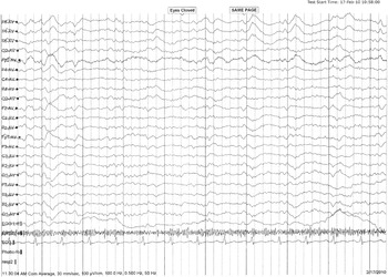
During the next few days, she was mute, staring lifelessly into space, not making eye contact, lying immobile in bed and unwilling to eat and was incontinent of bowel and bladder. Twelve days later, she experienced a generalised tonic/clonic seizure and was brought to the hospital. Examination revealed a patient of stated age who was unwashed, unkempt, drowsy and an oral temperature of 39 °C with nuchal rigidity. Her blood pressure and pulse were recorded within normal limits. No diaphoresis was evident. She was mute; eyes open but not making eye contact, along with negativism and waxy flexibility of her extremities. Her right ear exuded a clear discharge. A lumbar tap revealed a clear cerebrospinal fluid (CSF) at normal pressure with normal glucose and protein levels, and no organisms were cultured, CSF was negative for Cryptococcus and also negative for herpes simplex IgM antibodies. The total white cell count was elevated at 28 000 mm3; and comprised mostly a neutrophil leucocytosis. Creatinine phosphokinase levels were within normal limits, and a computed tomography scan of the brain was reported as normal. An electroencephalography (EEG) showed diffuse background slowing consistent with encephalopathy (Fig. 1). Following neurology and ENT (ear, nose and throat) consultations, a presumptive diagnosis of meningoencephalitis was made.
EEG showing background slowing suggestive of encephalopathy.

Intravenous valproate was commenced as well as intravenous ceftriaxone, an antibiotic, and acyclovir, an antiviral drug. Two days later, the patient was oriented and alert and the total white cell count had returned to normal. She was afebrile and no longer had neck rigidity. Her ear discharge cleared with a 7-day course of topical ofloxacin eardrops and the ENT consultant opined that this external infection was not contributory to the central nervous system (CNS) clinical picture. However, 4 days later, her psychiatric condition deteriorated, she began to refuse food and became socially withdrawn, electively mute and refused all medication. Haloperidol was started at a dose of 5 mg twice a day and was continued for 8 days with no improvement of the catatonic features. Haloperidol was then switched to amisulpiride 400 mg daily, and 2 days later, the patient was more alert and oriented, and her mental state had improved. Amisulpiride was increased to 600 mg and then to 800 mg by the fifth day with clear improvement in her mental state. A review of her previous relapses indicated none had involved catatonic features. The patient had presented with prominent positive psychotic symptoms in previous relapses. A repeat EEG prior to discharge showed significant improvement.
Discussion
This case illustrates a few pertinent clinical points. First, after years of clinical stability on clozapine maintenance treatment, the onset of a physical illness (i.e. early symptoms of meningoencephalitis) likely led to the abrupt cessation of clozapine. Second, the sudden withdrawal of clozapine most likely confounded the clinical picture, i.e. catatonic features combined with fever, headache, neck rigidity, bowel and bladder incontinence followed by a tonic clonic seizure raised the strong possibility of a CNS infection. An EEG report followed by a neurology consultation led to the diagnosis of meningoencephalitis. The rapid response of the neurological clinical features and laboratory parameters to a combination of parenteral antibiotic and antiviral treatment added to the confidence in the CNS diagnosis, though in the absence of a positive bacterial culture or Cryptococcus or elevated herpes simplex antibodies, it remains unclear if this CNS infection was bacterial, fungal or viral in origin. Nevertheless, she responded rapidly to a regime of intravenous valproate, along with antibacterial and antiviral treatment.
Although seizures are associated with clozapine treatment, it is noteworthy that this patient had been successfully treated for 7 years on the same dosage of clozapine (400 mg/day) without any seizures. In addition, the best available information is that she had stopped taking clozapine at least 2 weeks prior to the seizure, making it less likely this seizure was due to clozapine, but more likely related to the meningoencephalitis.
The abrupt cessation of clozapine has been associated with withdrawal symptoms and/or syndromes varying from catatonic features, neuroleptic malignant syndrome, fever, delirium, bowel and bladder incontinence, tardive dyskinesia and/or dystonias (Reference Yeh, Lee, Cheng, Wen and Chen1–Reference Stanilla, de Leon and Simpson4). These neuropsychiatric symptoms and syndromes often cloud the clinical picture already complicated by severe psychiatric decompensation because of clozapine discontinuation. Cholinergic, serotonergic or dopaminergic rebound or supersensitivity mechanisms have been invoked as possible explanations for clozapine withdrawal symptoms or syndromes (Reference Yeh, Lee, Cheng, Wen and Chen1,Reference Ahmed, Chengappa, Naidu, Baker, Parepally and Schooler3,Reference Baldessarini, Gardner and Garver5,Reference Meltzer6). Another possible explanation that may be unique to clozapine is the rapid elimination from the brain (Reference Baldessarini, Gardner and Garver5). Unlike several antipsychotic drugs, which are concentrated in lipid or other tissues and are released slowly from lipid stores or organs, the rapid extraction of clozapine may precipitate withdrawal symptoms in vulnerable individuals.
Suggestions for the use of anticholinergic drugs such as benztropine or trihexphenidyl and/or serotonergic agents such as ritanserin or cyproheptadine to mitigate sudden clozapine discontinuation appear reasonable but have not been tested in definitive trials (Reference Baldessarini, Gardner and Garver5–Reference de Leon, Stanilla, White and Simpson7). In this woman, the medical condition responded rapidly to the intravenous antibiotic and antiviral agent but her psychiatric condition did not respond to haloperidol but to amisulpiride instead. She was discharged on amisulpiride treatment, and a follow-up visit, months later indicated that she remained stable in the community receiving amisulpiride at a dosage of 600 mg/day. She has not required re-challenge with clozapine.
Clinicians need to increase their ability to recognise and manage clozapine withdrawal syndromes including those complicated by CNS infections. Consultations and collaboration with clinical colleagues may help uncover the organic aetiology quickly and appropriate management can then be instituted rapidly. Clinicians may also want to prepare patients and families for the symptoms of clozapine withdrawal including catatonia especially in the context of abrupt cessation of this agent.
Acknowledgements
We would like to thank the Nursing Staff, Department of Psychological Medicine, and Neurology and ENT Faculty, University Malaya, Kuala Lumpur, Malaysia and Ms Joan Spinogatti for typing the manuscript and coordinating the authors' contributions, corrections and submission.