Introduction
Angiostrongylus cantonensis is the most common infectious cause of eosinophilic meningitis or meningoencephalitis (EME) in areas of South-East Asia and many Pacific Islands (Hsieh, Reference Hsieh1959; Alicata, Reference Alicata1965; Maldonado et al., Reference Maldonado, Simões, Oliveira, Motta, Fernandez, Pereira, Monteiro, Torres and Thiengo2010; Wang et al., Reference Wang, Qi, Diao, Zheng, Li, Ma, Ji and Yin2010; Gelis et al., Reference Gelis, Spratt and Raidal2011). Chen discovered A. cantonensis in the pulmonary arteries of rats in 1935 (Chen, Reference Chen1935). Humans, acting as a non-permissive host, become infected after eating intermediate or parasitic hosts, or vegetables that contain the infective larvae (the third stage) of the worm. Once ingested, the infective larvae are digested from the vectors and invade intestinal tissue, thus causing human enteritis before passing through the liver (Yii, Reference Yii1976). When the worms move through the lungs, cough, rhinorrhoea, sore throat, malaise and fever may develop (Cross, Reference Cross1978). Finally, after about 2 weeks, larvae reach the central nervous system and cause eosinophilic meningitis with ensuing eosinophilic pleocytosis. Unlike its life cycles in rats, the worms spontaneously die in human meninges, brains or eyes to which they randomly migrate. Consequently, severe inflammation occurs in those affected organs and manifests as three main clinical forms: eosinophilic meningitis (EoM), eosinophilic encephalitis (EoE) and ocular angiostrongyliasis (Sawanyawisuth & Sawanyawisuth, Reference Sawanyawisuth and Sawanyawisuth2008).
Some effective anthelmintics, such as albendazole, have been used to treat A. cantonensis in humans. However, dead worms in the brain can evoke a severe immune response which results in brain damage (Wang et al., Reference Wang, Jung, Chen, Wong, Wan and Wan2006). Recently, albendazole has been used in combination with corticosteroids to prevent an inflammatory reaction (Tu & Lai, Reference Tu and Lai2006). However, the use of corticosteroid therapy is still controversial (Chen & Lai, Reference Chen and Lai2007). As EoE is fatal and no effective treatment is available (Sawanyawisuth & Sawanyawisuth, Reference Sawanyawisuth and Sawanyawisuth2008), it is necessary to develop some alternative therapies to treat these diseases.
Almost three-quarters of the Earth's surface is covered by seas and oceans. Their resources are varied and vast and are partly comprised of fish, shellfish, other aquatic animals, vegetation, algae, bacteria and fungi (Pietra, Reference Pietra1997; Faulkner, Reference Faulkner2002; Laird & van Altena, Reference Laird and van Altena2006). The study of marine fungi blossomed in the 1990s; more notably, since 1998 this study has so far described more than 200 compounds (Li et al., Reference Li, Lee, Choi, Kang and Son2003). Terrestrial fungi have provided many natural products with significant therapeutic value (Liberra & Lindequist, Reference Liberra and Lindequist1995; König & Wright, Reference König and Wright1996; Mayer & Gustafson, Reference Mayer and Gustafson2004). Since marine organisms live in a very different environment from those of terrestrial organisms, it is reasonable to suppose that marine fungi will differ considerably from their terrestrial counterparts. Recent investigations have shown that marine compounds may have broad anti-inflammatory effects and may modulate the immune and nervous systems (Ina et al., Reference Ina, Hayashi, Nozaki and Kamei2007; Bittencourt et al., Reference Bittencourt, Figueiredo, Mota, Bezerra, Silvestre, Vale, Nascimento, Sampaio, Nagano, Saker-Sampaio, Farias, Cavada, Assreuy and de Alencar2008; Courtois et al., Reference Courtois, Simon-Colin, Boisset, Berthou, Deslandes, Guézennec and Bordron2008; Dang et al., Reference Dang, Lee, Yoo, Shinde, Lee, Hong, Kim and Jung2008). Among them, m2-9 is one such marine compound and has been isolated from mangrove endophytic fungus by Dr Lin's group (unpublished).
In this study, we evaluated the efficacy of albendazole combined with m2-9 in the treatment of eosinophilic meningitis in a mouse model. Administration of the drugs was initiated on day 5 post-inoculation (PI). Efficacy parameters including body weight, worm recovery, neurological function and behavioural analysis, leucocytes and eosinophil counts, and histopathological examination were observed.
Materials and methods
Parasitological procedures
Angiostrongylus cantonensis has been maintained in our laboratory since 2006. It cycles through the snail Ampullarum crossean and Sprague–Dawley rats. Larvae were liberated from the minced snail tissue by pepsin (Sigma, St. Louis, Missouri, USA) digestion. The morphological criteria for identification of the third larval stage (L3) of A. cantonensis were carried out under microscopy. Twenty-five 5-week-old male BALB/c mice (weight 20–25 g, Grade III, Certificate 0070290) were purchased from the Laboratory Animal Center of Sun Yat-sen University.
Albendazole and candidate marine fungal extracts
Albendazole (Sigma) is a benzimidazole derivative. The molecular formula is C12H15N3O2S and the molecular mass is 265.34 (Hwang & Chen, Reference Hwang and Chen1988). Albendazole is widely used in veterinary medicine and has been found to be a safe broad-spectrum anthelmintic in animals and humans. It acts by binding to the parasite's β-tubulin, inhibiting its polymerization and thus impairing glucose uptake, and finally causing worm death (Venkatesan, Reference Venkatesan1998). m2-9, sourced from the secondary metabolites of mangrove endophytic fungus, was isolated by chromatographic technique and was supplied by the School of Chemistry and Chemical Engineering, Sun Yat-sen University. Its structure was identified by comprehensive physicochemical properties and spectral methods. The molecular formula is presumed to be C11H10O5 and the molecular mass is 222.19. It has moderate cytotoxicity (half-maximal inhibitory concentration (IC50) = 3 μg/ml for the cancer cell strains KB or BC-1), as well as antimalarial activity (IC50 = 4 μg/ml for the multi-drug resistant K1 strain of Plasmodium falciparum).
Experimental infection of mice and treatment
The animals, purchased a week before the experiment, were raised at a 12-h light/dark cycle, and were provided with solid chow and water ad libitum. Five mice were randomly selected as normal controls, and were orally dosed with distilled water for 7 days starting from day 5 of the experiment. The remaining mice were randomly divided into four groups (5 mice in each group). Each mouse was orally infected with 25 larvae. The mice in the four groups were treated with one of the following, daily for 7 days beginning on day 5 PI: (a) edible oil; (b) albendazole at 20 mg/kg/day; (c) m2-9 at 10 mg/kg/day (a solution of m2-9 in edible oil); or (d) a combination of albendazole (20 mg/kg/day) and m2-9 (10 mg/kg/day).
Body weight, worm recovery and neurological function/behaviour
The animals from the five groups were sacrificed by cervical dislocation on day 23 PI for the pre-planned analyses. Assessment of body weight and neurological examination were performed on days 0 and 23 PI. The mean body weight of each treatment group was compared with the edible oil treated infected group.
Each brain was shredded in 10 ml of 0.15 M phosphate buffered saline (PBS) and examined for viable larvae under 20 × magnification using a dissecting microscope. The mean number of larvae was compared with the edible oil treated infected group to evaluate the treatment efficacy.
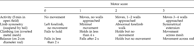
Neurological examination was performed as described previously (Parra et al., Reference Parra, McGirt, Sheng, Laskowitz, Pearlstein and Warner2002). A motor score (0–12) was derived from spontaneous activity, symmetry of limb movements, climbing, balance and co-ordination (see table 2). A sensory score (5–15) was derived from body proprioception, vibrissae, visual, olfactory and tactile responses (see table 3).
To evaluate the differences in learning and memory and action ability of the five groups, a series of behavioural experiments, the Morris water maze test and spontaneous locomotor activity test, were carried out.
The water maze consisted of a 120-cm diameter circular pool made of grey plastic. The pool was divided into four different quadrants (north, west, south and east). A clear, round platform (12 cm in diameter and 2 cm below the water surface) was placed in quadrant west. The water temperature was maintained at 20 ± 2°C. A digital video recorder was mounted above the maze and connected to a computer. A video tracking system (Ethovision 2.0, Noldus, Wageningen, The Netherlands) facilitated the tracking of mouse movements and was used to plot completed movement paths. Mice were acclimatized to the water for 60 s and then placed on the platform for 60 s. Subsequently, they underwent accustomization tests of two trials per day for 4 days (on days 20–23 PI) in which they searched for the submerged platform located in the centre of quadrant west. In each trial, the mouse was placed in the water, facing the edge of the pool, in one of the standard start locations (north, west, south and east). The start locations were assigned in a random order. The mouse was then allowed 60 s to locate the platform. The swimming path and time used to locate the platform (escape latency) were automatically calculated in each trial. Once the mouse located the platform, it was permitted to remain on it for 10 s. If the mouse did not locate the platform within 60 s, it was guided to it and allowed to remain on it for 10 s. The interval between trials was 15–20 min. Escape times of start locations on the first run of each day were recorded.
The horizontal (ambulation) and vertical (rearing, an animal stood on its hind legs) spontaneous locomotor activity of each mouse was measured using a ZZ-6 spontaneous activity apparatus (Chengdu Technology & Market Co. Ltd, China). The apparatus is composed of six boxes and a micro-computer controller, which can test the activities of six mice, simultaneously. The mice were gently placed into the boxes and the frequency of ambulation and rearing within 10 min were recorded.
Cell counts in cerebrospinal fluid and histopathology
On day 23 PI, the mice were sacrificed. The brains were placed in 35-mm dishes. The cranial cavity and cerebral ventricles (lateral, third and fourth ventricles) were rinsed with 1 ml of 0.15 m PBS (pH 7.0). Cerebrospinal fluid (CSF) was harvested and identified as ‘CSF-like fluid’ (Lee et al., Reference Lee, Chou, Chen and Lai2004; Chen & Lai, Reference Chen and Lai2007). The CSF-like fluids were centrifuged at 400 g for 5 min, and the sediments were resuspended with 30 ml PBS for a total leucocyte count. The cells were calculated with Wright–Giemsa staining of 5 ml for each smear.
The mouse brains were fixed separately in 10% neutral buffered formalin for 24 h, then dehydrated in a graded ethanol series (50, 75 and 100%) and xylene, and embedded in paraffin at 55°C for 24 h. Each specimen was cut into 4-mm sections. Paraffin was removed by heating the sections at 65°C for 5 min. The sections were dewaxed by washing three times for 5 min each in xylene, then rehydrated through 100, 95 and 75% ethanol for 5 min each, and finally rinsed with double-distilled water. After staining with haematoxylin and eosin, pathological changes were examined under 100 × magnification, using a light microscope.
Data analysis
Results in the different groups of mice were compared using a non-parametric Kruskal–Wallis test, followed by post-testing using Dunn's multiple comparisons of means. These results were presented as mean ± standard deviation (SD). P values of < 0.05 were considered as statistically significant.
Ethical statement
This study was carried out in strict accordance with the guidelines of the National Institutes of Health on animal care and ethics. The protocol was approved by the Animal Care and Use Committee of Sun Yat-sen University (Permit Number: SCXK (Guangdong) 2009-0011). All surgery was performed under sodium pentobarbital anaesthesia, and all efforts were made to minimize any pain and suffering in the subject animals.
Results
Treatment and mouse body weight and larval recovery
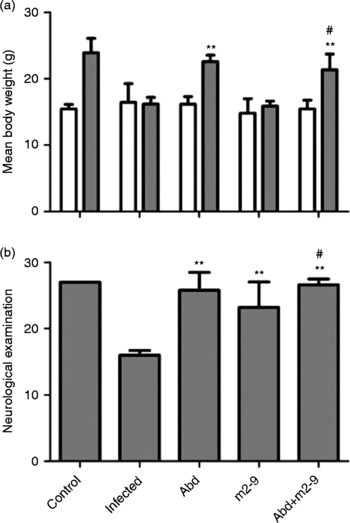
All mice gained weight except those in the infected group treated with edible oil (fig. 1a). In those mice treated with albendazole plus m2-9 or albendazole alone, the weight increased more than in the infected control group treated with edible oil (P < 0.01). There were also significant differences (P < 0.01) in the weight gain between the albendazole plus m2-9 group and the m2-9 treated group of infected mice.
(a) Mean body weight changes in mice. The white and grey columns represent the mean body weights of the mice on days 0 and 23 PI, respectively. Abd, m2-9 and Abd +m2-9 represent mice treated with albendazole alone, m2-9 alone and albendazole plus m2-9, respectively. **P < 0.01 compared to the edible oil treated infected mice. ##P < 0.01 compared to m2-9 treated infected mice. (b) Neurological examination score changes in mice. These values represent the mean neurological examination scores of the mice on day 23 PI. Abd, m2-9 and Abd +m2-9 represent mice treated with albendazole alone, m2-9 alone and albendazole plus m2-9, respectively. **P < 0.01 compared to the edible oil treated infected mice. #P < 0.05 compared to m2-9 treated infected mice.

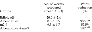
Recovered larvae were virtually absent in the brain tissue of mice treated with albendazole alone or albendazole and m2-9 co-therapy. There were also significant differences (P < 0.01) in worm recovery between the edible oil group and the m2-9 treated group of infected mice. The numbers of recovered larvae in the albendazole plus m2-9 group were obviously fewer than in the m2-9 treated group of infected mice (P < 0.01). The highest level of worm reduction was observed after co-therapy with albendazole and m2-9 (table 1).
The effect of treatment on A. cantonensis-induced meningitis in BALB/c mice over 7 days after inoculation on day 5.
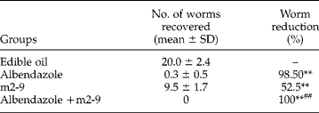
Larval recovery in mice on day 23 post-inoculation. **P < 0.01 compared to the edible oil treated infected mice. ##P < 0.01 compared to m2-9 treated infected mice.
Treatment and neurological function/behaviour
On day 23 PI, we found that uninfected mice maintained normal neurological function (27 points) (tables 2 and 3). The neurological functions in the other mice had deteriorated, as evidenced by reduced neurological scores. However, the neurological scores of the three drug-treated groups were significantly higher than that of the edible oil treated infected mice (fig. 1b). The neurological scores in the albendazole plus m2-9 group were obviously higher than in the m2-9 treated group of infected mice (P < 0.01).
Evaluation of neurological function in mice by motor score.
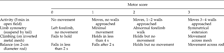
0, 1, 2 and 3 represent the respective scores a mouse acquired in an inspected item.
Evaluation of neurological function in mice by sensory score.

1, 2 and 3 represent the respective scores a mouse acquired in an inspected item.
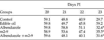
In the water maze task, the escape latency of the albendazole group, m2-9 group and albendazole plus m2-9 group was shorter compared with the edible oil treated infected mice on day 23 PI (table 4). However, there was no significant difference in escape latency between the albendazole plus m2-9 group and m2-9 alone or albendazole alone treated groups of infected mice on day 23 PI (P>0.05). No significant difference in escape latency was observed between the three drug-treated groups and the edible oil treated group of infected mice on days 20–22 PI (table 4). These results indicate that single or combination treated mice have decreased escape latency equal to that of uninfected mice.
Changes in the mean escape time (s) in mice on days 20–23 post-inoculation (PI).
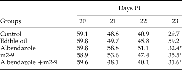
*P < 0.05, compared to the edible oil treated infected mice.
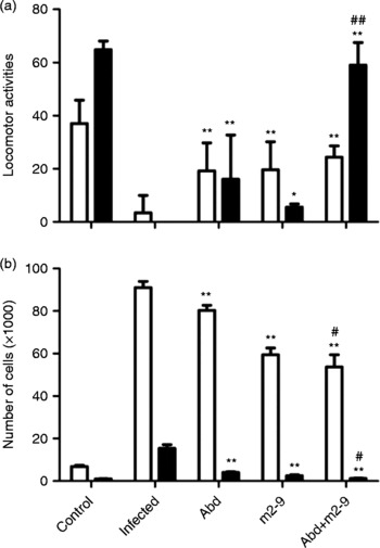
In the spontaneous locomotor activity test, the frequency of ambulation and rearing in the edible oil group were significantly less than those in the normal group (fig. 2a). There were significant differences in the frequency of ambulation and rearing between the three drug-treated groups and the edible oil treated infected group (fig. 2a). The combination of albendazole and m2-9 treatment significantly increased the frequency of rearing of the infected mice, compared to the m2-9 alone or albendazole alone treated infected mice (P < 0.01). These results also indicated that albendazole plus m2-9 treatments restored the spontaneous locomotor activities of infected mice back to normal.
(a) Spontaneous locomotor activity changes in mice. The white and black columns represent the mean frequency of ambulation and rearing of the mice on day 23 PI, respectively. Abd, m2-9 and Abd+m2-9 represent mice treated with albendazole alone, m2-9 alone and albendazole plus m2-9, respectively. *P < 0.05, **P < 0.01 compared to the edible oil treated infected mice. ##P < 0.01 compared to m2-9 alone or albendazole alone treated infected mice. (b) Leucocyte and eosinophil counts of surviving mice on day 23 PI. The white and black columns indicate number of leucocytes and eosinophils, respectively. Abd, m2-9 and Abd +m2-9 represent mice treated with albendazole alone, m2-9 alone and albendazole plus m2-9, respectively. **P < 0.01 compared to the edible oil treated infected mice. #P < 0.05 compared to m2-9 alone or albendazole alone treated infected mice.

Treatment and leucocyte counts and histopathology
CSF total leucocytes and eosinophil cell numbers were significantly reduced (P < 0.05) in the three drug-treated groups compared to the edible oil treated infected group (fig. 2b). Total leucocyte and eosinophil reduction were most obvious in the albendazole plus m2-9 combination group (fig. 2b).
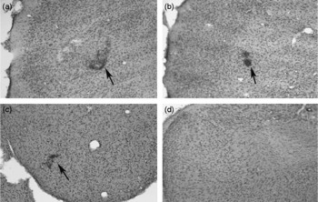
Light microscopic examination of tissues stained with haematoxylin and eosin revealed that eosinophilic meningitis was induced in mice by A. cantonensis infection. The large-scale infiltration of the cortex by leucocytes was evident on day 23 PI (fig. 3a). Leucocytes were reduced by treatment with albendazole or m2-9 (fig. 3b and 3c). However, the reduction was more significant in the combination therapy group (fig. 3d).
Haematoxylin and eosin staining of inflammatory cells (arrowed) in the cortex of treated infected mice (magnification × 100) with leucocyte accumulation following treatment with edible oil (a), albendazole (b) and m2-9 (c), and no leucocyte accumulation with albendazole plus m2-9 (d).

Discussion
Human and non-human primates are the unsuitable hosts for A. cantonensis. The parasite is unable to complete its development and usually dies in the central nervous system, causing eosinophilic meningitis or sometimes death (Wang et al., Reference Wang, Lai, Zhu, Chen and Lun2008). Therefore, more effective treatments for eosinophilic meningitis are urgently needed.
In recent years, more and more research on the chemistry of marine organisms has been pursued, in response to the need for compounds possessing bioactivity with possible pharmaceutical applications or other economically useful properties, such as fine chemicals, drugs, cosmetics and personal-care products (Andersen & Williams, Reference Andersen and Williams2000). Marine micro-organisms have become an important source of pharmacologically active metabolites. More than 272 new compounds have been isolated from marine fungi and the number is increasing (Bugni & Ireland, Reference Bugni and Ireland2004). Over 15 compounds of marine origin are currently in human clinical trials. Marine fungi have been shown to be a rich and promising source of novel anti-inflammatory and immunomodulatory agents. Oh et al. (Reference Oh, Gontang, Kauffman, Jensen and Fenical2008) purified a novel polyketide salinipyrone A from the marine actinomycete Salinispora pacifica, which moderately inhibited interleukin-5 (IC50 = 10 μg/ml) in a mouse splenocyte model of allergic inflammation, with low cell cytotoxicity. Ponomarenko et al. (Reference Ponomarenko, Kalinovsky, Afiyatullov, Pushilin, Gerasimenko, Krasokhin and Stonik2007) discovered that new diterpenoids isolated from a Northern Cook Islands marine sponge Spongia (Heterofibria) sp. moderately activated murine splenocyte lysosomes (apparent IC50 greater than 100 μg/ml). Aiello and colleagues established the molecular pharmacology of a novel bromopyrrole alkaloid, isolated from the Mediterranean sponge Axinella verrucosa. In a series of in vitro studies, the alkaloid was observed to display potent neuroprotective activity against the agonists serotonin and glutamate (Aiello et al., Reference Aiello, D'Esposito, Fattorusso, Menna, Muller, Perović-Ottstadt and Schröder2006).
Albendazole is a broad-spectrum anthelmintic drug, which acts by binding to parasite β-tubulin, inhibiting its polymerization and impairing glucose uptake, causing death of the parasite (Venkatesan, Reference Venkatesan1998). The drug can penetrate into the subarachnoid space (John & Martinez, Reference John and Martinez1975). The ability of albendazole to kill larvae of A. cantonensis has been demonstrated (John & Martinez, Reference John and Martinez1975; Lan et al., Reference Lan, Wang, Lai, Chen, Lee, Hsu and Lee2004; Tu & Lai, Reference Tu and Lai2006; Chen & Lai, Reference Chen and Lai2007). Albendazole exhibits marked larvicidal activity in angiostrongyliasis. However, sole treatment with albendazole in eosinophilic meningitis cannot completely inhibit the inflammatory reaction (Lan et al., Reference Lan, Wang, Lai, Chen, Lee, Hsu and Lee2004). The dead worms could cause more severe inflammation through activating eosinophils in the host brain (Wang et al., Reference Wang, Jung, Chen, Wong, Wan and Wan2006). In our present study, the number of larvae in mice treated with albendazole alone was reduced, but the drug did not improve the inflammatory reaction.
Our results showed that the combination of albendazole and m2-9 decreased leucocyte response and increased the frequency of rearing compared to monotherapy. Combination therapy reduced the number of leucocytes, increased body weight as well as improved neurological function of the infected mice. In severe infection, combination therapy with an anthelmintic drug (to kill larvae) and an anti-inflammatory agent (to reduce the inflammatory response to the organism breakdown products) may well be a better treatment approach (Lai et al., Reference Lai, Chen, Chang and Lee2008). We hypothesize that in combination treatment albendazole kills the worms, while m2-9 reduces the inflammatory reaction evoked by the dead worms. However, further investigations, including examination of the inflammatory cytokines in the mouse brain and the immunomodulatory effect of m2-9, are needed to confirm this hypothesis.
Acknowledgements
We thank Li-Si Ou yang technical assistance for conducting histological observations at Sun Yat-sen University. The study was funded by grants from the National Basic Research Program of China (grant no. 2010CB530004) and the Chinese Important Scientific Research Project on Infectious Diseases (grant no. 2008ZX10004-011).