Association of aortic atresia and interrupted aortic arch is usually lethal due to coronary hypoperfusion, unless coronary flow is provided by anastomosis with pulmonary vesselsReference Careddu, Oppido, Ialonardi and Gargiulo 1 – Reference Yew, Coleman and Calder 4 . A girl had prenatal diagnosis of conoventricular ventricular septal defect, aortic atresia, and probable aortic arch interruption. Neonatal echocardiography confirmed prenatal diagnosis, with large ductus arteriosus vascularising descending aorta. During the first day of life, she presented myocardial ischemia, with troponin elevation at 3884 ng/L.
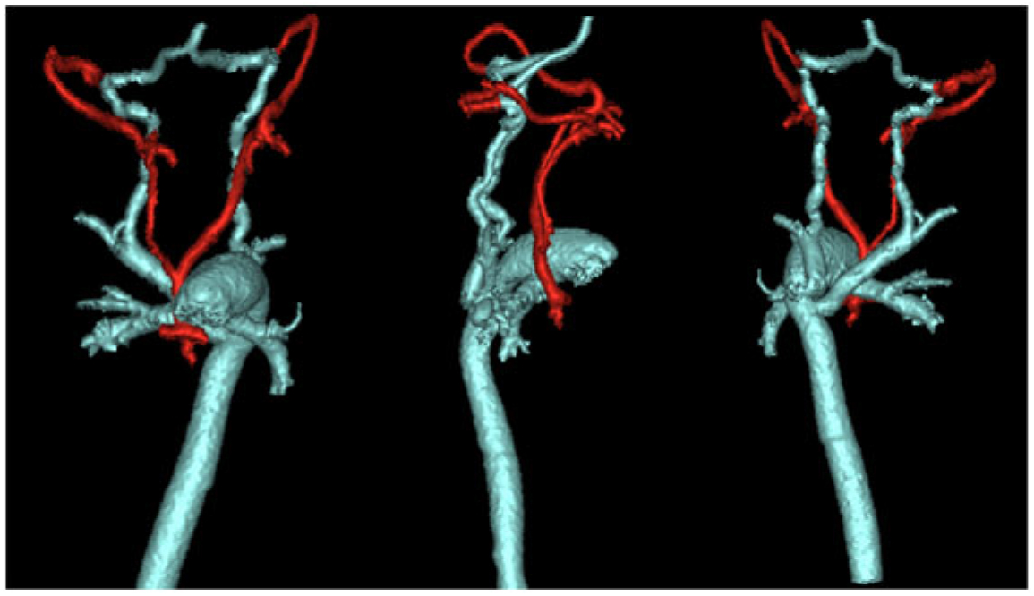
Cervico-thoracic angio-CT scanner displayed aortic valve atresia with hypoplastic ascending aorta connected to common carotid arteries. Aortic arch was interrupted after carotid arteries. Left-sided descending aorta was connected to a large pulmonary trunk through a large ductus arteriosus. Right and left subclavian arteries arise from descending aorta, right subclavian artery having retro-esophageal course, without Kommerell diverticulum. Large right and left vertebral arteries arise from subclavian arteries. On both sides, they vascularised large collateral arteries arising from the junction of their V3 and V4 segments, which were connected to the external carotid arteries. Therefore, coronary arteries were supplied by retrograde flow in common carotids arteries coming from subclavian arteries through an anastomosis of vertebral arteries and external carotid arteries (Fig 1).
3D reconstruction of arterial system. Anterior (left image), right (middle image), and posterior (right image) view of aortic, supra-aortic, and pulmonary arteries. In red, hypoplastic ascending aorta is connected to common carotid arteries, which terminates as external carotid arteries without internal carotids arteries. In blue, pulmonary artery is connected with descending aorta through a large patent ductus arteriosus; descending aorta is connected with subclavian arteries which continue as two large vertebral arteries. Anastomoses between these two systems are located on both sides at the junction of V3 and V4 segments of vertebral arteries, allowing a retrograde flow within carotid arteries and ascending aorta.

At 7 days of age, the patient underwent surgical repair, with pulmonary homograft and Damus–Kaye–Stansel procedure to reconstruct aortic arch, surgical connection of right ventricle to pulmonary arteries using a prosthetic conduit.
We report here the first case of bilateral extracranial anastomosis between distal vertebral arteries and external carotid arteries, providing retrograde flow to carotids arteries, ascending aorta, and finally to coronary arteries.
Author ORCIDs
Volodia Dangouloff-Ros 0000-0003-1260-5886
Acknowledgements
None.
Financial Support
This research received no specific grant from any funding agency, commercial or not-for-profit sectors.
Conflict of Interest
None.
Ethical Standards
The authors assert that all procedures contributing to this work comply with the ethical standards of the relevant national guidelines on human experimentation and with the Helsinki Declaration of 1975, as revised in 2008, and has been approved by the institutional committees.