Introduction
Embryo transfer (ET) technology was initially developed to optimize the genetic and economic resources of herds. It reached its peak in popularity in the 1970s (Hasler, Reference Hasler2006). Nowadays, embryos are massively produced and recovered at early stages of development. Females of high commercial value produce from 10 to 15 calves per year; boosting herd improvement and achieving up to 50% genetic enhancement (Christensen, Reference Christensen1991). However, ET has not had the same success in tropical regions. The reproductive efficiency of ET programs in Zebu cattle (Bos indicus) is not as high as with Bos taurus due to generally lower percentages (32–50%) of pregnancy (Donaldson, Reference Donaldson1984; Nogueira et al., Reference Nogueira, Barros and Teixeira2002; Montiel et al., Reference Montiel, Galina, Rubio and Corro2006) compared with the European breeds (Wright, Reference Wright1981; Hasler, Reference Hasler1992; Spell et al., Reference Spell, Beal, Corah and Lamb2001) which are generally between 60–70%.
Besides this problem, the number of good quality transferable embryos obtained per donor is less in Bos indicus (Donaldson Reference Donaldson1984; Aguilar et al., Reference Aguilar, Galina, Merchant, Montiel, Canseco and Marquez2002; Neto et al., Reference Neto, Sanches, Perri, Sedena and Garcia2004; Márquez-Alvarado et al., Reference Márquez-Alvarado, Galina, Moreno, Ruiz and Merchant2005) than in Bos taurus (Barrios et al., Reference Barrios, Romge, Harms, Blake and Kraemer1982, Hasler et al., Reference Hasler, McCauley, Schermerhorn and Foote1983; Hasler Reference Hasler1992; Neto et al., Reference Neto, Sanches, Perri, Sedena and Garcia2004). Aguilar et al., (Reference Aguilar, Galina, Merchant, Montiel, Canseco and Marquez2002) showed that embryo grading may vary in approximately 30% between routine stereoscopic evaluation and the more invasive electronic microscopy. Another important parameter to be considered is the time of the year at which embryo evaluation takes place; previous studies have shown that embryo quality is better during the rainy season than the dry season (Bastidas & Randel, Reference Bastidas and Randel1987; Márquez-Alvarado et al., Reference Márquez-Alvarado, Galina, Moreno, Ruiz and Merchant2005).
The aim of the present study, therefore, was to compare evaluations performed with the stereoscopic microscopy (SM) and inverted phase contrast microscopy (PCM) to determine if one method offers an advantage over the other in the evaluation of embryo quality. A secondary objective was to assess if either of the two methods were better in grading embryos produced in the rainy or the dry season as it has been shown that embryo evaluation is more inaccurate in the dry season.
Material and methods
Embryo production
Twenty Brahman (Bos indicus) cows aged between 3 and 7 years were synchronized and superovulated at two different times of the year: in May (dry season, 1500 mm/year) www.elocal.gob.mx/work/templates/enciclo/veracruz/municipios/30183a.htm and September (rainy season, 2,245.5mm/year) www.emexico.gob.mx/work/EMM04/Veracruz/.../30023a.htm
Two injections of 25 mg of PGF2α (dinoprost tromethamine; Lutalyse-Pfizer) with an interval of 14 days between treatments were used to synchronize estrus (day 0). Successfully induced cows continued with the superovulation programme. Total dose per donor was 240 mg of follicle stimulating hormone (Folltropin) administered in decreasing doses during days 9, 10, 11, and 12 of the estrous cycle (Lindsell et al., Reference Lindsell, Murphy and Mapletoft1986). In the afternoon of day 11, and morning of day 12, cows received 25 mg of PGF2α (dinoprost tromethamine; Lutalyse-Pfizer). Artificial insemination was performed in cows that displayed overt signs of estrus. Donors were artificially inseminated at 12 and 24 h after estrus onset with Brahman bull semen. Embryo were collected by non-surgical uterine flushing 7 days after the onset of estrus
Embryo evaluation
Embryos were evaluated and classified by a qualified technician using the conventional method of SM (Stereo Zoom®) with the ×15 eyepiece and subsequently with an inverted PCM (Olympus CKX 31) with the ×40 eyepiece by a different technician, unaware of the previous evaluation. Both evaluations were done according to the guidelines established by the International Embryo Transfer Society (IETS) handbook (levels 1, 2 and 3). In total, 118 fresh embryos were evaluated with the two microscopes at both times of the year: 78 during the rainy season and 40 during the dry season. A subsample of 52 embryos from the rainy season and 35 from the dry season was processed by the TUNEL (terminal deoxynucleotidyl transferase dUTP nick-end labelling) technique (Roche Diagnostic Kit. Indianapolis, IN, USA) to evaluate the extent of apoptosis.
Evaluation of apoptosis
The apoptotic cells were visualized using the TUNEL technique (Roche Diagnostic Kit. Indianapolis, IN, USA) as described by Contreras et al. (Reference Contreras, Galina, Ávila, Asprón and Moreno2008). TUNEL-positive cells score was used to measure the degree of programmed cell death (apoptosis) as a criterion to define embryonic quality and for comparing the two methods of evaluation.
Statistical analysis
Analysis of embryo morphological features classified as good, moderate and poor quality was performed by descriptive statistics. Cohen's kappa test was used to determine agreement between results obtained by SM and PCM, SM and TUNEL, and PCM and TUNEL. The same test was used to compare embryos collected during the two seasons. The following concordance levels were used to determine the degree of significance, k < 0.20, insignificant; from 0.21–0.40, poor; 0.41–0.60, moderate; 0.61–0.80, good and from 0.81–1.0, very good.
Results
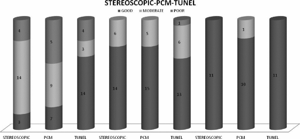
Evaluations for the total of 118 embryos, 78 during the rainy season and 40 during the dry season, are given in Figure 1
Absolute value of embryos with different quality evaluated by stereoscopic microscopy, phase contrast microscopy and TUNEL (terminal deoxynucleotidyl transferase dUTP nick-end labelling) at different developmental stages.

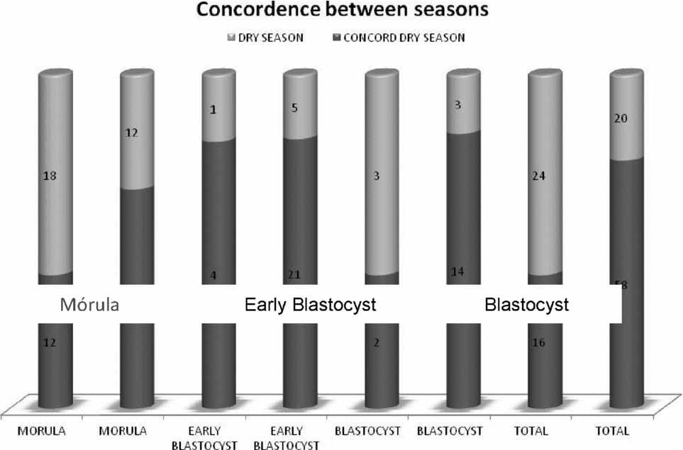
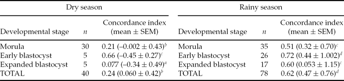
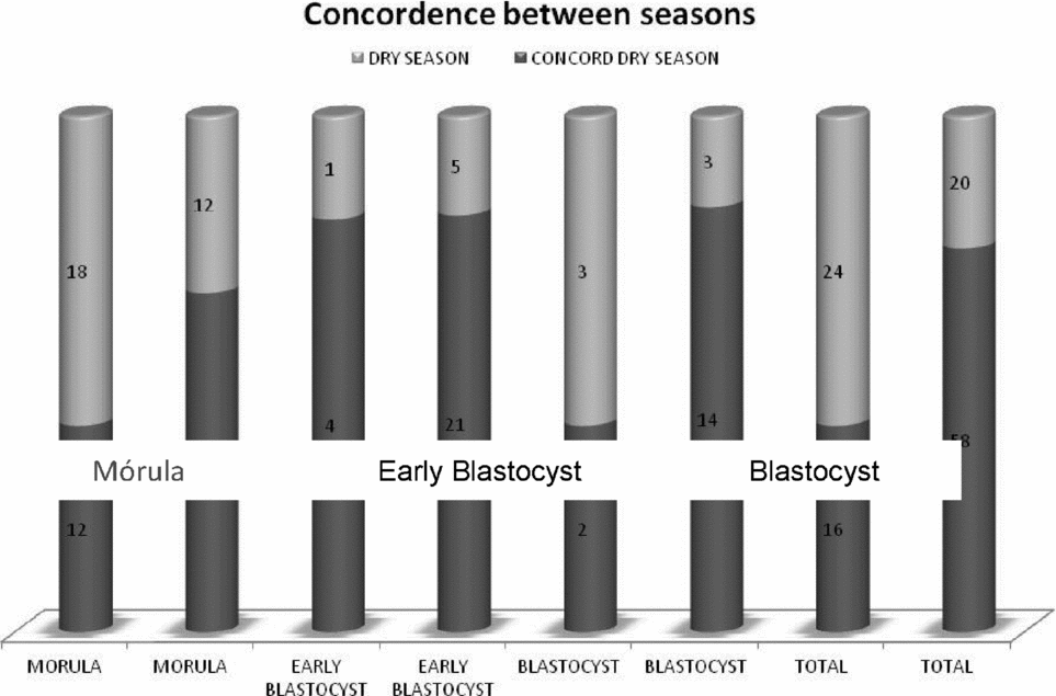
Global concordance between SM and inverted PCM in the two seasons of the year showed higher kappa values in the rainy season than in the dry season. Concordance, regarding developmental stage was high for the early blastocyst (k = 0.66), whilst for other stages they were poor or insignificant (Table 1). A tendency was found for the embryonic developmental stage evaluations to show increasing levels of agreement during the rainy season as the developmental stage progressed, a pattern that did not occur during the dry season. Comparison of absolute values between the two seasons, dry and rainy, showed that the best concordance for the different developmental stages was shown by early blastocysts and blastocysts during the rainy season (Fig. 2)
Comparison of absolute values obtained in the dry and rainy seasons according to the developmental stage (morula, early blastocyst or blastocyst).
Concordance index (Cohen's kappa index) shown by embryos according to their developmental stage (morula, early blastocyst or expanded blastocyst) in the dry or rainy season of the year
aInsignificant, bpoor, cmoderate, dgood, every good. SEM, standard error of the mean.
Comparison between morphological evaluation and TUNEL
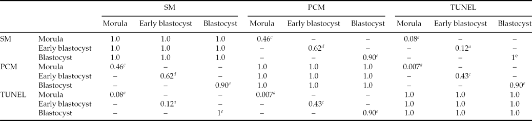
Fifty-two embryos were collected during the rainy season and evaluated by the two morphological methods (SM and PCM) and then by the TUNEL technique. It should be mentioned that 30 out of the 40 embryos collected during the dry season did not resist the procedure for the TUNEL test; thus, embryos obtained were not enough to be included in the present study. Statistical analysis was therefore performed only on embryos in the rainy season. Regarding quality level and according to the kappa coefficient (k = 0.61), the first two methods showed good agreement. However, notice should be taken of the observed tendency of the kappa index to increase as the developmental stage progressed, regardless of agreement between the methods (Table 2)
Kappa concordance index among evaluation methods for each developmental stage.
aInsignificant, bpoor, cmoderate, dgood, every good.
PCM, phase contrast microscopy; SM, stereoscopic microscopy; TUNEL, terminal deoxynucleotidyl transferase dUTP nick-end labelling.
Interestingly, 67% of the morulas were classified as having moderate quality, whilst early and expanded blastocysts were good quality in 70 and 100% of cases, respectively. The same tendency was found with the PCM evaluation where 43% of morulas were found to have regular quality. Concordance between the two methods changed from good to moderate for this developmental stage (k = 0.46). On the other hand, most blastocysts were found to be of good quality and agreement between methods was good, and for expanded blastocysts, very good. The number of embryos classified as good (n = 28) by the first method, 96%, was classified similarly by the second method.
As for the optical assessment methods, no important differences were found with TUNEL evaluation regarding good and poor quality embryos. However, of the moderate quality embryos, specifically at the morula stage, nine out of 14 qualified as moderate by SM, and seven out of eight by PCM were identified as having good quality by TUNEL. Agreement between the two morphological evaluation methods (SM and PCM) and TUNEL was poor (Fig. 3)
Concordance among different developmental stages (morula, early blastocyst, expanded blastocyst) and quality levels: 1, 2 and 3 among the different evaluation methods. Embryos were classified as: G = good, M = moderate or P = poor. PCM, phase contrast microscope; SM, stereoscopic microscope; TUNEL, terminal deoxynucleotidyl transferase dUTP nick-end labelling. (See online for a colour version of this figure.)
Discussion
The aim of the present study was to compare two methods (SM and inverted PCM) for assessment of Bos indicus embryos which were evaluated morphologically in the morula and blastocyst stage according to their optical features of size, form, symmetry, color and general appearance of embryonic cells. At present embryonic evaluation based exclusively on morphological observation of the structures is imperfect. However, even in programmes of assisted reproduction in humans this is still the most frequently used method for selecting embryos (Alikani et al., Reference Alikani, Sadowy, Cohen and Van Soom2002).
Agreement between evaluation methods was good during the rainy season (74%) as opposed to the dry season when it was poor (40%). Several studies have reported a seasonal effect on the success of ET. Bastidas & Randel (Reference Bastidas and Randel1987) reported the autumn and winter as the worst times of the year for programmes of ET in Bos indicus cattle, whilst in a previous paper, Randel (Reference Randel1984) indicated that the best time of the year was the summer. This difference could be due to more favourable climatic conditions and better food availability but this assumption remains to be tested. Additionally, Molina (2000) and Márquez-Alvarado et al. (Reference Márquez-Alvarado, Galina, Moreno, Ruiz and Merchant2005) showed that embryo quality of Zebu cows is higher during the rainy season than during the dry season, improving evaluation accuracy and confirming the results of the present study. In other assays (Hasler et al., Reference Hasler, McCauley, Schermerhorn and Foote1983; Ryan et al., Reference Ryan, Prichard, Kopel and Godke1993) evaluated Bos taurus embryos and found that a higher percentage of transferable embryos was forthcoming during the cold season, suggesting that embryo quality might be differently affected by climate in each species.
In the present study, good agreement was found between the two optical evaluation methods (SM and PCM) in quality level for the 52 embryos evaluated during the rainy season. However, concordance with the TUNEL technique was poor according to Cohen's kappa coefficient. As mentioned above, dry season embryos were not included in this analysis because they did not resist the procedure for the TUNEL test. Production of follicles with low fertilization possibility has been explained in several ways. Recently, Bridges et al. (Reference Bridges, Mussard, Burke and Day2009) reported that proestrus duration and follicular diameter may affect fertility. Alternatively, embryos may suffer during the freezing and thawing processes, and collapse, considering that they are fragile cells (Márquez-Alvarado et al., Reference Márquez-Alvarado, Galina, Moreno, Ruiz and Merchant2005). Fertility in frozen embryos is known to be less than 10% when compared with non-frozen cells (Dobrinsky, Reference Dobrinsky2002; Spell et al., Reference Spell, Beal, Corah and Lamb2001). Accordingly, dry season embryos are probably capable of developing, but are extremely fragile when frozen. Further studies are needed in this important area of reproductive biotechnology.
Stereoscopic evaluation of morula collected during the rainy season revealed 67% of moderate quality embryos, whilst more advanced stages of early and expanded blastocysts showed a higher percentage classified as good in 70 and 100%, respectively. However, when evaluated by the TUNEL technique, 67% of morula was deemed to be of good quality. According to Cohen's kappa coefficient, this observation shows poor agreement with observations by SM (k = 0.169) and by PCM (k = 0.217). This finding indicates that evaluation by morphological methods only, gives little assurance on quality level. Rondeau et al. (Reference Rondeau, Guay, Goff and Cooke1995) evaluated embryo metabolism reporting that 47% of embryos classified as being of good quality had abnormal metabolic activity due to defects which cannot be observed with ordinary microscopy.
In contrast, expanded blastocysts evaluated as having good quality level by morphological methods, showed good or very good concordance when evaluated with the TUNEL technique. These results agree with observations by Callesen et al. (Reference Callesen, Lǿvendahl, Bak and Greve1995), who reported that embryos evaluated at a more advanced, as opposed to early developmental stages, have better quality and are easier to assess with precision. In this context, Antunes et al. (2008) reported that in vitro produced embryos show lower quality level from the 9-cell stage to the morula as opposed to the blastocyst stage, when evaluated by TUNEL. This disagreement in evaluations has also been found in other studies. Aguilar et al. (Reference Aguilar, Galina, Merchant, Montiel, Canseco and Marquez2002) reported up to 30% variability in evaluation criteria being the highest in embryos judged as being of moderate quality. López-Demián et al. (Reference López-Demián, Galina, Merchant, Cedillo-Peláez and Aspron2008) found in Bos taurus embryos that the highest uncertainty level appeared when assessing embryos of moderate quality. In the present study, variability for good quality embryos was 10%. These differences may be due to the evaluation method and to the observer. Farin & Farin (1995) mentioned that consistency between evaluators for correct classification of quality level is approximately 60% in in vivo embryos. This finding suggests that routinely produced embryos are evaluated with some degree of error; implying that there is still currently no precise, objective and non–invasive method of evaluation of embryo viability.
The inaccuracies found in the present study in the evaluation of embryos judged as being of moderate quality have been observed by several authors (Farin et al., 1995; De Leeuw, Reference De Leeuw1996; Van Soom et al., Reference Van Soom, Ysebaert, Vanhoucke-De Medts, Van de Velde, Merton, Delval, Van Langendonckt, Donnay, Vanroose, Bols and de Kruif1996; Lopes et al., Reference Lopes, Ramsing, Larsen, Räty, Peippo, Greve and Callesen2005). In these studies, agreement between evaluators has been considered poor, specifically when evaluating embryos of moderate quality. López-Demián et al. (Reference López-Demián, Galina, Merchant, Cedillo-Peláez and Aspron2008) also reported the highest percentage of errors when moderate quality embryos were evaluated. However, in their study these types of embryos were judged as being of lower quality, whilst in the present study, moderate quality embryos were subjected to the TUNEL technique and assessed as being of good quality. A possible explanation is that, in previous studies, inclusion criteria to evaluate embryos by SM were less strict, and once embryos were analyzed using a more precise method, their quality level tended to fall. Moreover, Farin et al. (Reference Farin, Slenning and Britt1999) stated that an appropriate evaluation is important to foresee the outcome of pregnancies as discrepancies between evaluations may give up to 16% difference in gestation rates. In the present study, TUNEL-positive cells were found in all embryos. This finding agrees with results by Gjørret et al. (Reference Gjørret, Knijn, Dieleman, Avery, Larsson and Maddox-Hyttel2003), Márquez-Alvarado et al. (Reference Márquez-Alvarado, Galina, Castilla, León and Moreno-Mendoza2004), and Contreras et al. (Reference Contreras, Galina, Ávila, Asprón and Moreno2008), who detected TUNEL-positive cells in embryos evaluated at all developmental stages. Previous studies have reported that, although good quality embryos may be found by routine methods (SM), their metabolic activity is not always normal, and may show functional defects which go undetected when embryos are evaluated prior to freezing or transfer. These embryos will be non-viable when transferred (Aguilar et al., Reference Aguilar, Galina, Merchant, Montiel, Canseco and Marquez2002; Márquez-Alvarado et al., Reference Márquez-Alvarado, Galina, Castilla, León and Moreno-Mendoza2004). This issue could be critical for massive embryo-production programmes.
The detection of apoptosis in embryos at early stages is based on the morphological observation of typical processes such as condensation and nuclear fragmentation (electron microscopy and fluorescence staining) or evaluating specifically the presence of DNA degradation (TUNEL test; Betts & King, Reference Betts and King2001). The present study showed that embryos that were classified as being of good quality had a smaller number of TUNEL-positive cells than those of moderate or poor quality, which agrees with results published previously (Márquez-Alvarado et al., Reference Márquez-Alvarado, Galina, Castilla, León and Moreno-Mendoza2004; Contreras et al., Reference Contreras, Galina, Ávila, Asprón and Moreno2008). However, in the present study, embryos classified as having moderate quality had 60% of TUNEL-positive cells, a per cent similar to that of embryos of good quality. In this context, studies have been performed based on embryo fragmentation, one of the most significant criteria used to classify them according to their morphology. Hardy & Spanos (Reference Hardy, Spanos and Van Soom2002) mentioned that cytoplasmic fragmentation may be a non-invasive marker of apoptosis-type programmed cell death, especially of the inner cell mass (ICM). Antunes et al. (2008) considered the apoptosis index to be a better parameter than fragmentation to measure embryonic quality. However, in humans, the relationship between fragmentation and apoptosis has been described as subjective (Hardy et al., Reference Hardy, Handyside and Winston1999; Jurisicova et al., Reference Jurisicova, Antenos and Vermussa2003). Also, Ikeda et al. (Reference Ikeda, Prendes, Alonso-Montes, Rodriguez, Díez, Kitagawa, Imai and Gómez2006) have suggested that embryonic fragmentation is due to cellular division asymmetry, more than to the apoptosis process. The index of apoptotic cells remained the same in the morula stage, whilst the fragmentation index was 50% higher; indicating that apoptosis and fragmentation may be independent processes (Van Soom et al., Reference Van Soom, Mateusen, Leroy and De Kruif2003). This finding suggests that one of the errors of morphologic evaluation may be the measurement of the fragmentation percentage, especially in the morula stage, which appears morphologically to have lower quality (moderate or poor) and therefore may be evaluated wrongly. Byrne et al. (Reference Byrne, Southgate, Brison and Lesse1999), Contreras et al. (Reference Contreras, Galina, Ávila, Asprón and Moreno2008) and Godínez (Reference Godínez2009) showed that those blastocysts with more apparently apoptotic cells have less development potential when cultured; proving that when the percentage of cell death reaches a critical level there is damage to embryo development.
Based on these results, embryo production in Zebu cows in tropical countries should be carried out in the rainy season to guarantee a given percentage of accuracy during embryo evaluation, and the subsequent transfer or freezing processes.