Introduction
Hypertrophic cardiomyopathy is a complex genetic cardiovascular disorder characterised by thickening of the heart muscle, with a prevalence of 1 in 500 in the general population according to the disease phenotype.Reference Litt, Ali and Reza1 A higher prevalence (1:200) is recorded when researchers take into consideration subclinical cases, genotype-positive occurrences of sarcomeric genes, and familial transmission.Reference Maron, Desai and Nishimura2 Hypertrophic cardiomyopathy is a well-known cause of sudden cardiac death in athletes and young adults. Understanding the genetic underpinnings of hypertrophic cardiomyopathy is crucial for early diagnosis and personalised treatment. Hypertrophic cardiomyopathy is predominantly attributable to mutations in genes that encode sarcomeric proteins, notably MYBPC3, MYH7, TNNI3, TPM1, TNNT2, ACTC1, MYL3, and MYL2, with MYBPC3 and MYH7 being the most prevalent.Reference Hong, Xi and Yang3 Most patients have variants of the eight core genes that encode sarcomeric proteins. These mutations can lead to abnormal heart muscle structure and function, contributing to the development of the condition.Reference Olalekan, Bakare and Okwute4
In recent years, pathogenic mutations in non-sarcomeric genes have been identified, including, but not limited to, in proteins associated with calcium-handling genes (JPH2, TNNC1, PLN, and RYR2), the Z-disk (VCL, ACTN2, and TCAP), and the proteasomal components (TRIM63).Reference Litt, Ali and Reza1 These genes, like sarcomeric genes, also exhibit both inheritance models, with autosomal dominant inheritance being more common.Reference Walsh, Offerhaus, Tadros and Bezzina5 Nevertheless, these genes only affect 0.06% to 8.7% of hypertrophic cardiomyopathy patients. A synthesis of published data includes genes with strong, moderate, and weak causation evidence, as well as genes associated with hypertrophic cardiomyopathy based exclusively on functional data.Reference Hespe, Waddell and Asatryan6 Thus, evidence shows that up to 50% of hypertrophic cardiomyopathy patients lack a pathogenic variation.Reference Jaouadi, Morel and Martel7 Currently, the unexplained heritability is increasingly attributed to the observation that patients who are genotype-negative and sarcomeric-negative (Sarc-negative) exhibit a non-Mendelian or virtually Mendelian illness pattern, and this means the disease manifests as if it were caused by a single gene. This condition arises from the combined influence of hereditary and non-hereditary variables.Reference Bonanni, Del Franco and Setti8
Rare variants are considered more likely than common variants to cause missing heritability and oligogenic inheritance. It is anticipated that variations with potent effects will be maintained under selective pressure and continue to occur in the population with frequencies that are low to very low.Reference Rout, Ramu and Mariana9 Due to variable expressivity, incomplete penetrance, and absent heredity in hypertrophic cardiomyopathy, further investigation into the genetic underpinnings of hypertrophic cardiomyopathy is essential for a more thorough understanding of its pathophysiology.
Whole-exome sequencing facilitates an extensive analysis of the protein-coding regions inside the genome. It provides a powerful tool for identifying pathogenic variants associated with hypertrophic cardiomyopathy, aiding in accurate diagnoses and risk stratification. Interpreting variants identified through whole-exome sequencing presents challenges, including distinguishing between pathogenic and benign variants, as well as understanding the significance of variants of uncertain significance (VUS). Whole-exome sequencing insights can alter the clinical management of hypertrophic cardiomyopathy. They allow for a precise genetic diagnosis, risk prediction, and the potential for developing personalised therapeutic interventions based on an individual’s genetic profile.Reference Ireland and Ho10
The first national study in Egypt that analysed sarcomeric and non-sarcomeric variants in a cohort of idiopathic paediatric hypertrophic cardiomyopathy patients using targeted next-generation sequencing was reported by Darwish et al. 2020; it identified 10 rare variants in 8 patients, of which only 2 variants were pathogenic. Our current study aimed to identify significant rare genetic variants and enhance our understanding of pathogenic mutations in eight patients aged 14 to 50 years with hypertrophic cardiomyopathy who underwent testing with whole-exome sequencing (genome-wide test).Reference Darwish, Haghighi and Seliem11
Materials and methods
DNA extraction and ethical approval
The clinical indications and peripheral blood samples were obtained from hypertrophic cardiomyopathy patients at the National Heart Institute in Egypt. The clinical diagnosis of hypertrophic cardiomyopathy was made based on current guidelines.Reference Elena, Protonotarios and Gimeno12 Patients were selected consecutively from the outpatient clinic if they met the following criteria: (1) echocardiographic evidence of unexplained left ventricular hypertrophy (maximal wall thickness ≥15 mm, or ≥13 mm with positive family history); (2) absence of other causes of hypertrophy (e.g., hypertension, aortic stenosis, infiltrative diseases); and (3) willingness to undergo genetic testing and provide informed consent. No exclusion based on age, sex, or disease severity was applied to capture the full phenotypic spectrum.
Total genomic DNA was isolated from whole blood collected in Ethylenediaminetetraacetic acid tubes using the QIAamp DNA Blood Mini Kit (Qiagen, Germany) according to the manufacturer’s instructions. The Institutional Review Board at the General Organization for Teaching Hospitals and Institutes approved our study with IHc00084, and written informed consent was secured from each patient.
Sample preparation and whole-exome sequencing
Whole-exome sequencing was conducted on eight hypertrophic cardiomyopathy patients aged 14 to 45 years and analysed for research purposes. Exome capture was conducted utilising the xGen Exome Research Panel v2 (Integrated DNA Technologies, Coralville, Iowa, USA), and sequencing was executed with the NovaSeq 6000 system (Illumina, San Diego, CA, USA). A total of ±9,601,998,207 bases of sequence were constructed and uniquely aligned to the Genome Reference Consortium Human Build 37 (GRCh37) and Revised Cambridge Reference Sequence (rCRS) of the mitochondrial genome. This resulted in a mean depth of coverage of ±131.53X within the 34,366,187 bases of the captured region, which accounted for ±99.3% of the RefSeq protein-coding region. We attained ∼98.90% of the targeted bases with coverage at a depth of 20x or higher.
All samples were submitted in BioProject: PRJNA1053668 at National Center for Biotchnology Information (NCBI); the raw data can be accessed at NCBI SRA, with accession numbers SRR27236368, SRR27236367, SRR27236366, SRR27236365, SRR27472026, SRR27472025, SRR27472024, and SRR27472023.
Quality control of raw sequencing data
To guarantee the accuracy of the following bioinformatics analysis, raw sequencing data were filtered to produce high-quality sequences. The raw reads underwent quality control using FastQC and SeqPrep.Reference He, Zhu and Yang13 The methods involved (1) removing adapter sequences from reads, (2) trimming bases with quality values of less than 20, (3) discarding reads with an N ratio exceeding 10%, and (4) eliminating sequences of less than 20 bp in length.
Whole-exome sequencing pipeline analysis
BWA-MEM 0.7.8 was used with the default settings to align the filtered reads to the hg19 human reference.Reference Youngmok and Dongsu14 Then, the SAM (systolic anterior motion) format was converted into a binary alignment map file by using Samtools 0.1.19.Reference Chen, Dong and Yi15 The MarkDuplicate function in Picard v1.9.2 (https://broadinstitute.github.io/picard/) was employed to eliminate polymerase chain reaction duplicate readings. We conducted delRealigner and BaseRecalibration using GATK v2.3.9Reference Ebbert, Wadsworth and Staley16 to enhance the accuracy of variant calling. We detected single-nucleotide variations using Strelka software, along with deletions (indels) and insertions.
Annotation of variants
We utilised snpEff v4.3iReference Cingolani, Platts and Wang17 to annotate the types and genomic locations of variations, classifying them into categories such as intronic, splice-site variant, missense, insertions/deletions, stop gain/loss, and synonymous. Variants were further annotated and analysed using the following resources: Genomize (v6.14.4), Interval, Franklin, VarSome, OMIM, ClinVar databases, and PubMed.
Variant interpretation
Initially, we filtered the whole-exome sequencing data to detect genomic variations in genes previously associated with hypertrophic cardiomyopathy (HP:0001639). A comprehensive list of 291 genes attributed to hypertrophic cardiomyopathy in the Human Phenotype Ontology (HPO) database was used as an initial broad filter. Subsequently, to enhance the clinical specificity of our analysis, we prioritised a refined set of 29 genes with definitive, strong, or moderate evidence for causing hypertrophic cardiomyopathy or isolated left ventricular hypertrophy, as recently established by the Clinical Genome Resource (ClinGen) Hereditary Cardiovascular Disease Gene Curation Expert Panel.Reference Hespe, Waddell and Asatryan6 Afterwards, we considered genomic variants only in these 29 genes to enhance clinical specificity and selected variants that had an allele frequency of 0.01 or lower. Pathogenicity predictions were generated using computational algorithms, and variants were categorised as LP variants, VUS, or P variants according to American College of Medical Genetics and Genomics (ACMG) guidelines.Reference Liu, Yang and Gong18 In silico assessments of variant pathogenicity were further performed using bioinformatics prediction tools, including SIFT, PolyPhen-2, and Mutation Taster.
Results
The study enrolled eight patients diagnosed with hypertrophic cardiomyopathy at the National Heart Institute in Egypt; they were 14 to 45 years of age, with 37.5% being female (n = 3) and 62.5% male (n = 5). Whole-exome sequencing was performed on their peripheral blood DNA, achieving high coverage with about 78 million reads per sample, a mean coverage of 131.53X, and 98.90% of target regions covered at 20X.
Variants were identified using advanced methods of bioinformatics and were filtered based on the specific criteria of (a) a sequencing depth of less than 10x, (b) an allele frequency of less than 0.01 in the gnomAD database, and (c) a combined annotation dependent depletion (CADD) score of more than 10 to prioritise the most relevant variants.
A total of 21 rare variants were identified in 10 genes after filtering, including 15 missense variants (71.4%), 3 intron variants (14.2%), 1 stop gained variant (4.7%), and 2 splice region variants (9.5%). In the 8 Egyptian hypertrophic cardiomyopathy samples, 19 substitutions of single nucleotides were found, with the most common being C→T (n = 5). Each patient carried a median of approximately two variants.
Variant identification in the definitive hypertrophic cardiomyopathy genes
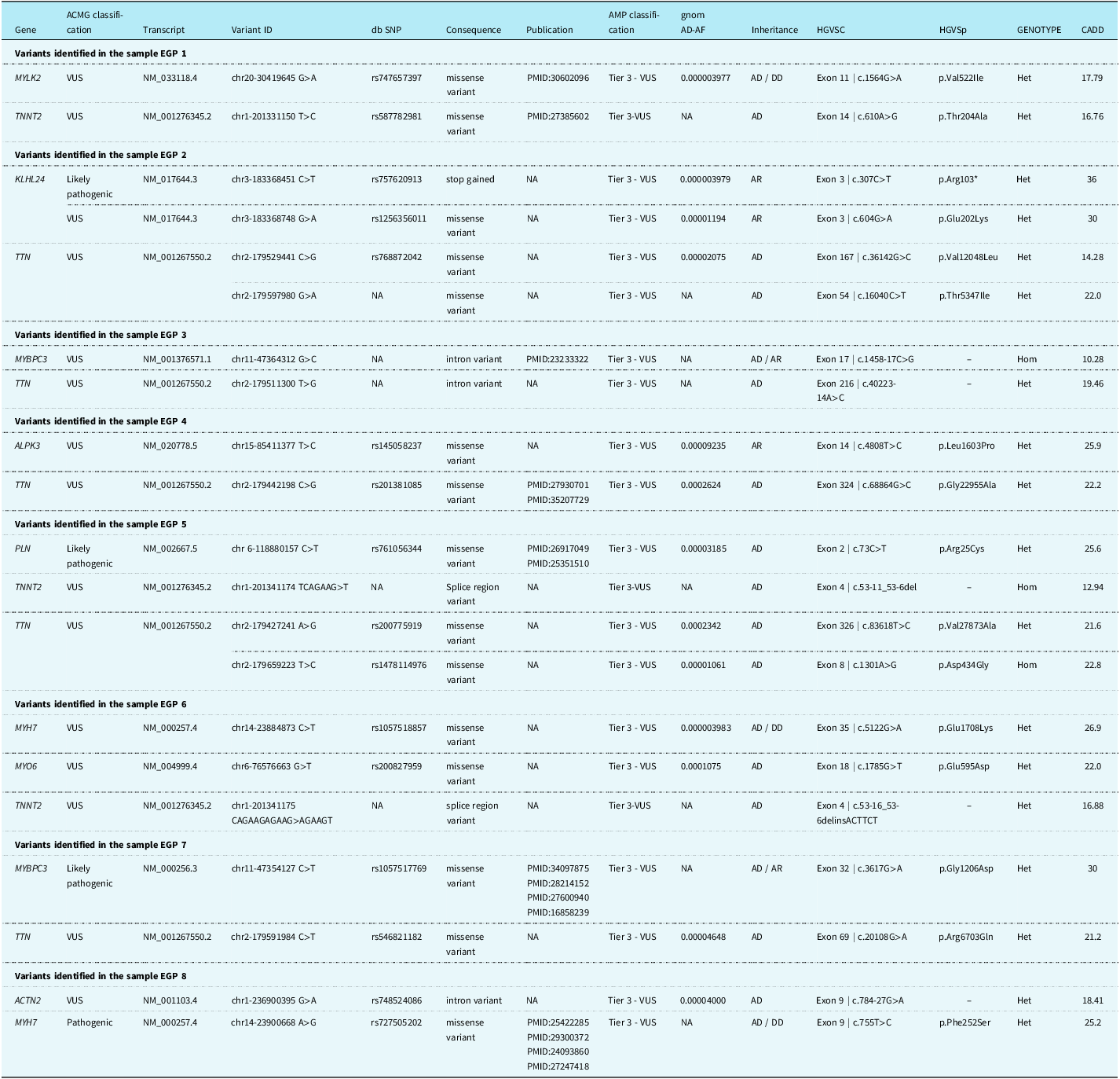
This study identified the variants associated with hypertrophic cardiomyopathy across 10 relevant genes: ACTN2, ALPK3, KLHL24, MYBPC3, MYH7, MYLK2, MYO6, PLN, TNNT2, and TTN. Table 2 provides the specific coordinates of the variants in these genes.
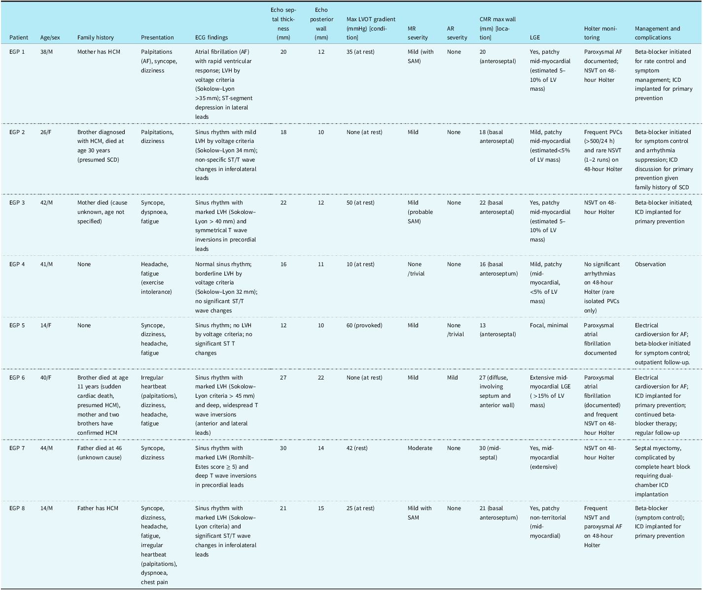
Clinical characteristics, diagnostic findings, and management of Egyptian patients with HCM

Table 1. Long description
The table has 8 rows and 15 columns. The columns are labeled as Patient, Age/sex, Family history, Presentation, ECG findings, Echo septal thickness (mm), Echo posterior wall (mm), Max LVOT gradient (mmHg) [condition], MR severity, AR severity, CMR max wall (mm) [location], LGE, Holter monitoring, and Management and complications. The rows are labeled with patient identifiers EGP 1 to EGP 8. Each row provides detailed information on the respective patient’s age, sex, family history, presentation, ECG findings, echo septal thickness, echo posterior wall thickness, max LVOT gradient under specific conditions, MR severity, AR severity, CMR max wall thickness and location, presence of LGE, Holter monitoring results, and management and complications. Notable trends include variations in family history, presentation symptoms, ECG findings, and management strategies across different patients.
LVOT = left ventricular outflow tract; SAM = systolic anterior motion; PVC = premature ventricular complex; NSVT = non-sustained ventricular tachycardia; LGE = late gadolinium enhancement; CMR = cardiac magnetic resonance; ICD = implantable cardioverter-defibrillator; AF = atrial fibrillation; HCM = hypertrophic cardiomyopathy; ECG = electrocardiogram; LVH = left ventricular hypertrophy; LV = left ventricle; SCD = sudden cardiac death; AR = aortic regurgitation.
Characteristics of variants identified in 10 genes associated with HCM in the Egyptian patients

Table 2. Long description
The table lists genetic variants identified in 10 genes associated with hypertrophic cardiomyopathy in Egyptian patients. It includes columns for gene, ACMG classification, transcript, variant ID, db SNP, consequence, publication, AMP classification, gnomAD frequency, inheritance, HGVSC, HGVSp, genotype, and CADD score. The table is divided into sections for variants identified in samples EGP 1 through EGP 8. Each section lists specific genes, their classifications, transcript IDs, variant IDs, db SNP identifiers, consequences, publication references, AMP classifications, gnomAD frequencies, inheritance patterns, HGVSC and HGVSp notations, genotypes, and CADD scores. For example, in sample EGP 1, the MYLK2 gene has a VUS classification with a missense variant identified by rs747657397, and the TNNT2 gene also has a VUS classification with a missense variant identified by rs587782981. Each row provides detailed information about the specific variants found in each gene for the respective samples.
ACMG = American College of Medical Genetics and Genomics; Het = heterozygous; Hom = homozygous; NA = no data available; CADD = combined annotation-dependent depletion scores; AF = allele frequency; AD = autosomal dominant; AR = autosomal recessive; DD = digenic dominant; HCM = hypertrophic cardiomyopathy; VUS = variants of uncertain significance; SNP = single nucleotide polymorphism; HGVSC = human genome variation society coding (C) namenclature; HGVSp = humman genome variation society protein (P) namenclature; AMP = association for molecular pathology.
Validated sarcomeric genes
In our study, we identified variants in three of eight sarcomeric genes (ACTC1, TNNI3, MYH7, TPM1, MYBPC3, TNNT2, MYL2, and MYL3) associated with hypertrophic cardiomyopathy that had strong evidence of disease aetiology.Reference Park, Packard and Levin19 They were the MYBPC3, MYH7, and TNNT2 genes.
In the MYBPC3 gene, two variants were identified in two out of eight patients. The identified variants included one missense variant and one intron variant. According to the ACMG classification, there was one VUS variant and one likely pathogenic variant. The likely pathogenic variant from patient EGP 7 was submitted to ClinVar (accession SCV004562018). Patients with MYBPC3 variants also had additional variants in lesser-known hypertrophic cardiomyopathy genes including TTN.
Two patients (EGP6 and EGP8) had distinct variants in the MYH7 gene, including two missense variants, classified as one VUS and one pathogenic variant. The pathogenic variant from patient EGP8 was submitted to ClinVar (accession SCV006082319). MYH7 and MYBPC3 are the most common genes with pathogenic or likely pathogenic variants per ACMG/AMP (Association for Molecular Pathology) criteria. Additionally, three variants in the TNNT2 gene were found in three patients (EGP1, EGP5, and EGP6), with three classified as VUS according to ACMG guidelines.
Variant detection in lesser-known hypertrophic cardiomyopathy genes
Several genes associated with hypertrophic cardiomyopathy encode both sarcomeric and non-sarcomeric proteins, which contribute to the genetic aetiology of hypertrophic cardiomyopathy to a small extent.Reference Walsh, Offerhaus, Tadros and Bezzina5 The study identified variants in seven minor genes associated with hypertrophic cardiomyopathy, including rare and harmful variants in genes such as ACTN2, ALPK3, KLHL24, MYLK2, MYO6, PLN, and TTN. Notably, an intron variant in the ACTN2 gene was classified as a VUS. In the KLHL24 gene, one VUS variant (p.Glu202Lys) and one likely pathogenic variant (p.Arg103*) were identified based on ACMG and AMP classifications. The likely pathogenic variant was submitted to ClinVar under accession SCV006082313. In the ALPK3 gene, one missense variant was detected, and it was classified as a VUS variant by ACMG.
A missense variant (p.Val522Ile) in the MYLK2 gene was classified as a VUS. The TTN gene encodes titin, an essential sarcomeric protein that is crucial in passive force transmission and vital for sarcomeric organisation, elasticity, and cell signalling.Reference Jolfayi, Kohansal and Ghasemi20 Seven variants were found in the TTN gene and categorised as seven VUS variants. A missense variant (p.Arg25Cys) in the PLN gene was classified as likely pathogenic and submitted to ClinVar under accession SCV006082320. Additionally, one missense variant was identified in the MYO6 gene: one VUS (p.Glu595Asp).
Our analysis yielded 21 rare variants, including 2 in the MYBPC3 gene. One variant was detected in each of the ALPK3, MYLK2, MYO6, PLN, and ACTN2 genes. The TNNT2 gene had three variants, while the MYH7 gene and the KLHL24 gene each contained two variants. Additionally, seven variants were identified in the TTN gene.
Genotype–phenotype correlations
Integration of the clinical characteristics Table 1 with the molecular findings Table 2 revealed clinically meaningful genotype–phenotype associations. Patients carrying pathogenic or likely pathogenic variants in core sarcomeric genes exhibited a more severe phenotypic expression. Specifically, EGP8, harbouring a pathogenic MYH7 p.Phe252Ser variant, presented with a malignant phenotype characterised by marked hypertrophy (septal thickness 21 mm), significant left ventricular outflow tract obstruction (25 mmHg), paroxysmal atrial fibrillation, frequent non-sustained ventricular tachycardia, and required implantable cardioverter-defibrillator implantation. Similarly, EGP7, with a likely pathogenic MYBPC3 p.Gly1206Asp variant, displayed extreme septal hypertrophy (30 mm), a high left ventricular outflow tract gradient (42 mmHg), moderate mitral regurgitation, extensive late gadolinium enhancement, and non-sustained ventricular tachycardia, ultimately undergoing septal myectomy complicated by complete heart block. EGP5, carrying a likely pathogenic PLN p.Arg25Cys variant, experienced syncope and a high provoked left ventricular outflow tract gradient (60 mmHg) despite her young age.
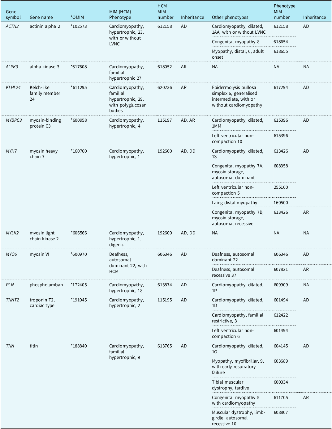
In contrast, patients with VUS in non-sarcomeric or minor genes tended to have milder clinical courses, as observed in EGP4 who carried ALPK3 and TTN VUS and was managed conservatively. Notably, all three patients with TNNT2 variants (EGP1, EGP5, and EGP6) exhibited arrhythmic manifestations (atrial fibrillation or non-sustained ventricular tachycardia), supporting the established role of TNNT2 as a marker for increased arrhythmic risk. Furthermore, the presence of multiple rare variants in several patients may contribute to the observed phenotypic heterogeneity in this cohort, particularly in those with a strong family history of sudden cardiac death. The identified genes and associated Online Mendelian Inheritance in Man (OMIM) database-based phenotypes are shown in Table 3.
A list of genes identified in the samples along with their corresponding OMIM phenotypes is provided

Table 3. Long description
The table presents genes identified in samples along with their corresponding OMIM phenotypes. It has 10 rows and 7 columns. The columns are Gene symbol, Gene name, OMIM MIM (HCM) Phenotype, HCM MIM number, Inheritance, Other phenotypes, Phenotype MIM number, and Inheritance. The table lists various genes such as ACTN2, ALPK3, KLHL24, MYBPC3, MYH7, MYLK2, MYO6, PLN, TNNT2, and TNN, along with their respective phenotypes and inheritance patterns. Each row provides detailed information about the gene, its associated phenotypes, and the mode of inheritance.
NA = no data available; AD = autosomal dominant; AR = autosomal recessive; DD = digenic dominant; HCM = hypertrophic cardiomyopathy; LVNC = left ventricular non-compaction; OMIM = Online Mendelian Inheritance in Man.
Discussion
This study utilised whole-exome sequencing on eight Egyptian patients with hypertrophic cardiomyopathy to identify and report prioritised variants, focusing on rare variants in hypertrophic cardiomyopathy-associated genes. This investigation represents the first exploratory genetic characterisation of hypertrophic cardiomyopathy in an Egyptian cohort using whole-exome sequencing, addressing a significant gap in the cardiovascular genomics literature, which has historically underrepresented populations from North Africa and the Middle East. By identifying 21 rare variants across 10 hypertrophic cardiomyopathy-associated genes in this small cohort, these preliminary findings offer an initial contribution to a more global understanding of hypertrophic cardiomyopathy’s genetic architecture. They highlight the importance of diversifying genomic datasets; however, larger studies are essential to determine the true prevalence and spectrum of these variants in the Egyptian population. Our sample size (n = 8) restricts the generalizability of our observations. Larger, population-based cohorts with appropriately ethnically matched controls are required to confirm these preliminary findings and to determine the true prevalence and spectrum of these variants in the Egyptian population.
According to the American Heart Association (AHA), cardiomyopathies are classified into three categories: genetic, non-genetic (acquired), and mixed (genetic and non-genetic).Reference Corrado, Graziano and Bauce21 In hereditary hypertrophic cardiomyopathy, a complex genetic architecture is connected to approximately 290 genes (https://hpo.jax.org/). After stringent filtering for rarity and predicted pathogenicity, we identified 21 rare variants in 10 genes with definitive or moderate evidence for hypertrophic cardiomyopathy causation, as defined by the ClinGen Hereditary Cardiovascular Disease Gene Curation Expert Panel. These variants were classified as pathogenic, likely pathogenic, or of uncertain significance (VUS) according to ACMG/AMP guidelines, with at least three in silico prediction tools consistently indicating a deleterious effect and an elevated CADD score.
A methodological constraint of this study is the exclusive reliance on in silico predictive algorithms (SIFT, PolyPhen‑2, MutationTaster, and CADD) and ACMG/AMP classification criteria, without the corroborative support of functional assays or familial segregation analyses. This is particularly critical for the numerous variants classified as VUS (e.g., in TTN, ACTN2, ALPK3, and others), which constitute the majority of our findings. The true contribution of these VUS to hypertrophic cardiomyopathy pathogenesis remains speculative, and we urge caution in their interpretation. We have therefore revised our discussion to explicitly emphasise this uncertainty, especially for non‑sarcomeric genes and VUS findings. Furthermore, we did not have access to an ethnically matched control population from Egypt, which would have strengthened the interpretation of variant frequencies and pathogenicity.
Crucially, these acknowledged constraints do not diminish the substantive contribution of our research. Rather, they serve as a framework for responsible data interpretation and highlight the inherent complexities of genotypic–phenotypic correlation in hypertrophic cardiomyopathy. Our findings represent a vital foundational genomic survey of an understudied population, providing a critical baseline of rare variant prevalence that will inform and necessitate future prospective investigations. The identification of these VUS, particularly within non-sarcomeric genes, offers compelling candidate targets for downstream mechanistic studies and underscores the urgent need for population-specific reference datasets. Consequently, we frame these limitations not as weaknesses, but as essential directives for the next phase of validation and discovery within this demographic.
Patients with hypertrophic cardiomyopathy most frequently have mutations in the genes that code for sarcomeric proteins. Early guidelines caused the classification of this cardiomyopathy as a disease affecting the sarcomere. Most sarcomeric diseases follow an autosomal dominant pattern of inheritance, with variations in the age of onset and the level of penetration.Reference Lorenzini, Norrish and Field22 Sarcomeric gene mutations are linked to a range of clinical manifestations (genetic pleiotropy), including cardiomyopathies such as restrictive, non-compaction, and dilated cardiomyopathies, as well as, infrequently, structural cardiomyopathies and congenital cardiac conditions such as Ebstein anomaly or septal defects.Reference García-Hernández, De la Higuera and Ochoa23
The MYBPC3 (myosin-binding protein C3) gene was found to have variants in two patients in our study. The leading cause of hypertrophic cardiomyopathy is mutations in MYBPC3. One of the primary mechanisms driving hypertrophic cardiomyopathy associated with MYBPC3 is haploinsufficiency.Reference Josephs, Roberts and Theotokis24 Consequently, whether they are evaluated with high or low confidence, non-truncating variants (i.e., stop-loss variants, missense variants, a protein-truncating variant anticipated to evade nonsense-mediated decay, and in-frame indels) are given precedence, irrespective of their pathogenic predictions. Two rare MYBPC3 variants were found in our data; they were an intron variant, a missense variant, and, in one patient (EGP7), a heterozygous missense variant (p.Gly1206Asp), which is classified as a likely pathogenic variant in the MYBPC3 gene.
In contrast to the MYBPC3 gene, which primarily causes disease through allelic imbalance and haploinsufficiency, the missense variations of the MYH7 (myosin heavy chain 7) gene function via a negative dominant effect, indicating that MYH7 is tolerant to loss-of-function mutations.Reference Park, Packard and Levin19 Giving priority to the MYH7 missense variant, which is predicted to have a deleterious effect by various in silico algorithms, could enhance the detection of actionable variants. Since rare and likely causal variants are often clustered in specific gene regions to enhance the outcomes of genetic testing in hypertrophic cardiomyopathy, a novel ACMG rule based on the aetiological fraction for rare missense MYH7 variants has been proposed.Reference Kim, Kim and Kim25
Two rare MYH7 variants found in this analysis were missense variants, one of them a pathogenic variant (p.Phe252Ser) and the other a VUS (p.Glu1708Lys) with a predicted deleterious effect, and with a gnomAD frequency of 0.000003983. Mutations in the MYH7 gene were related to considerable left ventricular hypertrophy, a hallmark of hypertrophic cardiomyopathy, as well as an adverse prognosis, compared with patients with mutations in other genes associated with hypertrophic cardiomyopathy.Reference Mathew, Zahavich and Lafreniere-Roula26
Our analysis reflects the distinct pathological mechanisms employed by different genes. The MYBPC3 variants suggest haploinsufficiency as a primary driver, whereas the MYH7 missense variants are indicative of a dominant-negative effect. This mechanistic distinction is crucial for understanding genotype–phenotype correlations.
Three variants of the sarcomeric gene TNNT2, which encodes for cardiac troponin T2, a protein that regulates the complex of thin-filament troponin in the sarcomere and is essential for contractile function, have been identified.Reference Pham, Giudicessi, Tweet, Boucher, Newman and Geske27 Notably, all three patients with TNNT2 variants (EGP1, EGP5, and EGP6) exhibited arrhythmic manifestations, including atrial fibrillation and non-sustained ventricular tachycardia, as documented in Table 1. This consistent association supports the established role of TNNT2 as a marker for increased arrhythmic risk and sudden cardiac death, reinforcing the clinical utility of identifying such variants for arrhythmic risk stratification.
The identification of rare, prioritised variants in core sarcomeric genes such as MYBPC3, MYH7, and TNNT2 aligns with the well-characterised molecular basis of hypertrophic cardiomyopathy. The presence of both pathogenic and VUS missense variants in MYH7, a gene associated with a severe phenotypic prognosis, underscores its critical role and the need for careful variant interpretation.
The cardiac sarcomere functions as part of a complex network within the myocyte, interconnected with the cell membrane and other sarcomeres. Structural and Z-disk proteins, and intermediate filaments that link the Z-disk to the cell membrane via costameres, are important components of its function. Additionally, several of the genes in this network are involved in calcium management, sarcomeric protein turnover, and transcription regulation. While a lot of genes have been studied as potential candidates in hypertrophic cardiomyopathy, only a few have a confirmed association with the phenotype.Reference Walsh, Offerhaus, Tadros and Bezzina5
According to the ACMG/AMP categorisation, we found a single patient to have a rare missense mutation in the PLN gene (p.Arg25Cys), which is likely pathogenic and has an expected deleterious impact. The PLN gene encodes phospholamban, which is essential for controlling calcium reuptake in the sarcoplasmic reticulum. A particular kind of arrhythmogenic/dilated cardiomyopathy and, less frequently, variants of the null type in simple heterozygosity and incomplete penetrance to hypertrophic cardiomyopathy have been associated with mutations in this protein.Reference Vafiadaki, Chaudhari and Soliman28 The main causes of PLN-induced cardiomyopathy are abnormal Ca2+ handling and SERCA2 dysfunction, which significantly affect the heart’s contraction and relaxation processes.Reference Brand, Baumgarten and Denzinger29 There are just a few known variants of PLN since it is a diminutive gene with only one coding exon, with an expected contribution to hypertrophic cardiomyopathy that is small—less than 1%, to be exact.
One patient (EGP2) had two heterozygous variants of the KLHL24 gene: Exon 3: c.307C>T (p.Arg103) and Exon 3: c.604G>A (p.Glu202Lys). The (p.Arg103) variant has a deleterious (fs/PTC) effect and is likely pathogenic according to the ACMG classification. KLHL24 encodes the Kelch-like protein 24, an ubiquitin ligase substrate receptor that contributes to the stability of intermediate filaments. Hypertrophic cardiomyopathy recessive, which presents with early-onset disease, a rising incidence of arrhythmias, and neuromuscular issues, potentially caused by desmin abduction or glycogen accumulation, is associated with pathogenic null mutations in this gene.Reference García-Hernández, De la Higuera and Ochoa23
The ALPK3 gene, which codes for alpha-protein kinase 3, was found to have a heterozygous missense mutation in Exon 14: c.4808T>C (p.Leu1603Pro). This nuclear protein is crucial for the early differentiation of cardiomyocytes, as it is involved in transferase and protein kinase activity and has widespread expression. Pathogenic variants in both alleles are associated with Noonan-like syndromic characteristics and paediatric-onset cardiomyopathy in over 80% of cases, and in more than 50% of cases, a prolonged QT interval is present. While missense variations can arise as compound heterozygotes with one loss-of-function mutation or as homozygotes, most autosomal recessive instances involve a null ALPK3 variant.Reference Andrade, Stephan and Furquim30 There is strong evidence that the ALPK3 gene causes an autosomal recessive form of infant-onset hypertrophic cardiomyopathy. Nonetheless, mild-to-moderate symptoms and an autosomal dominant inheritance pattern associated with adult-onset hypertrophic cardiomyopathy have also been connected to heterozygous loss-of-function mutations. Recently, a rare missense variant has been identified in Asian hypertrophic cardiomyopathy patients.Reference Chumakova and Baulina31 In our Egyptian study, the (p.Leu1603Pro) variant was classified as a VUS by ACMG, with predictions indicating potential deleterious effects on protein function.
The study successfully implicates genes beyond the canonical sarcomere in the Egyptian hypertrophic cardiomyopathy population. The discovery of a likely pathogenic variant in the PLN gene, a key regulator of calcium cycling, points to dysregulated calcium handling as a contributory pathway to the disease. Furthermore, the identification of variants in the KLHL24 gene and the ALPK3 gene underscores the role of proteins involved in sarcomeric stability, ubiquitination, and cardiomyocyte differentiation, expanding the genetic spectrum of hypertrophic cardiomyopathy to include developmental and proteostatic pathways.
Our analysis expands the known mutational spectrum of hypertrophic cardiomyopathy by identifying variants in both well-established sarcomeric genes and lesser-known or emerging genes, including KLHL24, PLN, ALPK3, and TTN. The detection of a likely pathogenic stop-gained variant in KLHL24 (p.Arg103*) and a likely pathogenic missense variant in PLN (p.Arg25Cys) adds to the growing body of evidence implicating these genes in hypertrophic cardiomyopathy pathogenesis. The submission of these novel variants to ClinVar (accessions SCV004562018, SCV006082319, SCV006082313, and SCV006082320) provides a valuable resource for the international scientific community and aids in future variant classification efforts.
Alpha-actinin 2, which is encoded by the ACTN2 gene, is an essential structural protein in the Z-disk that senses sarcomeric distention and mechanical traction, transmits force, and anchors the contractile apparatus. With substantial evidence supporting its familial inheritance in an autosomal dominant pattern, the current evidence linking ACTN2 to hypertrophic cardiomyopathy is based on a few missense variants.Reference Rodriguez, Schmeckpeper and Landim-Vieira32 Exemplary hypertrophic cardiomyopathy findings, such as enhanced calcium sensitivity in myofilaments, myocyte hypertrophy, hypercontractility, myofibrillar disarray, and extended relaxation, were observed in functional tests conducted using cardiomyocytes derived from human-induced pluripotent stem cells.Reference Guo, Wang and Li33 Even within families, disparate patterns of hypertrophy, left ventricular dilatation, obstructive and non-obstructive illness, atrial and ventricular arrhythmias, and conduction disorders make genotype–phenotype associations in hypertrophic cardiomyopathy unconvincing. Since the gene’s worldwide incidence in hypertrophic cardiomyopathy is modest, large case–control studies do not reveal any global enrichment of rare variations, indicating that only certain variants are associated with the disease.Reference Santoro, Mango and Mallardi34 In analyses of the ACTN2 gene, one patient was found to be a carrier of a VUS according to the ACMG classification. The variant is found at a low frequency in gnomAD.
The TTN gene encodes the titin protein, which extends from the M to Z lines of the sarcomere and is found only in skeletal muscles and cardiac myocytes. It can tolerate missense variations but is intolerant to loss-of-function variants. Truncating mutations in TTN are the most frequent cause of sporadic and familial dilated cardiomyopathy and are rare in hypertrophic cardiomyopathy.Reference Panwar, Singh and Mathur35 In our study, we found seven TTN variants, all of which are missense or intronic variants (i.e., non-truncating) and are categorised as VUS per ACMG guidelines. This aligns with the current understanding that truncating TTN variants are not a major contributor to hypertrophic cardiomyopathy pathogenesis. The role, if any, of these specific non-truncating VUS remains unclear. The high burden of TTN VUS (identified in 7 out of 8 patients) and their variable phenotypic impact—ranging from mild (EGP4) to severe (EGP6 and EGP7)—raises the hypothesis that TTN may act as a disease modifier rather than a primary driver. This is purely speculative at this stage, given the VUS classification of these variants, and warrants further investigation, including family segregation studies and functional assays, to determine if and how they might modify sarcomeric function. With respect to potential oligogenic effects, the co‑occurrence of multiple rare variants (e.g., in EGP6 and EGP7) is intriguing but remains speculative given the VUS status of many of these alleles.
The detection of a variant in ACTN2, encoding essential Z-disk proteins, reinforces the concept of the sarcomere as an integrated mechanosensory unit. Their involvement suggests that defects in force transmission and structural integrity can precipitate the hypertrophic cardiomyopathy phenotype. A significant proportion of the prioritised variants were classified as VUS, particularly in genes like TTN, ACTN2, and ALPK3. This highlights a common challenge in clinical genetics. It is crucial to emphasise that while we discuss the potential biological roles of these genes, the VUS status of these specific variants means their contribution to the patients’ phenotypes remains unconfirmed. This underscores the necessity of functional studies and population-specific data to reclassify these VUS and clarify their true pathogenic potential.
Myosin VI is involved in intracellular vesicular and organelle movement, where it is the unconventional actin-based molecular motor. It is involved in two stages of endocytic trafficking, being recruited to clathrin-coated pits and the subsequent uncoated endocytic vesicles.Reference Naccache, Hasson and Horowitz36 One variant was identified in the MYO6 gene, and the MutationTaster tool predicted a deleterious effect for one variant, c.1785G>T (Glu595Asp).
The MYLK2 gene encodes the calcium/calmodulin-dependent enzyme myosin light chain kinase (skMLCK) in fast-twitch fibres in adult skeletal muscle.Reference Wiebke, Andreas and Ottmar37 Calmodulin, which binds calcium, undergoes a conformational change upon calcium saturation, allowing it to interact with skMLCK, enhancing calcium affinity and promoting sustained muscle action. The MYLK2 gene variations are associated with mid-ventricular hypertrophic cardiomyopathy.Reference Ran, Li and Tan38 In our study, only one 38-year-old patient had a missense variant in the MYLK2 gene, Exon 11: c.1564G>A (p.Val522Ile), which is found at a low frequency in gnomAD (allele frequency = 0.000003977).
The identified genes and their associated OMIM phenotypes are summarised in Table 3, illustrating the broad phenotypic spectrum linked to these loci, including dilated cardiomyopathy, left ventricular non-compaction, and skeletal myopathies.
Clinical implications and future directions
The genotype–phenotype correlations observed in this study (detailed in the Results section) underscore the clinical utility of integrating genetic data into routine hypertrophic cardiomyopathy management. Patients with pathogenic or likely pathogenic variants in core sarcomeric genes consistently demonstrated more severe phenotypes, including extreme hypertrophy, significant left ventricular outflow tract obstruction, extensive myocardial fibrosis (late gadolinium enhancement), and a high arrhythmic burden. This association directly influenced clinical decision-making, leading to implantable cardioverter-defibrillator implantation for primary prevention or septal myectomy. These findings reinforce the value of genetic testing as a powerful tool for risk stratification, complementing traditional clinical risk factors to identify patients who may benefit from more aggressive surveillance and intervention.
An intriguing finding of this study is the co-occurrence of multiple rare variants in several patients, suggesting that oligogenic inheritance may contribute to the phenotypic heterogeneity of hypertrophic cardiomyopathy. For instance, EGP6, harbouring three VUS in MYH7, MYO6, and TNNT2, exhibited a particularly severe phenotype with diffuse hypertrophy and extensive late gadolinium enhancement, while her family history was already ominous. This challenges the traditional monogenic model of hypertrophic cardiomyopathy and supports a more complex genetic architecture, consistent with recent literature on oligogenic inheritance,Reference Burns, Bagnall and Lam39 where multiple rare variants may collectively modulate disease expression.
The consistent association between TNNT2 variants and arrhythmic manifestations (atrial fibrillation and non-sustained ventricular tachycardia) across three unrelated patients aligns with previous reports and reinforces the value of this gene as a predictor of increased sudden cardiac death risk. This highlights the potential for gene-guided arrhythmic risk assessment. Overall, this study demonstrates the practical utility of genomics in hypertrophic cardiomyopathy care by showing how genetic findings can directly inform personalised management strategies, from the choice of medical therapy to decisions regarding implantable cardioverter-defibrillator implantation. It supports the paradigm shift towards precision medicine, where an individual’s genotype informs prognosis, surveillance intensity, and therapeutic choices.
Conclusion
In conclusion, this exploratory pilot study provides the first detailed genetic and clinical characterisation of hypertrophic cardiomyopathy in a small Egyptian cohort, offering preliminary evidence of a diverse genetic landscape that includes both sarcomeric and non‑sarcomeric gene variants. However, due to the very small sample size and lack of functional or segregation studies, all findings should be considered hypothesis‑generating rather than definitive. The identification of clinically relevant genotype–phenotype correlations, particularly the association of pathogenic sarcomeric variants with severe disease and TNNT2 variants with arrhythmic risk, underscores the potential role of genetic testing in risk stratification and personalised management, pending validation in larger studies. The observation of multiple rare variants in several patients raises the hypothesis of oligogenic influences and requires Confirmation testing. As the first exploratory whole-exome sequencing study in an Egyptian cohort, these findings provide an initial glimpse into the genetic architecture of hypertrophic cardiomyopathy in this population and expand the global mutational spectrum. This study primarily serves as a proof‑of‑concept that paves the way for larger regional studies, functional investigations, and family‑based segregation analyses to further elucidate the molecular underpinnings of this complex disease.
Acknowledgements
This article was previously presented at the ICETTEM Conference at Amity France on May 19, 2025.
Author contribution
REN, RMG, AAE, AAK, AFE, and ME contributed equally to the concept and design of the manuscript. All authors approved the submitted version.
Financial support
The authors declare that no funds, grants, or other support were received during the preparation of this manuscript.
Competing interests
The authors have no relevant financial or non-financial interests to disclose.
Ethical standard
This study was conducted in accordance with the ethical standards of the Institutional Review Board (IRB) at the General Organization for Teaching Hospitals and Institutes (GOTHI) in Egypt and with the 1964 Helsinki declaration and its later amendments. The research protocol was approved by the Ethics Committee of the GOTHI (Approval Number: IHc00084). Informed consent was obtained from all individual participants included in the study.