INTRODUCTION
Schistosoma mansoni is one of the 3 major species from the genus Schistosoma that cause schistosomiasis, a worldwide disease that, according to the World Health Organization, affected at least 243 million people in 2011 (WHO, 2013). Due to its complex life cycle, the parasite can be considered as a challenging biological system to study regulation of gene expression (El-Ansary and Al-Daihan, Reference El-Ansary and Al-Daihan2005; Jolly et al. Reference Jolly, Chin, Miller, Bahgat, Lim, DeRisi and McKerrow2007; Fantappie et al. Reference Fantappie, de Oliveira, de Moraes Maciel dos Santos, Mansure, Furtado, de Abreu da Silva and Rumjanek2008). Thus, characterization of the proteins involved in the control of gene expression is of utmost importance and this is corroborated by the growing number of publications in this field (Lu et al. Reference Lu, Wu, Niles and LoVerde2006; Wu et al. Reference Wu, Niles, Hirai and LoVerde2007, Reference Wu, Tak and LoVerde2008; Wu and Loverde, Reference Wu and Loverde2008; Drummond et al. Reference Drummond, Calzavara-Silva, D'Astolfo, Cardoso, Rajao, Mourao, Gava, Oliveira, Macedo, Machado, Pena, Kitten and Franco2009; Bitar et al. Reference Bitar, Drummond, Costa, Lobo, Calzavara-Silva, Bisch, Machado, Macedo, Pierce and Franco2012). Also, the functional characterization of these proteins could lead to the identification of new potential drug targets in the parasite. Interestingly, proteins that participate in mRNA processes (such as splicing) are being studied as targets for anti-cancer drugs (Allemand et al. Reference Allemand, Hastings, Murray, Myers and Krainer2007, reviewed by Rymond, Reference Rymond2007 and van Alphen et al. Reference van Alphen, Wiemer, Burger and Eskens2009).
The Y-box binding proteins (YBPs) were originally described in humans for their ability to bind to the Y-box element present in promoters of HLA class II genes (Didier et al. Reference Didier, Schiffenbauer, Woulfe, Zacheis and Schwartz1988). The binding site for the human YB-1 was determined as being the DNA sequence 5′ – CTGATTGGCCAA – 3′, that presents an inverted CCAAT box (underlined) (Mathis et al. Reference Mathis, Benoist, Williams, Kanter and McDevitt1983; Saito et al. Reference Saito, Maki, Clayton and Tonegawa1983; Dorn et al. Reference Dorn, Durand, Marfing, Le Meur, Benoist and Mathis1987). This sequence is also present at the promoter region of several other genes (Sakura et al. Reference Sakura, Maekawa, Imamoto, Yasuda and Ishii1988; Lipson et al. Reference Lipson, Chen, Koniecki, Ku and Baserga1989; Pearson et al. Reference Pearson, Nasheuer and Wang1991; Goldsmith et al. Reference Goldsmith, Madden, Morrow and Cowan1993; Sommerville and Ladomery, Reference Sommerville and Ladomery1996; Nie et al. Reference Nie, Aijaz, Leefa Chong San, Balda and Matter2009). YBPs are found in many eukaryotes, but absent in the budding yeast Saccharomyces cerevisiae (Evdokimova et al. Reference Evdokimova, Ovchinnikov and Sorensen2006). The YBPs contain a highly conserved nucleic acid-binding domain, named the cold-shock domain (CSD) which, although not involved in the cold shock response in eukaryotes, possesses great similarity to bacterial cold-shock proteins (Wistow, Reference Wistow1990). In addition to the CSD, proteins from this family have a variable C-terminal TAIL domain consisting of alternate regions of predominantly basic or acid amino acids, which are responsible for either nucleic acid-binding or protein–protein interactions (reviewed by Matsumoto and Bay, Reference Matsumoto and Bay2005).
YBPs are DNA- and RNA-binding proteins that present properties of a nucleic acid chaperone, and also interact with a great variety of other proteins (reviewed by Eliseeva et al. Reference Eliseeva, Kim, Guryanov, Ovchinnikov and Lyabin2011). Although the YBPs were first characterized as transcription factors binding to the Y-box element, various articles associate these proteins with other functions such as translational regulation, DNA repair and pre-mRNA splicing (reviewed by Eliseeva et al. Reference Eliseeva, Kim, Guryanov, Ovchinnikov and Lyabin2011; reviewed by Kohno et al. Reference Kohno, Izumi, Uchiumi, Ashizuka and Kuwano2003). In this context, YBPs are pleiotropic proteins capable of coupling transcriptional and translational regulation. Furthermore, these proteins are often related to proliferation and transformation of eukaryotic cells (reviewed by Matsumoto and Wolffe, Reference Matsumoto and Wolffe1998), being also correlated with tumour size, degree of invasion, lymph node metastasis and poor prognosis in certain cancer types (reviewed by Kohno et al. Reference Kohno, Izumi, Uchiumi, Ashizuka and Kuwano2003).
Prior studies from our group identified an S. mansoni cDNA encoding a 217 amino acid protein homologous to the human YB-1, which was named SMYB1 (Franco et al. Reference Franco, Garratt, Tanaka, Simpson and Pena1997). Using protein comparative modelling we suggested that the N-terminus of SMYB1 folds as a closed β-barrel in a similar conformation to the Escherichia coli (CspA) and Bacillus subtilis (CspB) cold-shock proteins (Saito et al. Reference Saito, Maki, Clayton and Tonegawa1983; Sakura et al. Reference Sakura, Maekawa, Imamoto, Yasuda and Ishii1988; Lipson et al. Reference Lipson, Chen, Koniecki, Ku and Baserga1989; Pearson et al. Reference Pearson, Nasheuer and Wang1991; Sommerville and Ladomery, Reference Sommerville and Ladomery1996). However, the C-terminal domain differs from other YBPs as there are abundant arginine and glycine residues, which probably result in a β-pleated conformation of this domain. We have previously shown that SMYB1 is capable of binding to single- and double-stranded DNA sequences and, in particular, to oligonucleotides containing the CCAAT motif (Valadão et al. Reference Valadão, Fantappie, LoVerde, Pena, Rumjanek and Franco2002). Moreover, the protein binds to RNA as well, but independently of the presence of the CCAAT motif (Valadão et al. Reference Valadão, Fantappie, LoVerde, Pena, Rumjanek and Franco2002). We also demonstrated that SMYB1 is able to interact both in vivo and in vitro with the multi-functional protein SmPUR-α, acting synergistically to bind preferentially to pyrimidine-rich sequences (de Oliveira et al. Reference de Oliveira, da Silva, Rumjanek, Valadao, Franco, Mesquita, da Silva-Neto and Fantappie2004).
According to Kohno et al. (Reference Kohno, Izumi, Uchiumi, Ashizuka and Kuwano2003), the pleiotropic functions of YBP proteins result from a diverse range of molecular interactions, and the elucidation of these interactions is crucial for a further understanding of the YBPs functions. We report herein the identification of a novel SMYB1-interacting partner, as well as the cellular localization of SMYB1 in different stages of the parasite life cycle.
MATERIALS AND METHODS
Immunolocalization of the SMYB1 protein
Anti-SMYB1 specific antibodies
Polyclonal rabbit serum was produced against preparations of recombinant MBP-SMYB1. Rabbits were inoculated three times, at 15-day intervals with 250 μg of MBP-SMYB1. Recombinant protein was formulated with Freund's adjuvant (Complete Freund's adjuvant for the first immunization and Incomplete Freund's adjuvant for the subsequent injections). Fifteen days after the last inoculation, rabbits were exsanguinated and the serum containing anti-SMYB1 antibodies was collected. Pre-immune serum was obtained before the first immunization.
Schistosoma mansoni immunohistochemistry assays
Schistosoma mansoni stages of the adult worm, egg, schistosomulum, miracidium and cercaria were used in immunohistochemistry assays. Adult worms were recovered from perfused mice, separated into adult male and female worms and then fixed with dimethyl sulfoxide (DMSO)-methyl alcohol (1∶5) solution. Schistosoma mansoni eggs present in fragmented livers of experimentally infected mice were fixed with DMSO-methyl alcohol (1∶5) solution. Fixed adult male and female worms and fragments of livers were processed for Paraplast (Sigma, St Louis, MO, USA) embedding and histopathological sections were cut using a microtome at 5 μm. Lung-stage schistosomula were prepared according to the method of Harrop and Wilson (Reference Harrop and Wilson1993). Cercariae were obtained from Biomphalaria glabrata by exposing the infected snails to light for 2 h to induce shedding of parasites.
Sections of Omnifix (AnCon Genetics Inc., Melville, NY, USA) fixed, Paraplast-embedded adult male or female worms and livers containing S. mansoni eggs were deparaffinized using xylol, hydrated with an ethanol series, washed in PBS and then incubated in a blocking solution (0·05% Tween 20, 10% (w/v) BSA (bovine serum albumin) in PBS, pH 7·2) overnight at 4 °C. Samples were reacted for 1 h with either the anti-SMYB1 or a control, pre-immune rabbit serum, both diluted 1∶30 in 10× diluted blocking solution. Sections were then washed in PBS and reacted for 1 h with a goat anti-rabbit IgG-Cy-5 conjugate (1∶400 – Jackson Immunoresearch Laboratories Inc., West Grove, PA, USA) in 10× diluted blocking solution, which also contained Alexa Fluor 488 phalloidin (1∶100 – Invitrogen, Carlsbad, CA, USA) to stain the actin microfilaments. Afterwards, samples were washed, incubated for 10 min with propidium iodide (1∶3000 – Sigma-Aldrich, St Louis, MO, USA) in 10× diluted blocking solution to stain the nuclei and were then washed with PBS.
For experiments using Omnifix-fixed cercariae, miracidia and lung-stage schistosomula, a whole-mount protocol was chosen. Samples were first treated with a permeabilizing solution (0·1% Triton X-100, 1% (w/v) BSA and 0·1% (w/v) sodium azide in PBS, pH 7·4) for 3 h at 4 °C under constant agitation. Subsequent immunostaining steps used the same solution and conditions. Samples were incubated overnight with the anti-SMYB1 antibody (1∶60), washed five times and reacted for 4 h with the goat anti-rabbit IgG-Cy-5 conjugate, diluted 1∶1000 in a solution containing Alexa Fluor 488 phalloidin (1∶200). Samples were then incubated for 20 min with propidium iodide (1∶5000), washed once more and prepared with a mounting solution (90% glycerol, 10% Tris-HCl 1 m, pH 8·0).
The fluorescence images were captured with a Carl Zeiss LSM 510 META confocal microscope using a 63× oil-immersion objective lens in the Center of Electron Microscopy (CEMEL-ICB/UFMG). Images were analysed with Zeiss LSM Image Browser software and edited with Adobe Photoshop CS. All research protocols involving mice used in the course of this study were reviewed and approved by the local Ethics Committee on Animal Care at Universidade Federal de Minas Gerais (CETEA – UFMG # 043/2008).
Yeast two-hybrid assays
Plasmid construction
The cDNA encoding the full-length SMYB1 protein, as well as the cDNA fragments encoding its CSD and TAIL domains, were PCR amplified using as template the clone SMPAC89, obtained from an S. mansoni adult worm cDNA library (Mathis et al. Reference Mathis, Benoist, Williams, Kanter and McDevitt1983). Amplifications were performed in a 100 μL final volume containing 50 ng of template DNA, 0·2 pmol/μL of each primer Y-boxFwd 5′ – CTGGAATTCACAATGGCGGACACTAGAC – 3′ and Y-boxRev 5′ – GACCTGCAGTCAAAATGCATATTTGATTACG – 3′, CSDFwd 5′ – CTGGAATTCATGGAAGAGCGTGTCAAAGG – 3′ and CSDRev 5′ – GACCTGCAGTTAAGGGCCT GTCACTTCGGA – 3′ or TailFwd 5′ – CTGGAATTCGGCAGTGTCTACGCAGCTTT – 3′ and TailRev 5′ – GACCTGCAGACGTTTCTCAGTATTTGATCAGAGA – 3′ (the Fwd and Rev primers contained Eco RI and Pst I restriction enzyme sites, respectively), 2 mm MgCl2, 200 μ m dNTPs and 5 U of Taq DNA polymerase (Phoneutria, Belo Horizonte, Brazil) in the appropriate buffer (50 mm KCl, 10 mm Tris-HCl, pH 9·0 and 0·1% Triton X-100). The fragments obtained after PCR amplification (SMYB1, CSD or TAIL), as well as the expression vectors pGAD424 (Clontech, Mountain View, CA, USA), pGBT9 (Clontech) and pAS2.1 (Clontech) were double digested with Eco RI and Pst I enzymes (Invitrogen, Carlsbad, CA, USA). The digested DNAs were then purified using a GeneClean Kit (BIO101, La Jolla, CA, USA) and used to generate the following constructs: pGAD-SMYB1, pGAD-CSD and pGAD-TAIL; pGBT-SMYB1, pGBT-CSD and pGBT-TAIL; and pAS-SMYB1. Ligation products were used to transform the E. coli DH5a strain and the rescued plasmids were sequenced using 10 pmol of appropriate primers (ADFwd 5′ – CAGGGATGTTTAATACCACATC – 3′ and ADRev 5′ – GCACAGTTGAAGTGAACTTGC – 3′ from constructions with pGAD vector, and BDFwd 5′ – TCATCGGAAGAGAGTAG – 3′ from constructions with pGBT vector), 4 μL of DYEnamic ET Dye Terminator Kit (GE Healthcare Life Sciences, Uppsala, Sweden) and 200 ng of DNA. The sequencing products were analysed in the MegaBACE 1000 DNA Sequencer (GE Healthcare Life Sciences). The pGAD-SmPUR-α construct was available (Fantappie et al. Reference Fantappie, Osman, Niles and LoVerde2000).
Protein–protein interaction assays
To assess protein–protein interactions involving SMYB1 and its domains, a yeast two-hybrid assay was used, in which the transcription of the LacZ and His3 reporter genes was examined (Matchmaker-Clontech). To perform this , competent yeast strains Y190 (MATα, trp1-901 his3Δ200, leu2-3, 112 ura3-52 ade2-101, gal4, gal80,URA3::GAL1-lacZ, LYS2::GAL1-HIS3) were prepared using the Frozen-EZ Yeast Transformation II kit (Zymo Research, Orange, CA, USA) following the manufacturer's instructions. The strains were then co-transformed with 2 μg of different pair combinations of the constructs pGAD-SMYB1, pGAD-CSD or pGAD-TAIL and pGBT-SMYB1, pGBT-CSD or pGBT-TAIL in order to determine protein–protein interactions. The plasmid pair pVA3-pTD1 was used as a positive control. Co-transformants were selected onto SD/-Leu -Trp -His (synthetic dextrose growth medium lacking leucine, tryptophan and histidine) containing 25 mm 3-amino-1,2,4-triazole (3-AT, a HIS inhibitor-competitor used to suppress background growth). The plates were incubated at 30 °C for 6 days and checked each day for growth. On day 7, selected colonies were restreaked onto SD/-Leu-Trp-His containing 25 or 35 mm 3-AT. Histidine prototroph clones were transferred onto nitrocellulose membrane and permeabilized by freeze/thaw cycles (liquid N2/37 °C). β-galactosidase activity assay was performed by overlaying the filters on Whatman 3MM paper soaked in Z buffer (60 mm Na2HPO4·7H20, 40 mm NaHPO4.H20, 10 mm KCl, 1 mm MgSO4·7H2O, 35 mm β-mercaptoethanol) containing 1 mg of X-gal (5-bromo-4-chloro-3-indolyl-β-D-galactopyranoside). The filters were incubated at 30 °C for approximately 6 h to develop the blue colour.
Library screening
The pAS-SMYB1 construct was used as bait for the screening of an S. mansoni adult worm cDNA library previously cloned into the pAD-GAL4-2 activation domain vector (Stratagene, La Jolla, CA, USA). The competent yeast strain Y190 was co-transformed with 10 μg of pAS-SMYB1 and 40 μg of pAD-GAL4-2 vector. Co-transformants were selected onto SD/-Leu-Trp plates to test the transformation efficiency and onto SD/-Leu-Trp-His plates containing 25 mm 3-amino-1,2,4-triazole to test the activation of His3 gene transcription. The plates were incubated at 30 °C for 6 days. On day 7, selected colonies were restreaked onto SD/-Leu-Trp-His plates containing 25 mm 3-amino-1,2,4-triazole. Histidine prototroph clones were assayed for β-galactosidase activity by a filter assay, as detailed previously. Positive clones for the two assays were grown in liquid YPD medium (2% peptone, 1% yeast extract, 2% glucose, pH 7·0) and the binding and activation domain plasmids were isolated using the Zymoprep Yeast Plasmid Miniprep Kit (Zymo Research, Orange, CA, USA). The plasmids were used to transform E. coli TOP10 cells (Invitrogen, Carlsbad, CA, USA) and then were rescued and sequenced using 10 pmol of the appropriate primers (ADFwd and ADRev, described above). The sequencing products were identified by NCBI BLAST searches (Altschul et al. Reference Altschul, Gish, Miller, Myers and Lipman1990). To confirm the results, the isolated plasmids were then co-transformed with the pAS-SMYB1 plasmid into strains Y190 and PJ69-4A (MATa trp1-901 leu2-3, 112 ura 3-52 his 3-200gal 4Δgal80ΔLYS2::GAL1-HIS3GAL2-ADE2met2::GAL7-lacZ – kindly provided by Dr Philip James, University of Wisconsin Medical School). After transformation, PJ69-4A yeast cells were spread onto SD/-Leu -Trp -His; SD/-Leu -Trp -ADE and SD/-Leu -Trp -His -ADE containing 2 mm 3-AT. To investigate which part of the SMYB1 protein was involved in the interaction, pGBT-CSD or pGBT-TAIL were co-transformed with pAD-vectors isolated by screening into the Y190 strain.
Pull-down assays
Cloning, expression and purification of recombinant GST proteins
The complete coding region of SMYB1 was PCR amplified using as template a pGAD/SMYB1 construct (described above). Amplification was performed as depicted in the section ‘Plasmid construction’ using the primers Y-boxFwd and Y-boxRev. The fragments obtained after PCR amplification were cloned into predigested pGEX 4T-1 (GE Healthcare Life Sciences) as described in that same section. Isolated plasmids were sequenced using the MegaBACE 1000 DNA Sequencer (GE Healthcare Life Sciences), 10 pmol of each primer (pGEXFwd 5′ – GTAAAACGACGGCCAGTGGGCTGGCAAGCCACGTTTGGTG – 3′ and pGEXRev 5′ – CAGGAAACAGCTATGACCCGGGAGCTGCATGTGTCAGAGG – 3′), 4 μL of DYEnamic ET Dye Terminator Kit (GE Healthcare Life Sciences) and 200 ng of DNA. Escherichia coli clones harbouring the empty pGEX 4T-1 plasmid (negative control) or the pGEX-SMYB1 construct was used for expression of the fusion proteins, induced by 0·6 mm IPTG (isopropyl-β-D-thiogalactopyranoside). The expression protocol was maintained for 4 h at 30 °C. Cells were then harvested by centrifugation at 4000 g for 20 min, resuspended in lysis buffer (25 mm Tris-HCl, pH 8·0, 1 mm EDTA, 10% glycerol, 50 mm NaCl, 0·1% Triton X-100, 1 mm phenylmethylsulfonyl fluoride (PMSF), 10 μ m leupeptin and 75 μ m aprotinin) and sonicated. The supernatant fraction, containing the soluble proteins, was collected after centrifugation at 20 000 g for 25 min at 4 °C and filtered through a 0·45 μ m filter. The GST and GST-SMYB1 proteins were then purified by passage over glutathione-Sepharose 4B beads (Pharmacia, Uppsala, Sweden) in binding buffer (25 mm Tris-HCl, pH 8·0, 1 mm EDTA, 50 mm NaCl, 10% glycerol). GST fusion proteins were eluted by addition of 10 mm reduced glutathione in 50 mm Tris-HCl, pH 8·0. Protein samples were analysed on 10% SDS-PAGE gels and visualized by Coomassie blue staining.
Construction of plasmids used in transcription-translation in vitro assay
The fragments containing either the SMYB1 complete coding region or the portions coding for the CSD and TAIL domains were removed, using the restriction enzymes Eco RI and Bgl II, from the vectors pGAD/SMYB1, pGAD/CSD and pGAD/TAIL (described above), respectively, and cloned into pCITE-4a-c(+) (Novagen, Darmstadt, Germany), forming the constructs pCITE-SMYB1, pCITE-CSD and pCITE-TAIL. The cDNA fragments isolated in yeast two-hybrid screening using pAS-SMYB1 as a bait (described above) were removed from pAD-GAL4-2 activation domain vector using the restriction enzymes Eco RI and Xho I and cloned into pCITE-4a-c(+). The constructs were used to transform E. coli TOP 10 strains (Invitrogen, Carlsbad, CA, USA) and 200 ng of each rescued plasmid were sequenced using the MegaBACE 1000 DNA Sequencer (GE Healthcare Life Sciences) and 10 pmol of each primer (pCITEFwd 5′ – CGAACGCCAGCACATGGACA – 3′ or pCITERev 5′ – CAGGAAACAGCTATGAC GCTAGTTATTGCTCAGGC – 3′). Plasmids showing the correct sequence were used in downstream pull-down experiments.
In vitro binding studies by pull-down assay
The GST, GST-SMYB1 and GST-SmPUR-α (available from Fantappie et al. Reference Fantappie, Osman, Niles and LoVerde2000) proteins were, individually, immobilized in gluthatione-Sepharose 4B resin. For each reaction, 150 μL of resin was centrifuged at 12 000 g and the pellet, containing the resin, was washed 3 times with PBS buffer 0·015 m, pH 7·2. After the last wash, the resin was incubated with approximately 10 μg of the appropriate protein for 2 h. The resin was then washed 3 times with PBS, to remove the non-binding proteins, and used in the pull-down assays. The cDNA inserts cloned into pCITE-4a-c(+) were transcribed and translated using the Single Protein System (Novagen) following the manufacturer's protocol for 35S methionine incorporation.
Physical association of proteins was tested by the GST pull-down assay. For this, 10 μL of each of the 35S labelled products were added to the suspension of gluthatione Sepharose beads immobilized with appropriate GST-fusion proteins (GST-SMYB1 or GST-TAIL) in 200 μL final volume of binding buffer (25 mm Tris-HCl, pH 8·0, 50 mm NaCl, 10% glycerol, 1 mm EDTA, 0·1% NP 40). After overnight mixing (4 °C), the protein-complexed beads were washed extensively with binding buffer in order to remove non-bound proteins and were then boiled with 50 μL of SDS loading buffer. Proteins were separated by 12% SDS-PAGE and detected by autoradiography.
RESULTS
SMYB1 is mainly localized in the cytoplasm of S. mansoni cells
It has been shown that SMYB1 is able to bind both DNA and RNA molecules (Valadão et al. Reference Valadão, Fantappie, LoVerde, Pena, Rumjanek and Franco2002) and, like other members of this group, it can be involved in transcriptional or post-transcriptional regulation mechanisms. Aiming to better understand SMYB1 functions we verified its subcellular localization throughout S. mansoni life-cycle stages.
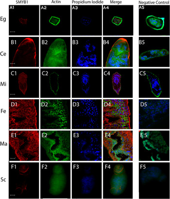
We carried out in situ immunohistochemistry experiments using an anti-SMYB1 antibody on S. mansoni samples from various life-cycle stages. The results show that SMYB1 is present in the cytoplasm of cells from eggs (Fig. 1 A1–A4), cercariae (Fig. 1 B1–B4), miracidia (Fig. 1 C1–C4), adult female (Fig. 1 D1–D4) and male (Fig. 1 E1–E4) worms, as well as in the schistosomula lung stage (Fig. 1 F1–F4). Supplementary Fig. 1S (in Online version only) shows a higher magnification image of male and female adult worms showing PI-stained nuclei surrounded by cytoplasmic labelled protein. Furthermore, it is interesting to note that the protein is accumulated in the subtegumental region in cercariae, miracidia and adult worms (see Supplementary Fig. 1S to observe this localization in male and female adult worms). While in cercariae and schistosomula the SMYB1 pattern of distribution seems to be anterior; in adult worms, miracidia and eggs the protein seems to be distributed all over the parasite body. Supplementary Fig. 2S shows the whole body of male and female worms demonstrating that there is no specific anterior or posterior distribution of the protein in adult worm bodies. No staining was observed in the negative controls (Fig. 1 A5, B5, C5, D5, E5 and F5) in which only the rabbit pre-immune serum was used.
Native SMYB1 displays cytoplasmic localization in various life-cycle stages of Schistosoma mansoni. Fixed parasites were incubated with a rabbit anti-SMYB1 antibody, and then with a Cy-5-conjugated anti-rabbit IgG in a solution containing Alexa Fluor 488 phalloidin to stain actin microfilaments. Samples were incubated with propidium iodide to visualize cell nuclei. Fluorescent images were obtained using a 63× oil-immersion objective lens and confocal microscopy (Carl Zeiss LSM 510 META). Images were analysed with the Zeiss LSM Image Browser software and edited with Adobe Photoshop CS. To help distinguish the individual fluorescence signals, the original fluorescence colours were digitally modified. In the figure, fluorescence from Cy-5 is shown in red, propidium iodide in blue and phalloidin-Alexa fluor 488 in green. For adult male worms a phase-contrast image is also presented. The following developmental stages were assayed: egg (A1–A4), cercaria (B1–B4), miracidium (C1–C4), adult female (D1–D4) and male (E1–E4) worms, and schistosomulum (F1–F4). Rabbit pre-immune serum was used as a control and was negative in all samples (A5, B5, C5, D5, E5 and F5). Bars = 20 mm.

The SMYB1 TAIL domain is responsible for protein–protein interactions
The CSD of SMYB1 is 64% identical to the cold-shock domain of other members of the Y-box binding protein family and shows 43% similarity to the E. coli (CspA) and 40% similarity to the B. subtilis (CspB) cold-shock proteins. The crystal structure of CspB reveals the formation of a homodimer stabilized by 6 hydrogen bonds linking 2 adjacent β4 strands (Saito et al. Reference Saito, Maki, Clayton and Tonegawa1983). Sequence analyses of S. mansoni SMYB1 revealed some structural characteristics that permit the dimerization interface required for protein–protein interaction as occurs in the B. subtilis CspB (Schindelin et al. Reference Schindelin, Marahiel and Heinemann1993).
To determine whether SMYB1 is capable of forming stable homo- and heterodimers, and to elucidate protein domains responsible for these interactions, we first performed in vitro pull-down assays using GST affinity chromatography. In order to perform the experiments, SMYB1 and its CSD or TAIL domains were translated in vitro and labelled with 35S. These proteins were then incubated with GST (negative control), GST-SMYB1 or GST-SmPUR-α immobilized on glutathione-Sepharose beads. After washing, proteins retained on the beads were separated by 12% SDS-PAGE and detected by autoradiography.
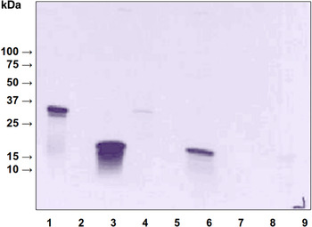
As shown in Fig. 2, SMYB1 was able to form a homodimer (Fig. 2, lane 1) and this interaction seemed to be via its TAIL domain (Fig. 2, lane 3). The CSD domain alone was not able to interact with the entire SMYB1 protein (Fig. 2, lane 2). As shown in Fig. 2, lane 4, it was confirmed that SMYB1 was able to interact with SmPUR-α (a single-stranded DNA/RNA binding protein), as previously reported (de Oliveira et al. 2004). Again, these interactions were mediated by the TAIL domain of SMYB1 (Fig. 2, lane 6), and the CSD domain was not involved in the interaction between SMYB1 and SmPUR-α (Fig. 2, lane 5). Negative controls, using only immobilized GST, were not able to retain any of the labelled proteins (Fig. 2, lanes 7–9).
The ability of SMYB1 to form homodimers and heterodimers with SmPUR-α. Autoradiogram showing the results of the pull-down assay performed in vitro to verify protein–protein interaction. GST-SMYB1, GST-SmPUR-α or GST were immobilized in a gluthatione-Sepharose 4B resin. After washing steps, the resin was incubated with SMYB1, CSD or TAIL labelled proteins. Non-bound proteins were removed and the bound ones were separated by 12% SDS-PAGE. Detection was performed by autoradiography. 1-GST-SMYB1/SMYB1; 2-GST-SMYB1/CSD; 3-GST-SMYB1/TAIL; 4-GST-SmPUR-α/SMYB1; 5-GST-SmPUR-α/CSD; 6-GST-SmPUR-α/TAIL; 7-GST/SMYB1; 8-GST/CSD; 9-GST/TAIL.

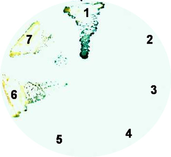
We next tested whether the homodimer formation could be confirmed in vivo by yeast two-hybrid assays. Yeast cells co-transformed with pGBT-SMYB1/pGAD-SMYB1 or pGBT-SMYB1/pGAD-CSD showed neither β-galactosidase activity nor histidine prototrophism (Fig. 3, sections 2–5). Nonetheless, when yeast cells were co-transformed with pGBT-SMYB1/pGBT-TAIL, a strong β-galactosidase activity was observed (Fig. 3 sections 6 and 7), comparable to that obtained with the positive control pVA3/pTD1 (Fig. 3, section 1), which encodes the interacting proteins p53 and SV40.
The ability of SMYB1 to form homodimers in vivo using the yeast two-hybrid assay. X-gal filter lift assay with the yeast strain 190 transformed with the pairs of vectors: 1- pVA3 (p53)/pTD1 (SV40); 2 and 3- pGBT-SMYB1/pGAD-SMYB1; 4 and 5- pGBT-CSD/ pGAD-SMYB1; 6 and 7- pGBT-TAIL/pGAD-SMYB1. Co-transformed cells were plated on SD/-Leu -Trp -His containing 25 mm 3-AT.

SmRNP is a novel SMYB1-interacting partner
In order to identify novel potential S. mansoni proteins capable of forming heterodimers with SMYB1, a yeast two-hybrid screening of an adult worm cDNA library was performed. The full-length SMYB1 was used as a bait protein fused to the GAL4 DNA-binding domain (pAS-SMYB1), and the library was fused downstream of the GAL4-activation domain. An initial screen for growth on selective medium yielded 42 positive colonies. Of these, however, only 19 showed β-galactosidase activity. These clones were rescued and the cDNAs sequenced for subsequent BLAST search at NCBI. After the BLAST analysis, only 2 out of the 19 clones were considered with potential biological relevance in terms of SMYB1 interaction. These clones had significant similarity with the Homo sapiens Ring box protein 1 (Rbx1) and with the Drosophila melanogaster SmD3 ribonucleoprotein. The S. mansoni SMYB1-interacting proteins were named SmRingBOX (SmRbx) and SmRNP, accordingly.
The other clones obtained were considered as ‘false positives’, which are very frequent in two-hybrid library screening. These false positive clones include proteins that interact with the bait in a partially specific manner (i.e. they will appear specific for the bait vs a small number of negative controls, but on subsequent analysis they will be found to interact with many partners) (Bruckner et al. Reference Bruckner, Polge, Lentze, Auerbach and Schlattner2009). Other false positives have been found to induce changes in the metabolic functioning of yeast, resulting in bias of the reporter assays used in the two-hybrid system (Serebriiskii et al. Reference Serebriiskii, Estojak, Berman and Golemis2000).
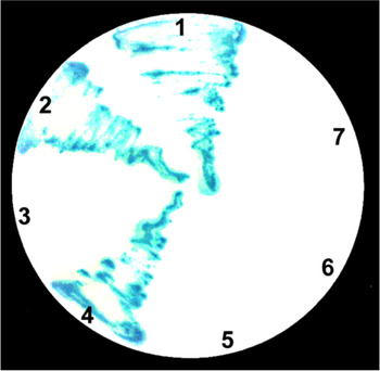
To investigate which part of SMYB1 is involved in the interaction with SmRbx or SmRNP, the isolated plasmids coding for these proteins (pAD-SmRbx or pAD-SmRNP) were co-transformed with plasmids encoding SMYB1, CSD and TAIL (pAS-SMYB1, pGBT-CSD and pGBT-TAIL, respectively) into the yeast strain Y190. After transformation, the yeasts containing the pAS-SMYB1/pAD-SmRNP and pGBT-TAIL/pAD-SmRNP grew on selective medium and were still blue on β-galactosidase assay (Fig. 4, sections 2 and 4). With this result we can conclude that the TAIL is the domain of the protein responsible for these protein–protein interactions. However, when assaying for SMYB1/SmRbx interactions, none of the constructs grew on selective media (Fig. 4, sections 5–7), which led us to conclude that SmRbx is a false-positive result of the library screening. The positive control of these experiments was able to grow on selective media, as well as to show a blue colour on the β-galactosidase assay (Fig. 4, lane 1).
Evaluation of the interaction of SMYB1 and its CSD and TAIL domains with the proteins SmRNP and SmRbx in vivo using the yeast two-hybrid assay. X-gal filter lift assay with the yeast strain 190 transformed with the pairs of vectors: 1-pVA3(p53)/pTD1 (SV40); 2-pAS-SMYB1/pAD-SmRNP; 3-pGBT-CSD/pAD-SmRNP; 4-pGBT-TAIL/pAD-SmRNP; 5-pAS-SMYB1/pAD-SmRbx; 6-pGBT-CSD/pAD-SmRbx; 7-pGBT-TAIL/pAD-SmRbx. Co-transformed cells were plated on SD/-Leu -Trp -His containing 25 mm 3-AT.

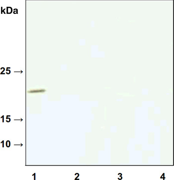
We next tested whether we could reproduce these interactions in vitro by GST pull-down assays. To perform this, the cDNAs coding to SmRNP and SmRbx were cloned into pCITE-4a-c(+) and transcribed and translated using the Single Protein System (Novagen). The labelled SmRNP and SmRbx were then incubated with GST-SMYB1 or GST (negative control) immobilized on glutathione-Sepharose beads. After washing, proteins retained on the beads were separated by 12% SDS-polyacrylamide gel electrophoresis and detected by autoradiography. Our pull-down data showed that SMYB1 could bind in vitro only to SmRNP (Fig. 5, lane 1), but not to SmRbx (Fig. 5, lane 3), confirming the results obtained with the two-hybrid assays. The GST protein alone showed no interaction with any of the assayed proteins (Fig. 5, lanes 2 and 4).
The ability of SMYB1 to form a heterodimer with SmRNP. Autoradiogram showing the result of the pull-down assay performed in vitro to verify protein–protein interaction. GST-SMYB1 or GST was immobilized in a gluthatione-Sepharose 4B resin. After washing steps, the resin was incubated with SmRNP- or SmRbx-labelled proteins. Non-bound proteins were removed and the bound ones were separated by 12% SDS-PAGE. Detection was performed by autoradiography. 1- GST-SMYB1/SmRNP; 2- GST/SmRNP; 3- GST-SMYB1/SmRbx and 4-GST/SmRbx.

DISCUSSION
It has been demonstrated already that, similar to other YBP proteins, SMYB1 is able to bind single- and double-stranded DNA, as well as RNA (Valadao et al. Reference Valadão, Fantappie, LoVerde, Pena, Rumjanek and Franco2002). Also, it was demonstrated that SMYB1 can form a heterodimer with SmPUR-α, a DNA-binding protein (Fantappie et al. Reference Fantappie, Osman, Niles and LoVerde2000).
The cellular localization of a protein is valuable information that can help to improve the knowledge about its biological functions. Thus, as an important step in SMYB1 characterization, we conducted immunohistochemistry analyses using an anti-SMYB1 antibody on different parasite developmental stages. We were able to show that SMYB1 is localized in the cytoplasm of S. mansoni adult female and male worms, eggs, miracidia, cercariae and schistosomula cells. The protein is present in all stages of the parasite life cycle studied, confirming previous results obtained by our group (Franco et al. Reference Franco, Garratt, Tanaka, Simpson and Pena1997) showing that the SMYB1 cDNA could be found in cDNA pools from different life-cycle stages. Also, a search for cDNAs annotated as Y-box binding protein at the UniGene database from NCBI retrieved sequences from the following S. mansoni library sources: adult, schistosomule larvae, miracidia, sporocyst, egg, cercariae and an unknown developmental stage. These data together suggest that SMYB1 might play important roles during all life-cycle stages. Also, its cytoplasmic localization, together with its ability to bind RNA (Valadao et al. Reference Valadão, Fantappie, LoVerde, Pena, Rumjanek and Franco2002), suggests a function in RNA metabolism.
It has already been shown that mRNA localization in the cell context is important for the spatial regulation of gene expression (Matsumoto et al. Reference Matsumoto, Uchiumi, Saito, Yagi, Takazaki, Kanki and Kang2012). The localization process is determined by elements in the mRNA molecules and by proteins that bind them. YB-1 proteins may exert an important role in this process (Matsumoto et al. Reference Matsumoto, Uchiumi, Saito, Yagi, Takazaki, Kanki and Kang2012). As such, SMYB1 localization on S. mansoni cells and tissues may give a hint to its role for parasite development. The presence of the protein very near to the tegument in adult worms may suggest an action on the translational regulation of tegument proteins.
The modulation of gene expression depends on a multitude of interactions between protein–DNA, protein–protein and protein–RNA (de Oliveira et al. Reference de Oliveira, da Silva, Rumjanek, Valadao, Franco, Mesquita, da Silva-Neto and Fantappie2004). It is well known that YBPs participate in almost all events of storing, reproduction and expression of genetic information (Lyabin et al. Reference Lyabin, Eliseeva, Skabkina and Ovchinnikov2011), such as DNA repair (Kuwano et al. Reference Kuwano, Oda, Izumi, Yang, Uchiumi, Iwamoto, Toi, Fujii, Yamana, Kinoshita, Kamura, Tsuneyoshi, Yasumoto and Kohno2004; de Souza-Pinto et al. Reference de Souza-Pinto, Mason, Hashiguchi, Weissman, Tian, Guay, Lebel, Stevnsner, Rasmussen and Bohr2009), replication, recombination, mRNA transcription (Skabkina et al. Reference Skabkina, Lyabin, Skabkin and Ovchinnikov2005), splicing (Raffetseder et al. Reference Raffetseder, Frye, Rauen, Jurchott, Royer, Jansen and Mertens2003) and post-transcriptional events that control the half-life of transcripts (Evdokimova et al. Reference Evdokimova, Ruzanov, Imataka, Raught, Svitkin, Ovchinnikov and Sonenberg2001).
Several studies with cytoplasmic YBPs demonstrated their ability to interact with mRNA molecules. Dong et al. (Reference Dong, Akcakanat, Stivers, Zhang, Kim and Meric-Bernstam2009) showed that the human protein YB-1 of MCF7 breast cancer cells binds preferentially to mRNA with a high GC content. Interestingly, YB-1 is able to bind to its own mRNA (Skabkina et al. Reference Skabkina, Lyabin, Skabkin and Ovchinnikov2005). Other YBP, MSY2 (a mouse germ cell protein), is predominantly localized in the cytoplasm and plays an important role in the translational repression and storage of paternal and maternal mRNAs in spermatids and oocytes, respectively (Yang et al. Reference Yang, Medvedev, Yu, Schultz and Hecht2006).
Protein–protein interactions are important events in all biological processes, since most proteins execute their functions through large supramolecular structures. Thus, several techniques have been developed and are currently available to demonstrate the formation of protein homo- or heterodimers. We used GST pull-down and yeast two-hybrid assays to test the interactions of SMYB1 with its putative protein partners and showed that SMYB1 is able to form homodimers via the TAIL domain. This result is in agreement with Izumi et al. (Reference Izumi, Imamura, Nagatani, Ise, Murakami, Uramoto, Torigoe, Ishiguchi, Yoshida, Nomoto, Okamoto, Uchiumi, Kuwano, Funa and Kohno2001) who showed, by pull-down and co-immunoprecipitation assays, that the human YB-1 protein of MCF7 cells is able to form homodimers, with the TAIL domain being the mediator of the homodimerization.
Although the YBPs have a conserved N-terminal domain responsible for nucleic acid interactions, the C-terminal (the TAIL domain) of these proteins is distinct and can perform protein as well as DNA and RNA interactions (Izumi et al. Reference Izumi, Imamura, Nagatani, Ise, Murakami, Uramoto, Torigoe, Ishiguchi, Yoshida, Nomoto, Okamoto, Uchiumi, Kuwano, Funa and Kohno2001). Thus, the different functions of many YBPs may be mediated by protein–protein interactions performed by their C-terminal domain. It has been demonstrated by independent studies that YB-1 can interact with the highly conserved zinc finger transcription factor CTCF (Chernukhin et al. Reference Chernukhin, Shamsuddin, Robinson, Carne, Paul, El-Kady, Lobanenkov and Klenova2000), Fragile X mental retardation RNA-binding protein (Ceman et al. Reference Ceman, Nelson and Warren2000), tumour suppressor p53 (Okamoto et al. Reference Okamoto, Izumi, Imamura, Takano, Ise, Uchiumi, Kuwano and Kohno2000), actin (Ruzanov et al. Reference Ruzanov, Evdokimova, Korneeva, Hershey and Ovchinnikov1999) and with the heterogeneous nuclear ribonucleoprotein K (Shnyreva et al. Reference Shnyreva, Schullery, Suzuki, Higaki and Bomsztyk2000). Each one of these complexes exerts different functions in the cellular context.
Aiming to increase our knowledge on SMYB1 functions in the parasite, we focused on finding its potential partners. Heterodimer formation with SmPUR-α, that had already been shown by de Oliveira et al. (Reference de Oliveira, da Silva, Rumjanek, Valadao, Franco, Mesquita, da Silva-Neto and Fantappie2004), was confirmed herein. We extended these studies and showed that the interaction between SMYB1 and SmPUR-α was mediated by the TAIL domain of SMYB1, once again highlighting the importance of this domain for protein-protein interactions. It is important to note that SMYB1–SmPUR-α and SMYB1–SMYB1 interactions could not be detected on the yeast two-hybrid assays, although they were confirmed by the pull-down experiments. This can be explained by the fact that the yeast two-hybrid conditions used in this work were very stringent, allowing the detection of very strong interactions only. It can also be speculated that these interactions would form in vivo only in a given physiological condition. PUR-α proteins are known to bind to several regulatory sequences present in both DNA and RNA and are involved in many events associated with gene transcription, RNA processing and transport and translation (Murray et al. Reference Murray, Schiller and Franke1992; Zambrano et al. Reference Zambrano, De Renzis, Minopoli, Faraonio, Donini, Scaloni, Cimino and Russo1997; Kelm et al. Reference Kelm, Elder and Getz1999). SmPUR-α interacts with specific regions of the schistosome female p14 gene promoter and activates transcription of the HIS3 reporter gene. Additionally, the nucleic acid-binding domain of SmPUR-α possesses a high degree of similarity with the human PUR-α (Fantappie et al. Reference Fantappie, Osman, Niles and LoVerde2000). Also the human counterpart of SmPUR-α strongly interacts with YB-1 and is able to increase its ability to bind target DNA oligonucleotides (Safak et al. Reference Safak, Gallia and Khalili1999).
We searched for other SMYB1 partners by screening a S. mansoni cDNA library using a yeast two-hybrid assay. This attempt resulted in the identification of two proteins, SmRNP and SmRbx, possibly able to interact with SMYB1. To confirm these interactions, the plasmids coding for SmRNP and SmRbx were isolated and used in other two-hybrid experiments. Also, pull-down assays were conducted using the immobilized GST-SMYB1 protein. Both analyses confirmed the SMYB1–SmRNP, but not SMYB1–SmRbx interaction. SmRNP is an S. mansoni protein similar to the SmD3 protein from D. melanogaster, which can form small nuclear ribonucleoprotein complexes (snRNPs) (Lehmeier et al. Reference Lehmeier, Foulaki and Luhrmann1990). These complexes are formed by small RNAs and specific proteins (Lehmeier et al. Reference Lehmeier, Foulaki and Luhrmann1990) and can constitute, for example, the spliceosomes, where they catalyse the processing of mRNA precursors. SMYB1 interaction with a protein involved in mRNA processing is very interesting and can suggest that it is a constituent of molecular machineries that regulate, transport and stabilize RNA molecules.
Interestingly, once again the TAIL seems to be the responsible domain for the SMYB1–SmRNP interaction. Like other proteins that bind RNA, the SMYB1 TAIL domain contains 20 RG (arginine and glycine) repetitions, from which three are RGG (Franco et al. Reference Franco, Garratt, Tanaka, Simpson and Pena1997). Such repetitions are also found in other YBPs such as DjY1, described in the Dugesia japonica planarian (Salvetti et al. Reference Salvetti, Batistoni, Deri, Rossi and Sommerville1998) and ApY1, a RNA-binding protein identified in the Aplysia californica mollusc (Skehel and Bartsch, Reference Skehel and Bartsch1994).
The work described herein showed that SMYB1 is present in the cytoplasm of cells of almost all S. mansoni stages. Importantly, we showed that SMYB1 is able to form homodimers (SMYB1–SMYB1) as well as heterodimers (SMYB1–SmPUR-α and SMYB1–SmRNP), and that the TAIL domain mediates these interactions. In conclusion, our work provided new findings towards a better understanding of the roles of SMYB1 in S. mansoni cell biology, but future studies are still required to unequivocally address the roles of SMYB1 in RNA metabolism.