Introduction
Transposition of the great arteries, also referred to as complete transposition, is a congenital cardiac malformation characterised by atrioventricular concordance and ventriculoarterial discordance. The incidence is estimated at 1 in 3500–5000 live births.Reference Martins and Castela 1 An association with syndromes or chromosomal anomalies is not known. There is a male preponderance 2:1.Reference Baumgartner, Bonhoeffer and De Groot 2
Transposition of the great arteries represents 5–8% of all congenital heart diseases and 20% of all cyanotic congenital heart diseases.Reference Kirzner, Pirmohamed, Ginns and Singh 3 About 50% of patients have isolated transposition of the great arteries. The remaining are patients with transposition of the great arteries and ventricular septal defect and complex transposition of the great arteries.Reference Van Praagh, Perez-Trevino and Lopez-Cuellar 4 , Reference Villafañe, Lantin-Hermoso and Bhatt 5 The prognosis for unoperated patients with isolated transposition of the great arteries is poor. The overall mortality rate for unoperated cases is 29% in the first week, 52% in the first month, and 89% in the first year. Unoperated transposition of the great arteries associated with atrial septal defect and/or ventricular septal defect has a better survival rate. The average life expectancy for patients with atrial septal defect or ventricular septal defect in addition to transposition of the great arteries is 9 or 22 months, respectively.Reference Martins and Castela 1 , Reference Galie, Torbicki and Barst 6 In the current era, the typical treatment is the arterial switch operation early in infancy; with this procedure, many patients reach adulthood.Reference Wijesekera, Babu-Narayan, Shore and Gatzoulis 7 , Reference Senning 8
Echocardiography is the primary imaging tool in the diagnosis and management of complex congenital heart disease in the adulthood. Serial quantitative assessment of valvular and ventricular function is one of the main uses of echocardiography in the long-term follow-up of complex congenital heart disease patients with chronic volume and pressure overload. Advanced cardiac imaging with magnetic resonance imaging and computed tomography is exceedingly useful in the new diagnosis of complex congenital heart disease, in addition to the evaluation of established or previously palliated complex congenital heart disease. The use of cardiac imaging should provide additional information rather than merely confirm abnormalities already diagnosed with another imaging modality. Echocardiography remains a cost-effective ubiquitous tool for cardiovascular imaging in the complex congenital heart disease patient. Because of moderately higher cost or the need for radiation exposure, cardiac magnetic resonance (CMR) and computed tomography (CT), respectively, are usually reserved for problem solving and specific indications.Reference Bhatt, Foster and Kuehl 9 – Reference Pujadas and Hidalgo 11
Case report
We present a 33-year-old woman with diagnosis of complex transposition of great arteries. The history of her illness began in February, 1985, at 3 months of age with the presence of dyspnea and ponderal hypodevelopment. She was taken to a hospital in her city, where a non-specific congenital heart disease was diagnosed. After this, the patient did not receive any medical attention until April, 2005, when she went to cardiologist for palpitations of sudden onset of 30 minutes duration that was accompanied by dyspnea on exertion, diaphoresis, and hypoxemia with oxygen saturation on room air of 61%. She received treatment with oxygen at home with improvement of her symptoms. The patient again lost her medical follow-up and in July, 2008, when she was 23 years old, was sent to our institution for management of her congenital heart disease. On physical examination, cyanosis grade II, digital clubbing, pansystolic murmur vertically irradiated to mesocardium, and second single intense heart sound were detected. Laboratory tests revealed a severe polycythemia with hemoglobin of 24.6 g/dl and hematocrit of 75%.
The electrocardiogram was in sinus rhythm, dextro-rotated, and vertical, with signs of biatrial enlargement and right ventricular hypertrophy with systolic overload of the right ventricle. Chest radiography showed cardiomegaly grade I, with main pulmonary artery posterior to the aorta and thick hilar vessels.
The echocardiographic study performed on 17 September, 2008 showed transposition of great arteries with atrial and ventricular septal defects and mixed (subvalvular and valvular) pulmonary stenosis with peak systolic gradient of 84 mmHg.
On February, 2009, the cardiac catheterisation demonstrated subvalvular gradient of 17 mmHg and valvular gradient of 83 mmHg and atrial and ventricular septal defects. In 2009, 2010, and 2011, recurrent episodes of deep vein thrombosis of the left and right femoral veins were detected in the angiography and Doppler ultrasound, respectively. In 2013, she presented a right parieto-occipital abscess that was drained and she stayed with left hemiparesis and seizures. Treatment with carbamazepine was started.
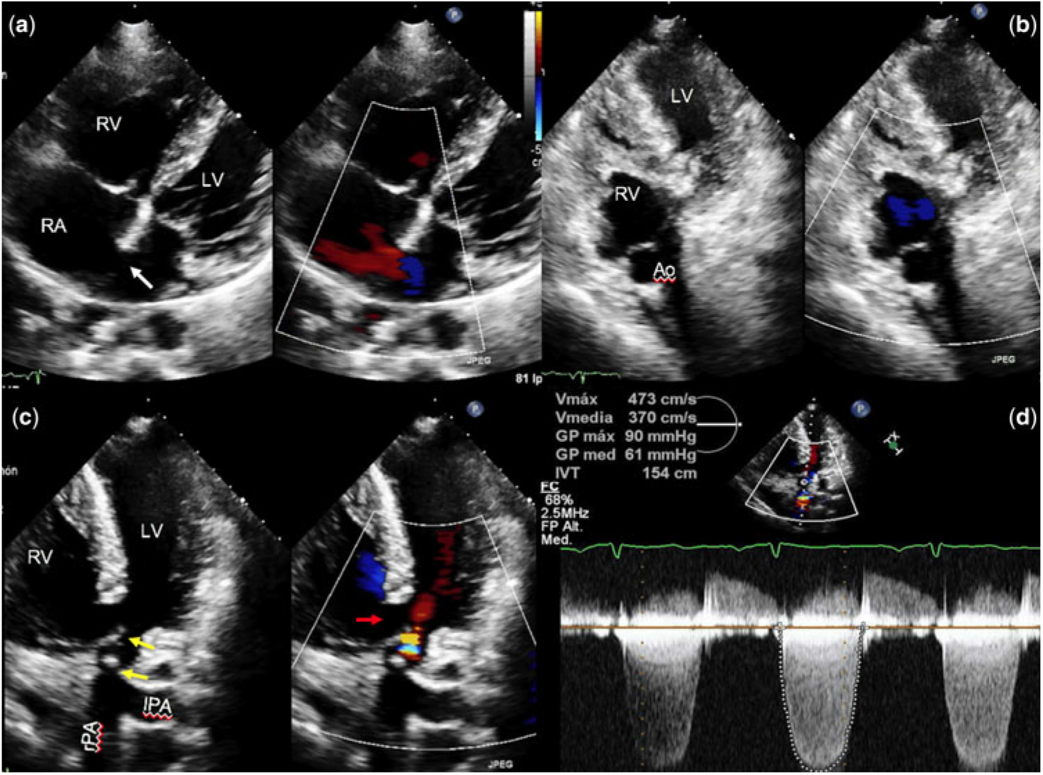
On 19 March, 2016, the transthoracic echocardiogram demonstrated situs solitus atrial, atrioventricular concordance and ventriculoarterial discordance with hypertrophy of the right ventricle, ostium secundum atrial septal defect of 8 mm, and ventricular septal defect of 17 mm. The biventricular systolic function was preserved with left ventricle ejection fraction of 68%, tricuspid annulus plane systolic excursion of 17 mm, tricuspid wave S of 10 cm/s and right ventricular shortening fraction of 43% and biventricular diastolic dysfunction with mitral E/A (peak filling wave/atrial contribution wave) ratio of 0.8 and tricuspid E/A ratio of 0.7. Severe valvular and subvalvular pulmonary stenosis with peak systolic gradient of 90 mmHg was documented (Fig 1, clips 1 and 2).
( a ) Transthoracic echocardiogram in four-chamber view showing atrioventricular concordance and atrial septal defect (white arrow). ( b , c ) Ventriculo-arterial discordance in bidimensional and color flow Doppler five-chamber view (clips 1 and 2). Subvalvular and valvular pulmonary stenosis and mild pulmonary regurgitation (yellow arrows) and ventricular septal defect (red arrow) are visualised. ( d ) In five-chamber view with continuous wave Doppler, a double pulmonary lesion was documented with severe pulmonar stenosis. RA, right atrium; RV, right ventricle; LV, left ventricle; Ao, aorta; rPA, right pulmonary artery; lPA, left pulmonary artery; Vmax, maximum velocity; Vmedia, mean velocity; GP max, maximum gradient; GP med, mean gradient; ITV, velocity time integral; FC, heart rate; FP, filling pressure

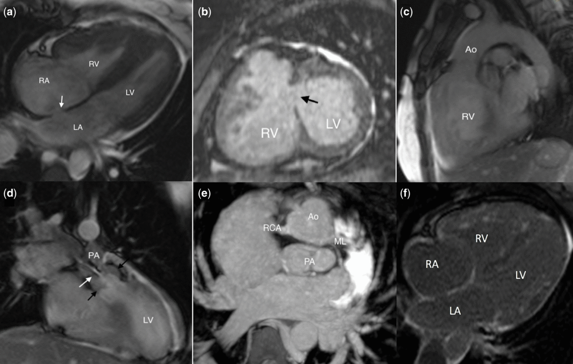
The CMR performed on 23 March, 2016 corroborated the presence of transposition of the great arteries with pulmonary valvular and infundibular stenosis. Pulmonary subvalvular, ventricular septal defect of 14 × 17 mm and ostium secundum atrial septal defect of 9 × 10 mm, atrial, bronchial, and abdominal situs solitus, dilatation of azygos vein, normal left ventricular volumes and biventricular function with left ventricular ejection fraction of 58% and right ventricular ejection fraction of 38.4%, and late enhancement in the four chambers with non-ischemic pattern corresponding to fibrosis were found. The aorta was anterior and right in relation to the pulmonary artery: D-transposition. The coronary pattern was normal; the left and posterior coronary ostium gave rise to the left anterior descending coronary artery and the left circumflex coronary artery, whereas the right and posterior coronary ostium gave origin to the right coronary artery. No intramural course was observed (Fig 2, clip 3).
( a ) Axial echo gradient acquisition shows four-chamber view, note ostium secundum atrial septal defect of 10 mm (white arrow). ( b ) CMR showing ventricular septal defect, central perimembranous type (black arrow), 17 × 10 mm diameter. ( c ) Axial echo gradient acquisition shows short axis of great arteries; note anterior aorta, right coronary artery originates from the right posterior sinus, and the main trunk of the left coronary artery from the left posterior coronary sinus. ( d ) Coronal echo gradient view, showing connection of left-sided right ventricle to aorta (ventriculo-arterial discordance), clip 3. ( e ) Sagittal echo gradient view, connection of right-sided left ventricle to pulmonary artery (ventriculo-arterial discordance), clip 4. The outlet tract of the left ventricle presents obstruction by fibromuscular component (black arrow) and acceleration jet stenosis flow at the valvular level (white arrow). Note ventricular septal defect. ( f ) Late enhancement in the four chambers with non-ischemic pattern corresponding to fibrosis. Abbreviations as in Figure 1. LA, left atrium; PA, pulmonary artery; ML, main left artery; RCA, Right coronary artery

On 4 April, 2016, she underwent for corrective surgery by Jatene procedure. Enlargement of the valvular ring of the neoaorta-type Nicks, valvular replacement of neoaorta by mechanical prosthesis, direct closure of the atrial septal defect and closure of the ventricular septal defect with bovine pericardium parch, and reimplantation of the coronary arteries to restore normal flow were performed. On 20 November, 2017, the control transthoracic echocardiography demonstrated only a slight generalised ventricular hypokinesia and mild dilatation of the right atrium.
After 2 years of follow-up, the patient is in NYHA functional class I, receiving medical treatment with Losartan and Acenocumarin.
Discussion
The ventriculo-arterial discordance is incompatible with life without communication between the two parallel circuits, which can be achieved through intracardiac (patent foramen ovale, atrial septal defect, or ventricular septal defect) and extracardiac shunts (patent ductus arteriosus or bronchopulmonary collateral circulation). The degree of mixing that occurs between the two parallel circulations will have the greatest impact on the stability of the patient.Reference Kirzner, Pirmohamed, Ginns and Singh 3 Our case represents a natural history of the complex transposition of the great arteries. In the literature, only a few cases have been reported with unrepaired transposition of the great arteries, whose survival is greater than 5 years;Reference AlAkhfash, Tamimi, Al-Khattabi and Najm 12 that is the reason our case is of great importance due to its hemodynamic stability despite its age. This patient presents atrial septal defect and ventricular septal defect, together with pulmonary stenosis, which surely increased her survival. In addition, the pulmonary arterial pressure, the ventricular volumes, the left ventricular, and the coronary pattern were normal. The presence of fibrosis was not quantified due to not having the respective software, but it corresponds to secondary changes of the pressure overload (due to pulmonary stenosis), but it was not a limitation for the repair. Cardiac catheterisation was not performed for pulmonary pressure and pulmonary resistance measurements, because non-invasive methods such as CMR and echocardiography found no signs of pulmonary hypertension.
CMR can provide detailed imaging of the vasculature, mediastinal structures, ventricular performance, and blood flow to the pulmonary and systemic circulation. This technique can also aid in the delineation of the coronary anatomy, given that coronary anomalies are frequently present in patients with transposition.Reference Fogel and Crawford 13
The main hematological finding in this group of patients is the erythrocytosis (increase of hemoglobin and hematocrit) secondary due to stimulation of the renal erythropoietin, as was found in our patient. Although these mechanisms are not yet clear, the hypoxemia is the main hyperproductive stimulation of erythropoietin in the juxtaglomerular apparatus. Erythropoietin stimulates erythropoiesis in the bone marrow and increases of erythrocytes in peripheral blood.Reference OliverRuiz, García and Elvira 14
Paradoxically, adults with cyanotic congenital heart disease show also higher incidence of thromboembolic complications, especially cerebrovascular events.Reference Berthrong and Sabiston 15 Although, the incidence of thrombotic stroke is much lower in adults than in children with complex congenital heart disease or in patients with polycythemia vera,Reference Perloff, Marelli and Miner 16 data from the Euro Heart Survey on Adult Congenital Heart Disease show that at least 10% of adults with complex congenital heart disease suffer cerebral thromboembolic events.Reference Engelfriet, Boersma and Oechslin 17
It is important to mention that our patient presented recurrent episodes of deep vein thrombosis of the left and right femoral veins and parieto-occipital abscess. Proposed pathogenic processes for abscess formation include chronic hypoxia predisposing to anaerobic infection, sluggish microcirculation, and passage of infected blood across right to left shunts.Reference Kumar 18 Cerebral abscesses comprise a distinct cause for neurodeficits in cyanotic congenital heart disease, and 25–46% of all cases of cerebral abscess can be attributed to underlying congenital cardiac disease.Reference Mehnaz, Syed, Saleem and Khalid 19 The most common cyanotic heart diseases associated with brain abscess are tetralogy of Fallot and transposition of the great arteries.Reference Fakhouri 20
Neurological complications constitute a large component of morbidity associated with cyanotic congenital heart disease, and can arise from a variety of mechanisms ranging from the acute global hypoxia of cyanotic spells to focal ischaemia due to arterial thrombosis, paradoxical embolisation, and cardioembolisation.Reference Kumar 18
CMR findings influenced the decision of the surgeons and the arterial switch surgery was performed. The arterial switch operation is aimed at normalizing ventriculoarterial concordance. It includes switching of the aorta and coronary arteries from the systemic right ventricle and of the pulmonary artery from the left ventricle and reattaching both great arteries to the appropriate ventricles. The coronary arteries are reimplanted into the neoaorta. The operation is typically performed during the first few weeks of life and has improved mortality rates from 90% in patients without surgical correction to less than 5%.Reference Kirzner, Pirmohamed, Ginns and Singh 3 , Reference Villafañe, Lantin-Hermoso and Bhatt 5 , Reference Jatene, Fontes and Paulista 21 The most common complications after the arterial switch operation include pulmonary (usually at the level of anastomosis and proximal branches) and pulmonary valve dysfunction, neoaortic root dilation with aortic regurgitation, stenosis of the coronary ostia, biventricular dysfunction, and pulmonary hypertension.Reference Khairy, Clair and Fernandes 22 , Reference Bonhoeffer, Bonnet and Piéchaud 23
The Jatene procedure, which has proven to be preferable in the treatment of transposition of the great arteries, has now left behind the Senning or Mustard procedure.Reference Wijesekera, Babu-Narayan, Shore and Gatzoulis 7 , Reference Senning 8 Both procedures create a baffle that rerouted systemic venous blood to the left ventricle and then to the pulmonary artery and directed pulmonary venous blood to the right ventricle and aorta. The right ventricle remained the systemic ventricle, the great vessels still arose from the “wrong” ventricle, but the pulmonary and systemic circulations were separated. The atrial switch procedures provide excellent midterm results (15-year survival, 77–94%; 20-year survival, 80%), with many patients able to lead fairly normal lives into their 3rd and 4th decades. Long-term complications with the atrial switch include atrial dysrhythmias, sinus node dysfunction, baffle leaks and obstruction, tricuspid regurgitation and dysfunction of the right (systemic) ventricle with heart failure, and risk for sudden cardiac death.Reference Deanfield, Camm and Macartney 24 – Reference Helbing, Hansen and Ottenkamp 26
Both procedures are valid and effective; at present, the decision regarding which surgery to perform is made according to the individual surgeon’s experience and characteristics of each patient.Reference Burkhart, Dearani and Williams 27
With the advent of newer and improved surgical techniques as well as postoperative intensive care, the scenario has completely changed, and very encouraging long-term survival rates almost achieving 90% at 15 years of age have been reported. The potentialities of the current corrective surgery modalities are also underlined by a low 10-year reintervention rate (6%) and a corresponding event-free survival of 88%.Reference Martins and Castela 1
Conclusion
In transposition of the great arteries patients, a high index of cases dies in the first month of life. Our case represents a natural history of the complex transposition of the great arteries. Non-invasive imaging studies are very useful for the diagnosis and follow-up of patients with transposition of the great arteries, especially echocardiography and CMR. In our case, the multimodality approach and the corrective surgery allowed her to survive.
Supplementary material
To view supplementary material for this article, please visit https://doi.org/10.1017/S1047951119000805
Acknowledgements
No acknowledgements.
Financial Support
This research received no specific grant from any funding agency, commercial or not-for-profit sectors.
Conflicts of Interest
None.
Ethical Standards
NA, because it is review of a case