Introduction
Suture otoplasty was originally described by Owens and Delgado (1955), and subsequently modified by Furnas, who placed non-absorbable mattress sutures between the conchal cartilage and mastoid fascia.Reference Janis, Rohrich and Gutowski 1 The correct anatomical placement of such sutures can sometimes prove challenging, particularly over the mastoid fascia, which is not always obvious as a precise landmark. Without the correct vector, reduction can be difficult and lead to malrotation.
The auricularis posterior muscle is apparent in dermofascial flap otoplasty as dissection proceeds posteriorly toward the mastoid. The muscle originates from the mastoid and inserts onto the ponticulus area on the concha.Reference Bendrihem, Vacher and Barbet 2 Its physiological function is to adduct the pinna. Clinical studies by Smith and Takashima (1978) have suggested that it has a role in the normal positioning of the auricle, and that dysfunction may cause protrusion.Reference Smith and Takashima 3
Materials and methods
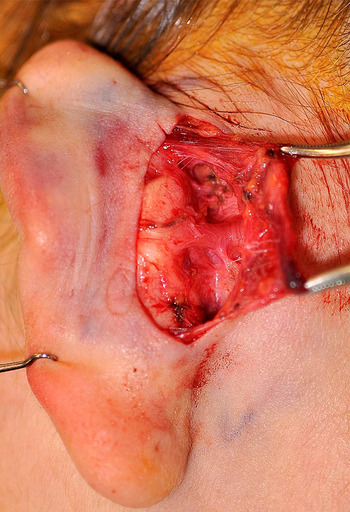
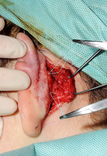
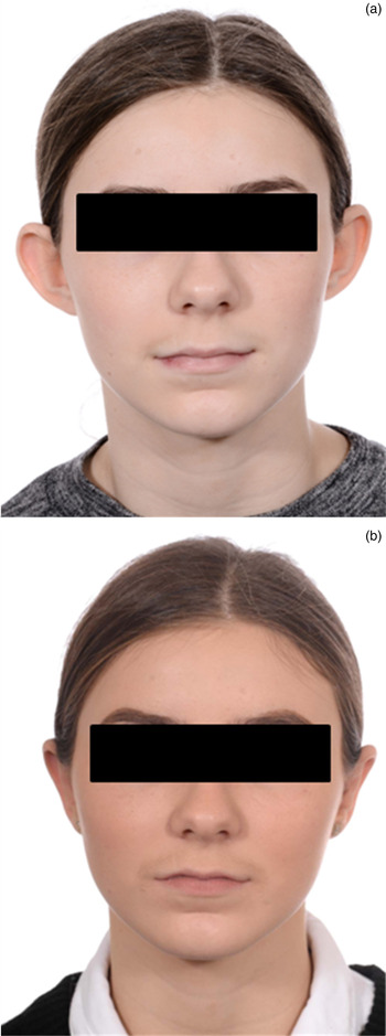
We have adopted the auricularis posterior as a marker for conchomastoid suture placement. Once exposed on the posterior concha, the muscle is dissected back to its origin from the mastoid (Figure 1). The mastoid fascia and posterior concha superior and inferior to the muscle are then dissected sufficiently to allow the placement of horizontal mattress sutures superior and inferiorly (Figure 2). Undyed 4-0 Prolene® sutures are used, clipping the first stitch to allow passage of subsequent stitches. Careful tensioning of the knots provides controlled, anatomical reduction of ear prominence and a natural aesthetic outcome (Figure 3).
Exposure and dissection of the posterior auricular muscle. Copyright of Medical Illustration Services, Perth Royal Infirmary. Reproduced with permission.

Placement of horizontal mattress sutures superior and inferior to the muscle (sutures have been highlighted electronically, but the photograph has not otherwise been modified). Copyright of Medical Illustration Services, Perth Royal Infirmary. Reproduced with permission.

Pre-operative (above) and post-operative (below) photographs.

Discussion
Nicolétis and Guerin-Surville first described the transposition of the posterior auricular muscle in otoplasty in 1978.Reference Nicolétis and Guerin-Surville 4 Guyuron and DeLuca described a linear relationship between the insertion of the posterior auricular muscle and ear projection.Reference Guyuron and DeLuca 5 More recently, techniques involving transposition of the insertion of the muscle have been described.Reference Azad, Edwin and Kumar 6 , Reference Scuderi, Tenna, Bitonti and Vonella 7 However, the employment of this muscle as a landmark in otoplasty (whilst leaving its insertion intact) has not previously been documented. We believe it will prove useful to the otoplasty surgeon.