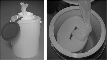
To prevent transmission of pathogens, environmental surface disinfection, especially of contaminated surfaces and surfaces with frequent contact to hands or skin, has become an important component in strategies to prevent healthcare-associated infections.Reference Dancer 1 – Reference Donskey 3 Adherence to local hospital policies, however, often has been shown to be low.Reference Havill, Havill, Mangione, Dumigan and Boyce 4 , Reference Carling, Von Beheren, Kim and Woods 5 To facilitate the adherence to surface disinfection protocols, reusable tissue dispensers have become more widely used and have been recommended by some authors (see Figure 1).Reference Voelz, Muller and Gillen 6 However, recent reports have suggested that the dispensers could be significantly contaminated with gram-negative bacteria—for example, Achromobacter spp., especially if surface-active ingredients, such as quaternary ammonium compounds, were used as compounds and reprocessing of the dispensers was inadequate.Reference Kampf, Degenhardt, Lackner, Jesse, von Baum and Ostermeyer 7 , Reference Kampf, Degenhardt, Lackner and Ostermeyer 8 Within the dispensers, theses isolates can adapt to the disinfectant solution, create biofilms, and even multiply.Reference Kampf, Degenhardt, Lackner, Jesse, von Baum and Ostermeyer 7 , Reference Kampf, Degenhardt, Lackner and Ostermeyer 8
We conducted a field study to investigate the frequency of contamination of tissue dispenser solution in the routine clinical setting, the kind of contaminating organisms, and potential association with nosocomial infection.
Therefore we investigated 30 quaternary ammonium compound solutions for the presence of bacterial contamination; these solutions were randomly retrieved out of reusable tissue dispensers from 4 different medical centers over a period of 4 weeks independent of the time since preparation of the dispenser solution or the reprocessing protocol. Potential contamination of disinfectant solutions was detected by membrane filter technique validated in our laboratory. In brief, at least 20 mL of each sample were drawn through a filter with 0.45-µm pore size, followed by flushing the filter 2 times with 100 mL neutralizing agent (3% Tween 80, 0.3% lecithin, 0.1% L-cysteine, 0.1% L-histidine). Membrane filters were then placed on Columbia agar with 5% sheep blood and incubated at 36°C for 48 hours. An additional 100 µL of each sample were plated directly on Columbia agar with 5% sheep blood. After incubation colonies were counted to calculate the number of colony-forming units per mL. Species identification of isolated bacteria was performed by matrix-assisted laser desorption ionization time-of-flight mass spectrometry (Bruker Daltonik). For typing of Achromobacter isolates a pulsed-field gel electrophoresis was performed using XbaI as restriction enzyme.Reference Tenover, Arbeit and Goering 9 In the same period Achromobacter spp. routinely isolated from clinical specimens of the 4 medical centers were collected and further identification and typing were performed as described above for the disinfectant solutions.
Despite the fact that in all 4 medical centers protocols for reprocessing of tissue dispensers were established, a surprisingly high proportion of 47% of the disinfectant samples (14 of 30) was contaminated with bacteria. The only bacterial species isolated in all contaminated samples was Achromobacter spp. Contamination ranged from 4 colony-forming units/mL up to more than 1×10⁴ colony-forming units/mL (9 samples). All Achromobacter isolates were typed using pulsed-field gel electrophoresis and 6 distinct strains were identified. Different strains were found in the 4 participating medical centers. Within individual medical centers different strains of Achromobacter were found as well as identical strains. In one institution all isolates (n=6) were genotypically identical. A wide genomic diversity for Achromobacter spp. in environmental sources has been described previously,Reference Amoureux, Bador and Fardeheb 10 thus the environment might be suspected as the primary source of the contamination. Noticing this, we found no clue that the contamination of the disinfectant solution was related to the manufacturing process of the disinfectant itself or the tissues used because we found different strains in identical disinfectants and different strains in identical tissues. Because all dispenser boxes were filled with disinfectant solutions taken from decentralized automatic dosing units, we also investigated samples of disinfectant solutions from dosing units to exclude these units as the source of the contamination. However, none of these samples showed bacterial growth. Furthermore, in the medical center with clonal dissemination of one Achromobacter strain, no epidemiologic link between contaminated dispenser solutions and healthcare workers or specific wards could be identified. Drinking water installations are an additional potential source of contaminations because Achromobacter spp. are frequently found in drinking water systems.Reference Amoureux, Bador and Fardeheb 10 However, using routine methods for drinking water examination in the participating medical centers, Achromobacter spp. was not isolated, so drinking water as the primary source of the contamination remains speculative and requires further investigation. In the period when the tissue dispenser solutions were sampled, 2 isolates of Achromobacter spp. were identified in routinely sampled clinical specimens in the medical center with the clonal distribution of one Achromobacter strain. One strain was isolated from tissue of an open leg fracture and was not classified as a healthcare-associated infection. The other strain was isolated from cerebrospinal fluid removed via a ventricular drainage on postoperative day 4 in a patient undergoing surgery due to a brain cyst. This isolate was classified as causing a healthcare-associated infection. Both clinical Achromobacter isolates were typed by pulsed-field gel electrophoresis as was the clonal strain of Achromobacter isolated from the disinfectant solutions. The strain from the healthcare-associated infection was identical with the isolates from the tissue dispenser solutions, whereas the strain from the open fracture was different. Achromobacter spp. can be isolated from many surfaces in the environment. However, isolation of an identical Achromobacter spp. strain in several tissue dispenser disinfectant solutions and a clinical specimen from the same institution indicates a potential safety hazard, especially considering the high levels of contamination.
In conclusion, disinfection solutions based on quaternary ammonium compounds from reusable tissue dispensers were frequently contaminated with gram-negative bacteria. Therefore, reprocessing of these dispensers according to the manufacturer’s recommendations seems to be crucial to prevent contamination of dispensers with the potential of transmission of pathogens and possible consecutive infections of patients.
Example of reusable tissue dispenser: left, complete dispenser ready for use; right, opened dispenser with tissue roll and disinfectant solution.

ACKNOWLEDGMENTS
Financial support. None reported.
Potential conflicts of interest. All authors report no conflicts of interest relevant to this article.