Introduction
Malnutrition is any disorder resulting from an inadequate or unbalanced diet, or a failure to absorb or assimilate dietary elements (Hughes & Kelly, Reference Hughes and Kelly2006). In developing countries, malnutrition is the most prevalent nutritional disorder among children (Passos et al., Reference Passos, da Fonte Ramos, Dutra, Mouco and de Moura2002) and parasitic infections are highly prevalent (McMichael, Reference McMichael2000). It is well recognized that the relationship between malnutrition and parasitic infection is an intimate one (Hughes & Kelly, Reference Hughes and Kelly2006). For instance, malnutrition hampers the outcome of the schistosome infection as well as adult worm growth (Ferreira & Coutinho, Reference Ferreira and Coutinho1999). Furthermore, it has been shown that feeding low-protein foods to mice not only leads to underdeveloped worms but also to reduction in the worm burden when compared to their well-fed counterparts (Dewitt, Reference Dewitt1957; Magalhaes et al., Reference Magalhaes, Guaraldo, Zanotti-Magalhaes, de Carvalho, Sgarbieri and de Alcantara1986).
A protein-deficient regional basic diet (RBD), based on the nutritional habits of the Brazilian north-eastern population, was produced and assayed in the acute phase of murine schistosomiasis (Coutinho et al., Reference Coutinho, de Freitas and Abath1992). This diet had a negative effect on growth, food intake, and protein absorption, and infected animals failed to develop hepatic periportal fibrosis (Coutinho et al., Reference Coutinho, de Souza, Silva, Cavalcanti, de Araujo, Barbosa Junior, Cheever and Andrade1997, Reference Coutinho, Barros, Barbosa, Oliveira, Silva, Araujo and Andrade2003; Oliveira et al., Reference Oliveira, Silva, Barbosa Junior, Ribeiro-Dos-Santos, Coutinho, Andrade and Soares2004). Over the past several years, we have focused on the effects of nutritional status on Schistosoma mansoni adult worms. Undernourished infected mice fed the RBD diet presented quantitative and qualitative alterations in the somatic development of adult worms (Neves et al., Reference Neves, Machado-Silva, Pelajo-Machado, Oliveira, Coutinho, Lenzi and Gomes2001, Reference Neves, Costa-Silva, Martinez, Biolchini, de, Lenzi, Gomes and Machado-Silva2003; Oliveira et al., Reference Oliveira, Barbosa, Gomes, Machado-Silva, Barros, Neves and Coutinho2003). This study identifies novel morphological alterations through light and confocal microscopy that may have implications for survival and reproduction of S. mansoni adult worms.
Materials and methods
Host–parasite model
Eighteen female Swiss Webster mice (3 weeks old) were obtained from the Laboratory Breeding Center (Oswaldo Cruz Foundation, Rio de Janeiro, Brazil) and were conventionally housed in a temperature (21 ± 1°C) and humidity (60 ± 10%) controlled room, exposed to 12 h light and dark cycles (artificial lights) and to an air exhaustion cycle. An S. mansoni–mouse model was used throughout the study.
Experimental diet
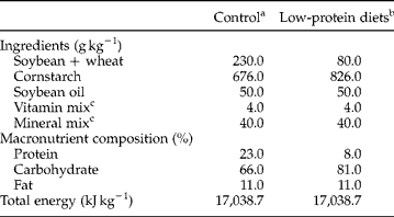
A total of 18 mice were split into the following groups: uninfected mice fed a standard diet (SD), uninfected mice fed a low-protein diet (LPD), infected mice fed a standard diet (ISD) and infected mice fed a low-protein diet (ILPD). In the experimental group, mice were fed a low-protein diet (8% protein, 81% carbohydrate, 11% lipids) beginning 2 weeks prior to experimental infection and continuing for the remainder of the experiment. The major ingredient proportions and total combustible energy (kJ kg− 1 and kcal kg− 1) of low-protein diets are shown in table 1 (de Moura et al., Reference de Moura, Lisboa, Custodio, Nunes, de Picoli Souza and Passos2007). The control groups were fed a standard commercial chow (Nuvilab, Paraná, Brazil) containing 11% fat, 23% protein, and 66% carbohydrate. Mice were housed in polypropylene cages with stainless-steel screen covers. All animals were given free access to food and water during the study. Food intake was recorded on a twice-weekly basis. All procedures were carried out in accordance with the conventional guidelines for experimentation with animals (Ellery, Reference Ellery1985). The experimental protocols used in the study were approved by the State University of Rio de Janeiro Ethics Committee for Animal Experimentation.
Composition of the control and low-protein diets.
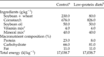
a Standard diet for rats (Nuvilab, NUVITAL Nutrients, Paraná, Brazil).
b The low-protein diet was prepared in our laboratory using the control diet and replacing part of its protein with cornstarch. The amount of the latter ingredient was calculated to make up the decrease in energy content due to protein reduction.
c Vitamin and mineral mixtures were formulated to meet the AIN-93G recommendation for a rodent diet and contain the recommended amount of iodine.
Parasites and infection protocol
Mice were infected subcutaneously with c. 80 S. mansoni cercariae (BH strain, Brazil) recovered from laboratory-reared Biomphalaria glabrata snails. The S. mansoni life cycle was maintained at the Malacology Department (Oswaldo Cruz Institute, Rio de Janeiro, Brazil), using B. glabrata snails and mice as intermediate and definitive hosts, respectively. The procedures for experimental infections have been described previously (Freire et al., Reference Freire, Rodrigues-Silva, Machado-Silva and Rey2003). For evaluation of malnutrition, body weight gain was monitored twice a week and serum albumin concentration was monitored only once.
Worm procedures
In brief, infected mice were euthanized at day 63 post-infection by cervical dislocation, and adult worms were recovered from the portal system and mesenteric veins and washed in saline. Schistosomes were processed for conventional bright-field microscopy as follows: specimens were fixed in AFA (alcohol, formalin and glacial acetic acid) at room temperature, stained with 2.5% hydrochloric carmine, dehydrated in a series of alcohols (70%, 90% and absolute), clarified in methyl salicylate with Canadian balsam (1:2), and preserved as whole-mounts on glass slides (Neves et al., Reference Neves, Pereira, de Oliveira, Gomes and Machado-Silva1998).
Morphological analysis
The whole-mounts were then separated into two groups: infected mice fed low-protein diet (ILPD, n = 114; 57 males and 57 females) and infected mice fed standard chow (ISD, n = 108; 55 males and 53 females). Each whole-mount contained only a single worm (male or female). The slides were examined microscopically (Olympus BX50, USA) to assess morphometry, using computer image analysis (Image Pro Plus, Media Cybernetics, USA) coupled to a Sony camera (640 × 480 pixels, RGB) using bright-field microscopy (Olympus BX50, USA) (Neves et al., Reference Neves, Pereira, de Oliveira, Gomes and Machado-Silva1998). The same whole-mounts were also used for further analysis by confocal microscopy (LSM 510, ZETA, Zeiss, Germany) in DIC and/or reflected mode, using a 543 He/Ne laser, with an LP 570 filter. The images were transferred from the LSM computer to Microsoft ImagerTM and Corel Draw 6.0TM for final contrast, brightness and gamma correction adjustments and then printed using a Codonics NP 1600TM printer (Machado-Silva et al., Reference Machado-Silva, Pelajo-Machado, Lenzi and Gomes1998). In both analyses, the reproductive system (testicular lobes and ovary) and tegument were focused. All available worms were submitted to morphometric analysis. In males, tegument thickness measurements were always taken from the region below the second testicular lobe, whereas, in females, measurements were taken from the area adjacent to the ventral sucker.
Data analysis
Data are presented as mean ± SEM. The statistical program SPSS 9.0 for Windows (SPSS Inc., Chicago, Illinois, USA) was used to facilitate calculations. For statistical analysis, the Student's t-test for unpaired samples was applied. Measurements with P values < 0.05 were considered statistically different.
Results
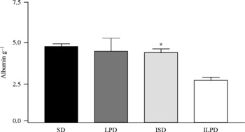
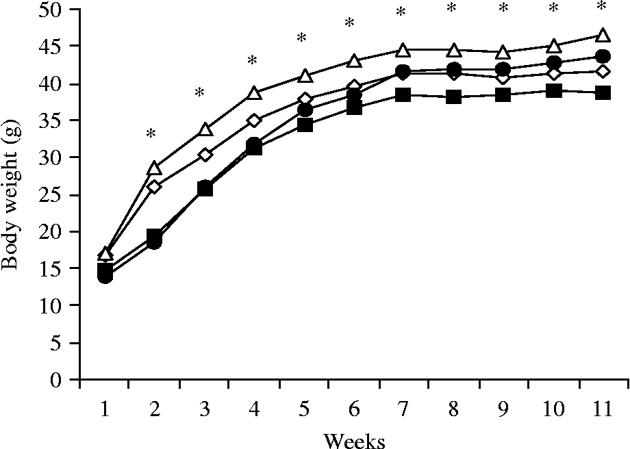
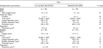
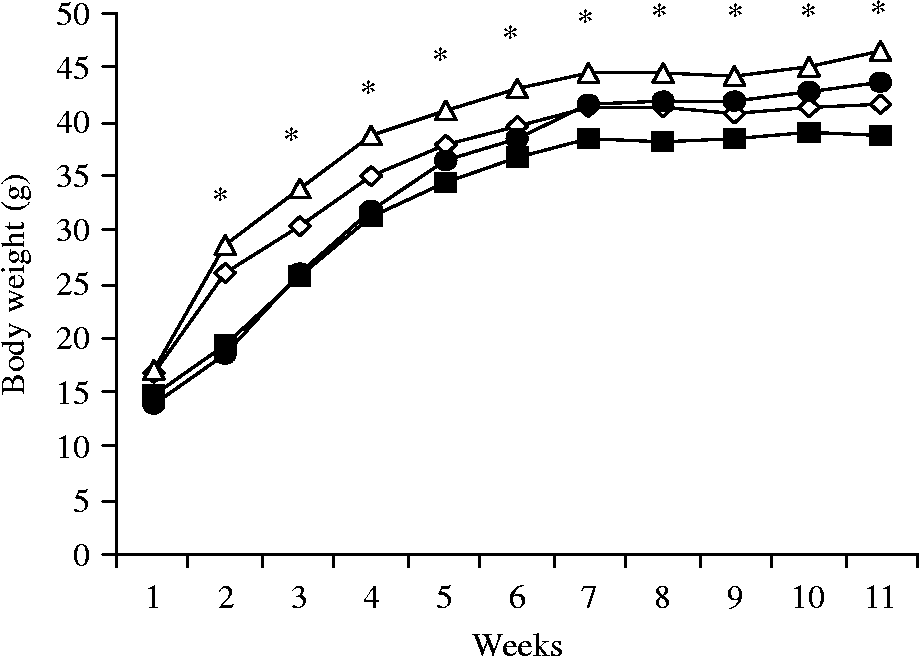
Animals showed varied average chow intake during the experiment. Animals fed the low-protein diet consumed relatively less than animals fed the standard diet. The serum albumin concentration was statistically (P < 0.05) lower in mice fed low-protein chow than that of the controls (fig. 1). The body weight gain, as shown in fig. 2, continuously increased for 7 weeks until the end of this study in uninfected, malnourished mice only. The change in body weight of infected malnourished mice decreased weekly after the infection was patent. The change in body weight became significant as of the second week when compared with that of control mice (P < 0.05). Light microscopy revealed some differences in the composition of the reproductive system between worms collected from each mouse group (table 2). In general, all morphometric values of specimens grown in malnourished mice were lower than those of controls, although sometimes the differences were not statistically significant. No significant difference (P >0.05) was seen in the length of the worms (7.6 ± 1.1 and 8 ± 1.2) or in the number of testicular lobes (8 ± 1) between each group. The mean values of testes area (23,224 ± 4243 μm2 and 26,078 ± 4967 μm2, P = 0.001) and larger diameter (304 ± 59 μm and 328 ± 57 μm, P = 0.03) were significantly different between worms from malnourished mice and controls, respectively. Additionally, morphological differences were mainly evidenced in female worms. Statistical analysis showed a significant decrease in length (9.2 ± 1.4 mm and 9.7 ± 1mm P = 0.03), ovary area (30,069 ± 7887 μm2 and 32,821 ± 3396 μm2, P = 0.02), larger diameter (369 ± 70 μm and 395 ± 50 μm, P = 0.03), smaller diameter (87 ± 17 μm and 90 ± 14 μm, P = 0.31), perimeter (893 ± 158 μm and 954 ± 98 μm, P = 0.02) and tegument thickness (4 ± 0.8 μm2 and 4.5 ± 1 μm2, P = 0.001) in the malnourished group when compared to controls, respectively. Average tegument thickness measurements (10 ± 2 μm) were similar between undernourished and well-fed male worms.
Serum albumin concentration of uninfected mice fed a standard (SD) and a low-protein (LPD) diet and infected mice fed a standard (ISD) and a low-protein (ILPD) diet. * P < 0.05 compared to SD, LPD and ISD.

Body weight gain among weanling mice fed a low-protein diet or standard diet for 11 weeks. Body weight of uninfected mice fed standard diet (SD, -△-), uninfected mice fed a low-protein diet (LPD,-●-), infected mice fed a standard diet (ISD, -⋄-) and infected mice fed a low-protein diet (ILPD, -■-). *P < 0.05 between the groups.
Light microscopy observations on the composition of the reproductive system in male and female Schistosoma mansoni adult worms collected from low-protein diet (ILPD) and standard diet (ISD) mice groups. Statistical significance (P <0.05).

A, P < 0.05; B, P >0.05.
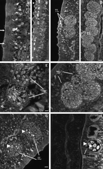
In addition, laser confocal microscopy also showed morphological details that differed between worms from mice in both groups. The tubercles were fewer in number on the dorsal surface of malnourished worms (fig. 3A), whereas worms from well-fed mice had higher concentration of tubercles (fig. 3B). Worms retrieved from undernourished mice underwent changes regarding the set of testicular lobes (fig. 3C), when compared to controls (fig. 3D). These organelles contained fewer differ-entiated germinal cells (fig. 3E) in comparison to those from the standard diet group (fig. 3F). Both the malnourished (fig. 3G) and control groups (fig. 3H) showed supernumerary testicular lobes with mature oocytes.
Confocal laser scanning microscope images of male S. mansoni stained with hydrochloric carmine. Malnourished mice (A, C, E, G) or well-fed mice (B, D, F, H). TL (testicular lobes), SL (supernumerary lobes). (A) Arrows indicate the tegument with few tubercles; scale bar 25 μm. (B) Normal tegument with large tubercles (arrowhead); scale bar 25 μm. (C) A specimen with only two testicular lobes; scale bar 25&thl;μm. (D) Normal set of testicular lobes; scale bar 25 μm. (E) Testicular lobes filled with few germinal cells; scale bar 50 μm. (F) Normal testicular lobes filled with germinal cells; scale bar 50 μm. Supernumerary testicular lobes with mature oocytes (arrowheads) observed in malnourished worms (G) or control (H), scale bars 25 μm.

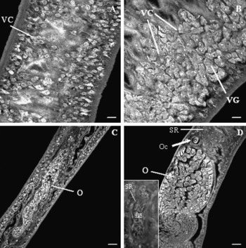
The female reproductive system was distinguished by the presence of an ovary, a seminal receptacle, oocytes, a vitelline follicle and vitelline glands (fig. 4A–D). Tomographic images showed that female worms from the malnourished group presented degenerate vitelline glands with few remaining follicles (fig. 4A) and ovaries lacking mature oocytes (fig. 4C). In contrast, well-fed female worms presented a vitelline follicle and vitelline cells (fig. 4B), an elongated ovary filled with mature oocytes and a well-developed seminal receptacle (fig. 4D).
Confocal laser scanning microscope images of the reproductive system of female S. mansoni stained with hydrochloric carmine. Malnourished mice (A, C) and well-fed mice (B, D). VC, vitelline cell; VG, vitelline glands; O, ovary; Oc, oocyte; SR, seminal receptacle; SPT, spermatozoa. (A) Degenerate vitelline glands with few remaining follicles; scale bar 50 μm. (B) Normal vitelline gland cells; scale bar 50 μm. (C) Ovary with an abnormal shape without mature oocytes; scale bar 25 μm. (D) Normal ovary composed of mature oocytes with an oocyte leaving the ovary close to the seminal receptacle (arrow); scale bar 25 μm. Higher magnification showing seminal receptacle with spermatozoa; scale bar 10 μm.

Discussion
In general, a low-protein diet produces a severe delay in laboratory animal growth (Console et al., Reference Console, Oyhenart, Jurado, Riccillo, Pucciarelli and Gomez Dumm2001). Our findings support previous experimental studies showing that a low-protein diet can affect both serum albumin and body weight in undernourished infected mice (Kanuft & Warren, Reference Kanuft and Warren1969; Akpom & Warren, Reference Akpom and Warren1975; Coutinho et al., Reference Coutinho, Silva, Barros, Araujo, Oliveira, Luna, Barbosa and Andrade2007).
Rapidly accumulating evidence shows that host nutritional factors can be decisive for the establishment and development of parasitic flatworms (Bansal et al., Reference Bansal, Bhatti and Sehgal2005). Diets deficient in B vitamins had a detrimental effect on the growth of Echinostoma caproni in ICR mice (Simpkins & Fried, Reference Simpkins and Fried2001) and E. trivolvis develops poorly in avian hosts lacking fat-soluble vitamins (Simpkins & Fried, Reference Simpkins and Fried1999). There is a significant growth retardation of E. caproni in ICR mice fed a protein-free diet (Rosario & Fried, Reference Rosario and Fried1999).
Schistosomes can remain paired for years, taking advantage of the high concentration of newly absorbed nutrients within nutrient-rich mesenteric veins of its host. In general, all morphometric values of specimens grown in malnourished mice were lower than those of controls, although sometimes differences were not statistically significant. However, the requirement of nutrients for reproduction in schistosomes under variable physiological conditions is poorly understood. To address this issue, we compared worms from mice that were made protein-deficient with those from control mice that were fed a normal diet.
In this experiment, we observed new diet-induced changes demonstrating that feeding mice a low-protein diet leads to gender-related differences. Quantitative and qualitative alterations were most evident in female parasites (Neves et al., Reference Neves, Machado-Silva, Pelajo-Machado, Oliveira, Coutinho, Lenzi and Gomes2001, Reference Neves, Oliveira, Machado-Silva, Coutinho and Gomes2002). This is not an unexpected finding since female schistosomes are metabolically more active than males (Tempone et al., Reference Tempone, Furtado, Gimba, Oliveira and Rumjanek2002). Although the schistosome obtains most of its dietary requirements by the ingestion of erythrocytes, the tegument also provides a significant source of nutrients (Braschi et al., Reference Braschi, Borges and Wilson2006). In addition, the tegumental outer surface is of crucial importance for modulation of the host response and parasite survival (Van Hellemond et al., Reference Van Hellemond, Retra, Brouwers, van Balkom, Yazdanbakhsh, Shoemaker and Tielens2006). Our studies have shown some evidence that malnutrition disrupts the full development of female teguments. The tubercles were fewer in number on the dorsal surface of malnourished worms. Such an alteration, particularly if it involves the molecular composition, may have profound effects on the physiology of the parasite, mainly in respect to the transport of nutrients. Although it cannot be concluded exclusively from the current data, it may be argued that malnourished worms had indeed received lower nutrients than those from mice maintained on the standard diet. However, protein content was not determined in this experiment. Laser microdissection microscopy could be used to evaluate protein content in schistosomes (Jones et al., Reference Jones, Higgins, Stenzel and Gobert2007). Haematocrit values were also not determined. Previous studies showed that schistosome-infected mice develop mild anaemia, expressed by reduced haemoglobin and lower haematocrit counts, compared with uninfected mice (Pyrrho et al., Reference Pyrrho, Lenzi, Ramos, Moura-Neto, Cachem, Silva, Takiya and Gattass2004). Therefore, the hypothesis that malnourishment leads to impairment of membrane bodies, which contribute to membrane turnover, was properly addressed. Our results showed that females were statistically smaller in undernourished mice than those from the control group. However, no information is available on the impact of a low-protein diet on female growth.
Successful reproduction is the ultimate purpose of parasitic helminths, and therefore it is essential to understanding the functional impact of physiological conditions on adult S. mansoni. Schistosomes have evolved a lifestyle that involves the cooperation of morphologically and transcriptionally distinct male and female forms. Male schistosomes are responsible for fecundation, maintaining female maturation and transporting them to the site of oviposition (Biolchini et al., Reference Biolchini, Neves, Hulstijn, Gomes and Machado-Silva2006). In contrast, females invest all of their metabolic energy in maximizing reproductive fitness and production of eggs (Morand & Muller-Graf, Reference Morand and Muller-Graf2000). This is expected since paired female schistosomes are egg-laying machines, producing several thousand vitelline cells per day (Erasmus & Davies, Reference Erasmus and Davies1979).
The gross anatomical changes in the genital system of female worms are recognized as useful methods in detecting effects of dietary deficiencies, at least in the acute phase (first 10 weeks of infection) of experimental schistosomiasis mansoni (Akpom, Reference Akpom1978). In the present experiment, malnourished females presented significant morphometric differences regarding the measurements and shape of the ovary. Atrophy of the ovary and of the vitelline glands is normally related to, and coincides with, a decrease in worm length (Hulstijn et al., Reference Hulstijn, Barros, Neves, Moura and Machado-Silva2003). Tomographic images of female worms also showed degenerate vitelline glands with few remaining follicles. Further work will be needed to establish whether protein synthesis during vittelogenesis is impaired in malnourished worms.
In contrast to control male schistosomes that undergo normal development, morphological changes were observed among specimens from malnourished mice. Unlike female worms, malnourished males showed only a few quantitative differences regarding the tegument and the reproductive system. The dorsal surface of male worms bore few large tubercles, whereas worms from control mice did not. This finding extends our previous studies in which flat tubercles and subtegumental vacuolated areas were found in worms from mice fed a protein-deficient diet (Neves et al., Reference Neves, Machado-Silva, Pelajo-Machado, Oliveira, Coutinho, Lenzi and Gomes2001). Another interesting observation is that only malnourished worms had a reduction in the number of testes when compared to worms from control mice. This result is not in line with previous experimental studies that showed a higher number of testes in worms recovered from mice fed a low-protein diet (Magalhaes et al., Reference Magalhaes, Guaraldo, Zanotti-Magalhaes, de Carvalho, Sgarbieri and de Alcantara1986). The lower production of cells in malnourished male worms is in agreement with a previous study that examined the organization of testicular lobes of schistosomes from malnourished mice (Neves et al., Reference Neves, Machado-Silva, Pelajo-Machado, Oliveira, Coutinho, Lenzi and Gomes2001). Curiously, a similar morphological alteration of the testicular lobes was observed in worms recovered from mice injected with streptozotocin, a drug used to induce diabetes mellitus (Hulstijn et al., Reference Hulstijn, Barros, Neves, Moura and Machado-Silva2003). Our data also showed that malnourished male worms exhibited supernumerary testicular lobes, which are identical in shape and arrangement to normal testicular lobes and also contain oocytes. Supernumerary lobes, whether containing male or female germinal cells, could be remnant from androecious or hermaphroditic ancestors that are occasionally expressed under the environmental pressure of the host (Hulstijn et al., Reference Hulstijn, Barros, Neves, Moura, Gomes and Machado-Silva2006). This structure seems to be frequent in schistosomes. To our knowledge, it was first described in a white mouse infected with S. mansoni from Egypt (Najim, Reference Najim1951). Our previous studies confirmed this atypical localization of testicular lobes in another host (the water rat Nectomy squamipes) (Machado-Silva et al., Reference Machado-Silva, Galvão, Presgrave, Rey and Gomes1994; Neves et al., Reference Neves, Pereira, de Oliveira, Gomes and Machado-Silva1998). Although other morphological changes were present among malnourished worms, male worm length was not significantly affected, suggesting that protein-deficient diets do not induce stunted male worms (Dewitt, Reference Dewitt1957; Magalhaes et al., Reference Magalhaes, Guaraldo, Zanotti-Magalhaes, de Carvalho, Sgarbieri and de Alcantara1986).
In summary, our findings indicate that nutrient deprivation leads to a hostile microenvironment with a deleterious effect on both male and female worms, culminating in morphological changes in the architecture of the tegument and the reproductive system.
Acknowledgements
Our thanks to Dr Lygia dos Reis Corrêa, Department of Malacology, Oswaldo Cruz Institute, for S. mansoni cercariae.