Introduction
Laryngocoeles result from upward dilatation of the laryngeal saccule with entrapped air. They are classified according to their relationship with the thyrohyoid membrane. An external laryngocoele is a symptomatic, cystic lesion occurring lateral to the membrane, whereas an internal laryngocoele is confined to the larynx and presents with dyspnoea. A combined laryngocoele lies both medial and lateral to the thyrohyoid membrane. The term laryngomucocoele denotes a dilated, cystic cavity filled with mucoid secretion, which is described as a laryngomucopyocoele when infected.
Supracricoid partial laryngectomy was first described in 1959 by Majer and Rieder for the treatment of laryngeal carcinoma.Reference Majer and Rieder1 Although the procedure was at that time indicated only for primary selected glottic and supraglottic carcinomas, it gained popularity over time and appeared suitable for a broader range of patients. The technique has reportedly been used as a salvage procedure instead of total laryngectomy after failure of radiation therapy.Reference Makeieff, Venegoni and Mercante2, Reference Spriano, Pelinli, Romano, Muscatello and Roselli3 Also, the advantage of resectability of one arytenoid or the upper part of the cricoid cartilage for the sake of complete removal of a tumour has allowed the technique to be used in advanced laryngeal cancers. Good local control and low complication rates have been reported in the literature.Reference Laccourreye, Brasnu, Perie, Muscatello, Menard and Weinstein4–Reference Farrag, Koch, Cummings, Goldenberg, Abou-Jaoude and Califano6
Supracricoid partial laryngectomy has several reported complications, including delayed decannulation, swallowing problems, intractable aspiration, wound infection and rupture of the pexy line. However, development of a laryngocoele following supracricoid partial laryngectomy has not adequately been discussed in the English language literature to date.
The current study aimed to evaluate the incidence, aetiology, clinical features and treatment of laryngocoele developing as a late complication of supracricoid partial laryngectomy.
Materials and methods
Before commencing, approval was obtained from the ethics committees of our institutions.
We reviewed retrospectively the medical records of 62 patients with laryngeal squamous cell carcinoma who had undergone supracricoid partial laryngectomy between January 1999 and July 2007 within our two institutions. Patients comprised 58 men and four women, aged from 37 to 68 years. Cricohyoidoepiglottopexy had been performed in 43 of the patients, while cricohyoidopexy had been conducted in the remaining 19. The incidence of laryngocoele formation, features of presentation, treatment modality and outcomes were evaluated. None of the patients had a laryngocoele before their operation. Computed tomography (CT) scans of the laryngocoeles were obtained.
Results and analysis
Of the 62 patients assessed, three developed a laryngocoele after supracricoid partial laryngectomy, giving an incidence of 4.8 per cent. All three had undergone cricohyoidoepiglottopexy. The interval between the initial operation and the diagnosis of laryngocoele was 12, 17 and 19 months, variously.
Two of the three patients had presented with a unilateral, soft, compressible cervical mass of two months' duration, together with a history of fever and subsequent dyspnoea. Endoscopic examination revealed partial immobilisation of the arytenoid cartilage on the involved side. In addition, one of these two patients had undergone direct suspension laryngoscopy due to suspicion of tumour recurrence.
The third patient had been admitted complaining only of a cervical mass which diminished with compression and swelled with coughing. Arytenoid cartilage movement was normal.
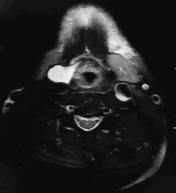
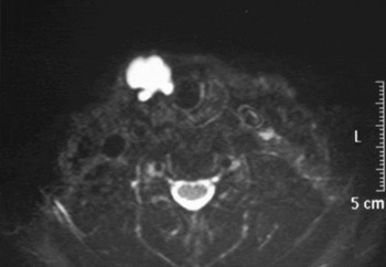
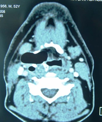
The diagnosis was established on CT scanning; the first (Figure 1) and second (Figure 2) patients were found to have a laryngomucopyocoele, while the third patient had a combined laryngocoele (Figure 3).
Axial computed tomography scan showing laryngomucopyocoele in the first patient.

Axial computed tomography scan showing laryngomucopyocoele in the second patient.

Axial computed tomography scan showing combined laryngocoele in the third patient.

All patients were treated with surgery via the transcervical approach. In addition, the first and second patients were treated by aspiration of infectious material and intravenous antibiotics commenced prior to surgery.
No recurrence of laryngocoele was detected over a mean of three years' follow up.
Discussion
It has long been known that an increase in intralaryngeal pressure is the most important aetiological factor in laryngocoele formation. It is also well known that glottic or ventricular tumours may enlarge the laryngeal ventricle size and cause laryngocoele formation by modification of airway patency. The tumour bulk may act as a one-way valve permitting air entrapment inside the laryngeal ventricle and blocking its escape. Micheau et al. reported an 18 per cent incidence of laryngocoeles coexisting with laryngeal cancers.Reference Micheau, Luboinski, Lanchi and Cachin7 Close et al. found this incidence to be 28 per cent in a series of 304 patients.Reference Close, Merkel, Burns, Deaton and Schaefer8 Individual variations in laryngeal ventricle anatomy may predispose to laryngocoele development. In an autopsy study, Broyles found larger than usual laryngeal ventricles in 7 per cent of normal larynxes.Reference Broyles9
The procedure for supracricoid partial laryngectomy includes removing the thyroid cartilage, bilateral vocal folds, bilateral laryngeal ventricles and paralaryngeal spaces. The need for resection of the epiglottis and one of the arytenoid cartilages depends upon the tumour extent. Preservation of the hyoid bone and the cricoid cartilage is mandatory. Laryngocoele formation after supracricoid partial laryngectomy may be caused by incomplete resection of the laryngeal ventricle during the procedure. The remaining ventricular mucosa, which may become entrapped at the pexy line after reconstruction, may present as a laryngocoele at a later date. Although the same surgical procedure was applied to all three reported patients, incomplete resection of the laryngeal ventricle in these patients may possibly have been due to enlarged laryngeal ventricle size, for the aforementioned reasons.
According to Dr O Laccourreye, incomplete resection of the laryngeal ventricle may easily be avoided at the time of surgery by checking the integrity of the resected laryngeal specimen (O Laccourreye, personal communication). This may be achieved by inserting a haemostatic clamp between the false and true vocal folds in order to look for a hole.
Supracricoid partial laryngectomy is widely used for the treatment of laryngeal cancer. However, laryngocoele development has previously been inadequately discussed in the English language literature as a late complication of supracricoid partial laryngectomy. Only two English language reports have addressed this situation. Laccourreye et al. conducted a retrospective study of 69 patients undergoing supracricoid partial laryngectomy, and found a laryngocoele incidence of 4.3 per cent.Reference Laccourreye, Brasnu, Perie, Muscatello, Menard and Weinstein4 Carrat et al. reported a case of laryngomucocoele as a late complication of supracricoid partial laryngectomy.Reference Carrat, Francois, Carles, Devars and Traissac10 In French language publications, Marlier et al. reported six cases in a series of 131 operations, and Thomassin et al. reported six cases of laryngocoele formation after supracricoid partial laryngectomy.Reference Marlier, Makieff, Gressillon, Crampette and Guerrier11, Reference Thomassin, Epron and Robert12
In our series, we found a laryngocoele incidence of 4.8 per cent. This result is similar to findings reported by Laccourreye et al. and Thomassin et al. Reference Laccourreye, Brasnu, Perie, Muscatello, Menard and Weinstein4, Reference Thomassin, Epron and Robert12 Two of our laryngocoele patients presented with a cervical mass, dyspnoea, infection and partial immobilisation of the arytenoid cartilage on the involved side. Cervical or local tumour recurrence was suspected in these patients. Unfortunately, one of these patients had undergone direct suspension laryngoscopy due to suspicion of tumour recurrence. The diagnosis of laryngocoele was made from the CT scan. Remobilisation of the arytenoid cartilage was achieved by transcutaneous aspiration of infected material and one week's intravenous antibiotic therapy. Clinicians should be aware of the possibility of a laryngocoele (in addition to the chance of tumour recurrence) in supracricoid partial laryngectomy patients admitted post-operatively with a cervical mass and dyspnoea, even years after the procedure.
• Laryngocoeles result from upward dilatation of the laryngeal ventricle with entrapped air, and are classified by their relationship with the thyrohyoid membrane
• This study sought to evaluate the incidence, aetiology, clinical features and treatment of laryngocoele occurring after supracricoid partial laryngectomy
• Remnants of the laryngeal ventricle may be the cause of such laryngocoele formation
• The integrity of the laryngeal ventricle in the resected specimen should be routinely checked in order to avoid this rare complication
Treatment of laryngocoele comprises three different modalities. Aspiration of the cyst with injection of a sclerosing agent constitutes a non-surgical option. The surgical options include an external approach and an endoscopic approach with or without carbon dioxide laser excision. The endoscopic approach should be used in cases of internal laryngocoele.Reference Frederick13–Reference Devesa, Ghufoor and Lloyd15 The external transcervical approach has the advantage of easy access and good exposure of the surgical field, and also overcomes dissection difficulties caused by the previous neck dissection. We preferred the transcervical approach in our patients, in which the skin incisions were made over a previous apron flap incision. The cyst mucosa was dissected from the surrounding structures up to the cricohyoidopexy line, and the cysts were removed completely. There was no recurrence a minimum of one year after the surgery.