Introduction
The life cycle of digeneans is accompanied by the alternation of several generations, each of which is capable of more or less intensive reproduction. The adult forms parasitizing the definitive hosts possess a complicated hermaphroditic reproductive system that often produces an immense number of eggs. The eggs hatch into ciliated miracidium larvae, which invade molluscs and use them as the first intermediate hosts (see review in Galaktionov & Dobrovolskij, Reference Galaktionov and Dobrovolskij2003). The offspring of sporocysts and rediae, the stages parasitizing the molluscs, develop from the germinal cells, which usually form in the germinal masses (see reviews in Dobrovolskij & Ataev, Reference Dobrovolskij, Ataev, Combes and Jourdane2003; Galaktionov & Dobrovolskij, Reference Galaktionov and Dobrovolskij2003; Ataev, Reference Ataev2017). The initial stages in the development of daughter individuals (daughter sporocysts, rediae or cercariae) also normally occur inside these specialized reproductive organs, and then the development continues in the brood cavity until the daughter parthenitae or cercariae escape through the birth pore or through a rupture in the body wall in some sporocysts.
Until fairly recently, the morphological aspects of the reproduction of sporocysts and rediae have been studied almost exclusively using light microscopy and conventional histological methods (see, for example, Van der Woude et al., Reference Van der Woude, Cort and Ameel1953; Cort et al., Reference Cort, Ameel and Van der Woude1954; Pearson, Reference Pearson1961; James & Bowers, Reference James and Bowers1967; Galaktionov & Dobrovolskij, Reference Galaktionov and Dobrovolskij1985; Dobrovolskij et al., Reference Dobrovolskij, Galaktionov and Ataev2000; Ataev et al., Reference Ataev, Isakova and Dobrovolskij2007; Isakova, Reference Isakova2011). Despite some notable progress, these studies have not fully resolved some contentious issues such as those concerning the structure and nature of the germinal cells, germinal masses and brood cavity. To further our understanding of these aspects of parthenital organization, we have conducted a comparative ultrastructural investigation of sporocysts and rediae in the digeneans representing different taxonomic groups (Podvyaznaya, Reference Podvyaznaya2007; Podvyaznaya & Galaktionov, Reference Podvyaznaya and Galaktionov2014, Reference Podvyaznaya and Galaktionov2018). The present study is a continuation of this research; it describes for the first time the ultrastructural organization of the germinal mass, brood cavity and birth canal in the rediae of a hemiuroid digenean, Bunocotyle progenetica. We have focused less attention on the ultrastructure of the germinal cells of these parthenitae, because they are almost identical to those in the rediae of Himasthla elongata and Tristriata anatis and in the sporocysts of Prosorhynchoides borealis (syn. P. gracilescens), which we had described earlier (Podvyaznaya, Reference Podvyaznaya2007; Podvyaznaya & Galaktionov, Reference Podvyaznaya and Galaktionov2014, Reference Podvyaznaya and Galaktionov2018).
Bunocotyle progenetica was chosen for this study not only because of our insufficient understanding of parthenital reproduction in hemiuroid digeneans, but also because of the special role that the rediae play in the life cycle of this species. The life cycle of B. progenetica is secondarily modified and occurs entirely within a single molluscan host (Markowski, Reference Markowski1936; Chabaud & Buttner, Reference Chabaud and Buttner1959; Stunkard, Reference Stunkard1973; Deblock, Reference Deblock1974, Reference Deblock1980; Levakin, Reference Levakin2005), the mud snails Peringia ulvae and Ecrobia ventrosa, which often share the same habitats. The snails become infected by ingesting the eggs of B. progenetica released into the environment from the dead infected molluscs. The eggs enter the intestine of the mollusc with food and hatch into miracidia that penetrate the gut wall and transform into mother sporocysts. The mother sporocysts give rise to rediae, which produce cercariae. The cercariae do not escape from the rediae, as in the majority of digeneans, but remain in the redial brood cavity, where they develop into sexually mature adults (Deblock, Reference Deblock1974; Galaktionov & Dobrovolskij, Reference Galaktionov and Dobrovolskij2003) and the adult worms also lay their eggs directly into the brood cavity. Therefore, in B. progenetica the development of daughter individuals of the hermaphroditic generation occurs almost entirely inside the rediae. One-host (monoxenous) life cycles, in which sexual adults develop in the first intermediate host, have been described in a few digenean species from several different taxa (see review in Poulin & Cribb, Reference Poulin and Cribb2002). There is evidence that such an extreme form of life-cycle modification can be associated with changes in morphology and development of the parasites, but the available information is limited and concerns only the hermaphroditic generation (see review in Galaktionov & Dobrovolskij, Reference Galaktionov and Dobrovolskij2003). The present study discusses the possible influence of the life-cycle modification on the organization of the redial reproductive apparatus in monoxenous B. progenetica.
Material and methods
Rediae of B. progenetica were extracted from the molluscs P. ulvae collected during the summer in the Sukhaya Salma Inlet of the Chupa Bay (White Sea). The specimens were fixed in 3% glutaraldehyde in 0.1 М cacodylate buffer (pH = 7.4) for 8–10 days and then post-fixed in 1% osmium tetroxide in the same buffer at 4°C. Osmolality of fixators and the buffer was adjusted to 760 mOsm by sucrose. The rediae were dehydrated and embedded in Epon-Araldite following the standard procedure (Mollenhauer, Reference Mollenhauer1964). Thin and semi-thin sections were cut on a Leica EM UC6rt (Wetzlar, Germany) ultratome. Thin sections were stained with aqueous uranyl acetate and lead citrate and examined at 80 kV on a Morgagni 268 (FEI, Eindhoven, Netherlands) transmission electron microscope. The specimens selected for transmission electron microscopy (TEM) of the germinal masses were young, actively developing rediae with the brood cavity containing early embryos (germinal balls); the brood cavity and birth canal were studied on rediae of different ages ranging from newly emerged to mature individuals. Different stages of post-embryonic development were also examined on live preparations using an Olympus CH-40 (London, UK) light microscope.
For confocal microscopy, rediae were fixed with 4% formaldehyde in 0.01 M phosphate-buffered saline (PBS) for 4 h at 4–6°C and transferred to 0.01 M PBS containing 0.1% (w/v) sodium azide at 4–6°C. The preparations were stained as whole mounts. The specimens were permeabilized for 2 h with 0.25% Triton X-100 in 0.01 M PBS, stained for 8 h with Alexa Fluor 546-conjugated phalloidin (1:150; ThermoFisher Scientific, Waltham, Massachusetts, USA; Cat# A22283), rinsed in 0.01 M PBS and then mounted on slides with Vectashield (Vector Laboratories Inc., Burlingame, California, USA; Cat# H-1000). Confocal images were collected on a Leica TCS SP5 (Wetzlar, Germany) confocal laser scanning microscope. All confocal images of phalloidin-stained whole mounts were presented as maximum-intensity projections of the Z-stacks.
Results
General morphology of the germinal mass
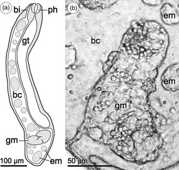
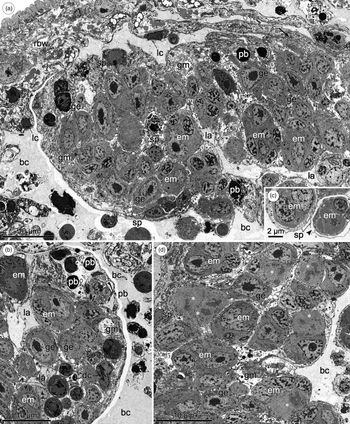
Young and mature rediae of B. progenetica have a relatively large (usually over 100 µm) germinal mass located in the posterior portion of the brood cavity behind the gut (fig. 1). The usually narrow base of the germinal mass, sometimes shaped like a stalk, is attached to the body wall of the rediae (fig. 1). The interior of the organ is penetrated by several deep, branching lacunae communicating with the brood cavity and filled with its fluid (figs 1a and 2a, b). The germinal mass of B. progenetica is typical in cellular composition and comprises a few undifferentiated cells, numerous germinal cells at different maturation stages and supporting cells, which constitute the interstitial tissue of the organ (figs 2a and 3a, c, e, f). It is noteworthy that an appreciable number of these cellular elements, mostly germinal cells, are involved in the degenerative processes that lead to the formation of pyknotic bodies (fig. 2b).
General view of the redial reproductive apparatus of Bunocotyle progenetica. (a) Schematic drawing of a young redia showing the germinal mass, brood cavity with early embryos and the birth canal in lateral view. (b) Light microscopic image of the germinal mass in a live mature redia. Abbreviations: bc, brood cavity; bi, birth canal; em, embryos; gm, germinal mass; gt, gut; ph, pharynx.

TEM micrographs showing the general organization of the redial germinal mass of Bunocotyle progenetica. (a) Posterior part of a germinal mass with a lacuna; note the connection of the germinal mass with the redial body wall (arrow). (b) Micrograph showing various stages of cell degeneration in a germinal mass. (c) Micrograph showing germinal balls in the peripheral area of the germinal mass; note a rupture (arrowhead) in the superficial layer of the supporting tissue. (d) Micrograph showing frequent mitotic divisions (asterisks) in the germinal mass. Abbreviations: bc, brood cavity; dc, degenerating cells; em, embryos; ge, germinal cell; gm, germinal mass; la, lacuna; lc, cavity lining cell; pb, pyknotic body; rbw, redial body wall; sp, supporting cells and their outgrowths.

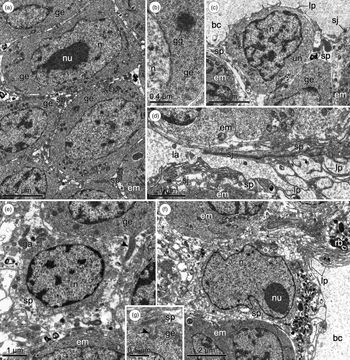
Ultrastructure of the cells composing the germinal mass of rediae of Bunocotyle progenetica. (a) Developing germinal cells separated by thin sheets of the supporting tissue. (b) Germinal cell with a germ granule. (c) Undifferentiated cell enveloped by the supporting tissue on the periphery of the germinal mass. (d) Lining of a lacuna with lamellar projections; note septate junctions connecting the supporting cells. (e) Perikaryon of a supporting cell in the central area of the germinal mass; note an outgrowth of a germinal cell (arrowhead) protruding into the supporting tissue. (f) Peripheral supporting cell with lamellar projections, numerous vesicles and residual bodies in the cytoplasm. (g) Protrusion of the supporting tissue (arrowhead) into a germinal cell. Abbreviations: bc, brood cavity; em, embryos; ga, Golgi apparatus; ge, germinal cell; gg, germinal granule; la, lacuna; lp, lamellar projections; n, nucleus; nu, nucleolus; rb, residual body; sj, septate junction; sp, supporting cells and their outgrowths; un, undifferentiated cell; v, vesicles.

In the rediae of B. progenetica, the germinal mass contains a large number of early embryos at the stages of naked cell aggregates and the newly formed germinal balls (fig. 2a–d). The germinal cells and embryos are positioned inside the organ without any apparent order, in different regions and layers. Electron micrographs show frequent mitoses (fig. 2d) in the embryos and individual cells (presumably, proliferating undifferentiated cells and mature germinal cells undergoing cleavage divisions).
The germinal balls, which are ready to leave the germinal mass and lie close to the brood cavity or lacunae, are usually separated from the surrounding supporting tissue by a slit-like electron-lucent space (fig. 2c). Occasionally, ruptures can be seen in this supporting tissue, which provide exit routes for the embryos into the brood cavity (fig. 2c).
Ultrastructure of the germinal mass
Undifferentiated and germinal cells of the germinal mass in the B. progenetica rediae show ultrastructural features typical of these types of cell. Small (4–5 µm) undifferentiated cells have a high nuclear/cytoplasmic ratio (fig. 3c). Their nuclei are rich in condensed chromatin; a thin layer of perinuclear cytoplasm contains primarily free ribosomes and a few small mitochondria (fig. 3c). Mature germinal cells and germinal cells at advanced stages of differentiation are markedly larger (7–9 µm) than undifferentiated cells (fig. 3a). Large euchromatic nuclei of these cells always contain a large nucleolus (fig. 3a). Their cytoplasm occupies a much greater volume than in undifferentiated cells and includes a greater number of cellular organelles (free ribosomes, small cisterns of sparse rough endoplasmic reticulum (EPR), Golgi apparatus, microtubules, mitochondria, elements of annulate lamellae). Germinal cells are also characterized by the presence of germ granules (fig. 3b). These structures have an appearance of small non-membrane-bound accumulations of electron-dense granular-fibrous material located close to the nuclear envelope or lying free in the cytoplasm.
The supporting cells of the germinal mass are few in number and are interconnected by septate junctions (fig. 3c, d). Their perikarya are normally located at the periphery of the germinal mass, but can also be found in deeper layers of the organ (figs 2a and 3e, f). The nuclei of the supporting cells usually have a well-developed nucleolus and the nucleoplasm of high electron density. The condensed chromatin always forms a layer closely associated with the nuclear membrane and some accumulations in the central part of the nucleus. The perinuclear cytoplasm contains the usual complement of organelles (Golgi apparatus, EPR, ribosomes and mitochondria); its characteristic feature is the presence of a large number of residual bodies and various vesicles, some of them resembling endosomes and lysosomes (fig. 3f). The extent to which these characteristics are present vary in different supporting cells, but in younger cells they tend to be less pronounced. Thin, long processes of the supporting cells branch out and form narrow sheets of interstitial tissue that separate undifferentiated cells, germinal cells and embryos (figs 2a, d and 3a, c). The cytoplasm of these processes is mostly electron lucent, with a small number of organelles (a few mitochondria, small cisterns of rough EPR, occasional residual bodies and small lysosome-like vesicles). The supporting cells often form short processes projecting into the cytoplasm of the germinal cells (fig. 3g); the latter also send cytoplasmic projections into the supporting tissue (fig. 3e).
In addition to these processes, the supporting cells form cytoplasmic layers that line the internal lacunae (fig. 3d) and cover the germinal mass externally to form an envelope on its surface (figs 2a–d and 3c, f). This envelope is often composed of several (2–8) closely packed cytoplasmic layers, but in some areas consists of a single cytoplasmic layer (figs 2c and 3c). On the side of the brood cavity, the outermost cytoplasmic layer gives rise to lamellar projections, which are quite numerous in some areas of the envelope (fig. 3c, f). The lining of the lacunae forms similar projections (fig. 3d).
Ultrastructure of the brood cavity and birth canal
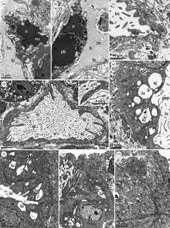
The youngest, newly emerged rediae, as well as more mature individuals, have a well-developed brood cavity that extends from the posterior end of the body to the anterior portion of the gut (fig. 1a). Along its entire length, the cavity has a well-pronounced intrinsic lining composed of sparsely spaced, highly flattened cells (figs 2a and 4a, b). We were unable to identify the boundaries between these cells and it cannot be ruled out that the lining is syncytial. The electron dense nuclei of the lining cells contain nucleoli and a heterochromatin layer adjacent to the nuclear membrane (fig. 4a, b). These nuclei are situated in the bulging protrusions of the lining, often filled with vesicles and residual bodies, among which there are intact pyknotic bodies (fig. 4a, b). The lining of the brood cavity forms lamellar projections on the luminal surface. The surface of the lining facing the redial body wall is irregular in shape because of various folds and invaginations. It is surrounded by parenchymal cells and their processes, or less often by other cellular elements of the redial body wall (fig. 4a, b). In the anterior region of the brood cavity, its lining is connected by septate junctions with the epithelial cells of the birth canal (fig. 4c).
TEM micrographs showing the brood cavity and the birth canal of rediae of Bunocotyle progenetica. (a, b) Brood cavity lining; note lamellar projections on its luminal surface and a pyknotic body in the perinuclear cytoplasm. (c) Connection of the brood cavity lining with the birth canal epithelium; note a septate junction. (d) Cross-oblique section through the proximal part of the birth canal. Insert shows an enlarged septate junction connecting epithelial cells. (e) Connection of the flattened epithelium of the proximal part of the birth canal with the epithelial lining of the middle part of the birth canal. (f) Oblique section through the middle part of the birth canal. (g) Micrograph showing the distal part of the birth canal lined by a modified tegument in a mature redia. (h) Cross-oblique section through the distal part of the birth canal lined by an unmodified tegument in a newly emerged redia. Abbreviations: bc, brood cavity; bin, basal invaginations; bl, basal lamina; dbi, distal part of birth canal; ip, irregular apical projections; lc, cavity lining cell; lp, lamellar projections; lu, duct lumen; mep, epithelium of middle part of birth canal; mte, modified tegument; mu, muscle; n, nucleus; nu, nucleolus; pa, parenchymal cell; pep, epithelium of proximal part of birth canal; rb, residual body; sj, septate junction; tc, tegumental cell; te, tegument.

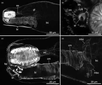
The birth canal is a duct that extends toward the anterior end of the redia and opens to the exterior near the mouth (figs 1a and 5a). It consists of three regions – proximal, middle and distal – which differ in thickness, length, structure of the epithelial wall and arrangement of muscle layers. The proximal region is rather long and wide and is lined by flat epithelial cells joined by septate junctions (fig. 4d, insert). The luminal surfaces of these cells give rise to long, irregularly shaped projections with irregular distensions along their length; these projections fill the whole space in the lumen of the canal. The canal becomes thinner as it approaches the birth pore and its lumen narrows down (fig. 5c). The epithelial cells lining the middle region of the birth canal lack long apical projections (fig. 4f). Their luminal surface bears small cone-shaped prominences and short lamellar processes lying parallel to the surfaces of the cells. The luminal surface is thrown into high and thick, closely packed folds, with parts of the lumen seen as vacuole-shaped spaces between the folds (fig. 4e, f). The basal and lateral surfaces of the cells in this area are also significantly increased by numerous folds (in case of the basal plasma membranes, by deep basal invaginations). The short distal region of the birth canal is lined with a continuation of the tegument and has a narrow lumen (fig. 4g, h); in mature rediae, this tegument is modified (fig. 4g).
Confocal images showing the musculature of the birth canal and brood cavity of rediae of Bunocotyle progenetica. (a) Longitudinal projection through the anterior region of the body; note that musculature is present only at the anterior end of the brood cavity. (b) Transverse projection through the anterior end of the body showing the pharynx and the middle region of the birth canal. (c) Longitudinal projection through the anterior end of the body. (d) Longitudinal projection through the anterior end of the brood cavity at the connection with the birth canal. Abbreviations: bc, brood cavity; bi, birth canal; bp, birth pore; cm, circular muscle fibres; es, esophagus; gt, gut; lm, longitudinal muscle fibres; mbc, muscle fibres of brood cavity; ph, pharynx; rm, radial muscle fibres; sr, sphincter.

The epithelial lining of the canal is underlain throughout its length by a thin basal lamina (at places highly plicate) (fig. 4c, e, f), which serves for attachment of the muscle fibres. In the proximal portion, the musculature of the canal consists of an inner layer of closely set, strap-like circular fibres (fig. 5c, d) and an outer layer of widely spaced, thin longitudinal fibres. In the middle and distal regions, the canal is surrounded by a single layer of longitudinal fibres (fig. 5b) and in the middle region there is an additional inner sphincter composed of 8–10 circular fibres (fig. 5b, c). This sphincter is connected to the outer layer of longitudinal muscles with thin radial fibres (fig. 5b). The anterior end of the brood cavity near its junction with the birth canal is surrounded by thin irregularly arranged muscle fibres (fig. 5c), probably derived from the longitudinal fibres of the canal; the rest of the cavity has no musculature (fig. 5a).
The brood cavity of young rediae contains primarily the germinal balls of different sizes (fig. 1a); in the brood cavities of more mature individuals there are numerous developing cercariae and metacercariae with a few occasional embryonic rediae. In both young and mature rediae, the brood cavity contains a large number of degenerating cells and pyknotic bodies (fig. 2a, b). The latter are very similar to the pyknotic bodies inside the germinal mass; some of these are enclosed in a layer of the cytoplasm resembling the surface layer of the supporting tissue in the germinal mass.
Discussion
Germinal mass
Light microscopy studies have identified several types of germinal masses distinguished by their number and arrangement inside the bodies of parthenitae (see reviews in Dobrovolskij & Ataev, Reference Dobrovolskij, Ataev, Combes and Jourdane2003; Galaktionov & Dobrovolskij, Reference Galaktionov and Dobrovolskij2003). These types include germinal masses embedded in the body wall at the posterior end of the parthenitae; germinal masses protruding into the brood cavity; germinal masses lying entirely in the brood cavity and connected to the body wall with a thin stalk; and germinal masses floating freely in the brood cavity. Although in most cases there is a single germinal mass, some parthenitae possess multiple, diffuse germinal masses (Ameel et al., Reference Ameel, Cort and Van der Woude1949). In other digeneans, the primary germinal masses become fragmented to produce smaller floating secondary germinal masses (Kniskern, Reference Kniskern1952; Kofiadi, Reference Kofiadi1995). Despite this diversity, the internal structure of the germinal masses has long been regarded as relatively evolutionarily conserved (Dobrovolskij & Ataev, Reference Dobrovolskij, Ataev, Combes and Jourdane2003; Galaktionov & Dobrovolskij, Reference Galaktionov and Dobrovolskij2003). The germinal masses were believed to be essentially similar in cellular composition and to show pronounced polarity in the arrangement of their constituent elements, with the early embryos and mature germinal cells located closer to the brood cavity than the undifferentiated cells and the early differentiation stages of the germinal cells.
Electron microscopy studies of four digenean species, including B. progenetica (Podvyaznaya, Reference Podvyaznaya2007; Podvyaznaya & Galaktionov, Reference Podvyaznaya and Galaktionov2014, Reference Podvyaznaya and Galaktionov2018; this study), have confirmed the conclusion about a uniform cellular composition of the germinal masses across different digenean groups. These studies have also shown a similarity in ultrastructure between undifferentiated proliferating cells, which we regard as oligopotent stem cells, and the germinal cells of the germinal masses in sporocysts and rediae of different digeneans. The germinal cells were shown to contain the organelles typical for the oocytes such as germ granules (or nuage) and annulate lamellae, which confirms that these cells belong to the germline. On the other hand, electron microscopy has revealed a significant diversity in ultrastructure of the germinal masses associated primarily with the structural variations in their supporting tissue and with different age spans and numbers of embryos in the germinal masses. In certain cases, some ultrastructural characteristics and combinations of characters are likely to be specific not only for species, but also for the taxa of family rank (Podvyaznaya & Galaktionov, Reference Podvyaznaya and Galaktionov2018).
Like previously studied parthenitae, the rediae of B. progenetica have germinal masses with a distinct set of characters. Judging from a rather large number of germinal cells and early embryos in the germinal masses and abundance of mitotic divisions, these organs appear to be highly reproductively active. At the same time, the large, highly proliferative germinal masses of B. progenetica, unlike those of many other digeneans including T. anatis (Podvyaznaya & Galaktionov, Reference Podvyaznaya and Galaktionov2018), do not show any evidence of polarity. In these rediae, many embryos develop in the central portion of the germinal mass, which complicates their release into the brood cavity. The germinal mass of B. progenetica has a relatively weakly developed supporting tissue, especially in its central portion. An important function of the supporting cells is the delivery of nutrients to the developing germinal cells and embryos (Podvyaznaya, Reference Podvyaznaya2007; Podvyaznaya & Galaktionov, Reference Podvyaznaya and Galaktionov2014, Reference Podvyaznaya and Galaktionov2018). In B. progenetica, this function is likely to have been partially taken over by branching lacunae that allow the nutrient-carrying fluid of the brood cavity to reach the deep layers of the germinal mass. The presumed adsorptive activity of the supporting cells in the area of the lacunae and in the surface layer of the germinal mass is further supported by the fact that their luminal surface is amplified by lamellar projections. The lacunae can also probably facilitate the release of the germinal balls from the central portion of the germinal mass into the brood cavity of the rediae.
Another characteristic feature of the redial germinal mass in B. progenetica is a relatively high intensity of the degenerative processes, which is indicated by the presence of numerous degenerating cells and pyknotic bodies. The pyknotic bodies have been observed in the germinal masses of other parthenitae (Dobrovolskij & Ataev, Reference Dobrovolskij, Ataev, Combes and Jourdane2003; Galaktionov & Dobrovolskij, Reference Galaktionov and Dobrovolskij2003; Podvyaznaya, Reference Podvyaznaya2007; Isakova, Reference Isakova2011; Podvyaznaya & Galaktionov, Reference Podvyaznaya and Galaktionov2014), but in actively functioning organs they were few in number (Isakova, Reference Isakova2011). As mentioned in the Introduction, cercarial embryos of B. progenetica develop into adult worms inside the redia. It is clear that highly proliferative germinal masses of this species can produce a significantly larger number of embryos than the redial brood cavity can contain and support. Active degeneration of the germinal cells in the germinal mass is likely to be one of the regulatory mechanisms that control the number of offspring. The resulting pyknotic bodies are apparently being gradually expelled into the brood cavity. It appears that some degenerating cells pinch off from the germinal mass along with small sections of its supporting tissue. Active degeneration of the germinal material similar to that of B. progenetica has been described by Kofiadi (Reference Kofiadi1995) in the daughter sporocysts of Hemiuridae gen. sp. According to Kofiadi, these degenerative processes control the intensity of reproduction in the course of sporocyst development and under unfavourable external conditions (for instance, during winter).
The germinal masses and their development in hemiuroid parthenitae have previously been studied using light microscopy in Halipegus eccentricus and Hemiuridae gen. sp. (Ameel et al., Reference Ameel, Cort and Van der Woude1949; Kofiadi, Reference Kofiadi1995). In very young rediae of H. eccentricus, Ameel et al. (Reference Ameel, Cort and Van der Woude1949) have described a prominent germinal mass in the posterior body region and several small germinal masses in front of it, attached to the body wall of the redia. In more mature parthenitae, these authors observed only the large caudal germinal mass. In the daughter sporocysts of Hemiuridae gen. sp., at the early stages of postembryonic development, Kofiadi (Reference Kofiadi1995) has described a single caudal germinal mass of a ‘typical structure’. According to Kofiadi, in the course of further growth and development of parthenitae, this germinal mass is replaced by another one, which is shaped like an elongated stolon. The latter buds off freely floating secondary germinal masses into the brood cavity. Kofiadi also noted that the germinal masses can shed not only early embryos but also individual germinal cells.
In the hemiuroid daughter sporocysts of Cercaria vaullegeardi (Lecithochirium furcolabiatum), which have been studied ultrastructurally by Matthews (Reference Matthews1980, Reference Matthews1982), no germinal masses have been observed. According to Matthews, the germinal cells in this species are arranged singly or in small groups inside the small cavities located in the cytoplasm of the cells lining the brood cavity in the posterior portion of the sporocyst body. Matthews asserted that further development of the germinal cells occurs after their exit into the brood cavity, where they divide and give rise to cercarial embryos (Matthews, Reference Matthews1982). By contrast, in the hemiuroid rediae of Tubulovesicula pinguas (see Stunkard, Reference Stunkard1980) and Cercaria·calliostomae (see Dollfus, Reference Dollfus1950), early embryos of cercariae have been reported in the body wall of the posterior body region of the parthenitae.
All of these studies provide evidence that hemiuroid digeneans, unlike some other digenean groups (Cort et al., Reference Cort, Ameel and Van der Woude1954; Podvyaznaya & Galaktionov, Reference Podvyaznaya and Galaktionov2018), show considerable variation in the organization and postembryonic development of the germinal material of daughter parthenitae. This conclusion is also supported by our results for the rediae of B. progenetica that have a combination of characters absent in the other hemiuroids studied. Significant differences in the architecture of reproductive structures in Hemiuroidea could be associated with the heterogeneity of this large taxon, which comprises several highly specialized families (Gibson & Bray, Reference Gibson and Bray1979; Gibson, Reference Gibson, Gibson, Jones and Bray2002), often with a markedly different body morphology of both hermaphroditic and parthenogenetic generations (for instance, in some hemiuroid digeneans, daughter parthenitae are represented by rediae, and in others by daughter sporocysts).
Brood cavity
Electron microscopy studies have shown that the rediae of B. progenetica differ from the rediae of T. anatis and H. elongata in the presence of the cellular lining around their brood cavity. In earlier ultrastructural studies (Reader, Reference Reader1975; Russell-Pinto et al., Reference Russell-Pinto, Bowers and James1996; Klag et al., Reference Klag, Niewiadomska and Czubaj1997; Podvyaznaya & Galaktionov, Reference Podvyaznaya and Galaktionov2012), similar specialized cells lining the brood cavity have been described in daughter sporocysts of Cercaria helvetica XII, Meiogymnophallus minutus, Diplostomum pseudospathaceum and in bucephalid sporocysts of P. borealis. It is notable that the lining of the brood cavity in digenean parthenitae often has characters that make it similar to an epithelial layer. For instance, in bucephalids the lining of the brood cavity is underlain (on the side of the sporocyst body wall) by a distinct basal lamina (Podvyaznaya & Galaktionov, Reference Podvyaznaya and Galaktionov2012). In B. progenetica, the luminal surface of this lining bears lamellar projections, which are common in the digenean epithelia.
The presence of numerous residual and pyknotic bodies in the cellular lining of the brood cavity of B. progenetica raises the possibility that this lining can participate in digestion of the cellular debris filling the brood cavity. A similar function of the lining cells has been reported by Klag et al. (Reference Klag, Niewiadomska and Czubaj1997) in the daughter sporocysts of D. pseudospathaceum, leading the authors to refer to them as ‘macrophages’.
Recently, the daughter parthenitae in some hemiuroid digeneans (Derogenes varicus, Lecithaster sp.) have been shown to possess well-developed muscles surrounding the brood cavity (Krupenko et al., Reference Krupenko, Krapivin and Gonchar2016). The contraction of these muscles probably pushes the slow-moving cystophorous cercariae toward the birth pore through which they escape the mother individual. In B. progenetica, the cercariae remain inside the rediae, which could explain an almost complete absence of musculature around their brood cavity: irregular muscle fibres are present only at the anterior end of the cavity near its junction with the birth canal.
Birth canal
The birth pore and canal in the rediae of B. progenetica are essentially identical in shape and arrangement to those of a closely related Bunocotyle cingulata, which have been described using light microscopy by Chabaud & Biguet (Reference Chabaud and Biguet1954). In B. cingulata, like in most digeneans, the rediae produce the cercariae that escape through the birth pore. Bunocotyle progenetica lacks this function, but the birth canal and pore are well developed and do not show any apparent signs of reduction. It is possible that highly mobile daughter rediae that are occasionally found in the redial brood cavity can use the birth canal and pore as a way to escape the mother organism. The ability of rediae to produce daughter rediae along with cercariae has been noted on many occasions in other digenean species (see, for example, Jamieson, Reference Jamieson1966; Galaktionov et al., Reference Galaktionov, Podvyaznaya, Nikolaev and Levakin2015; Gonchar & Galaktionov, Reference Gonchar and Galaktionov2017). One of these examples is the hemiuroid digenean Parahemiurus bennettae, which is similar to B. progenetica in having a monoxenous life cycle and rediae that retain individuals of the hermaphroditic generation in their brood cavity (Jamieson, Reference Jamieson1966).
The birth pore and canal have previously been studied at the ultrastructural level in the daughter sporocysts of Cercaria vaullegeardi and some microphallids (Matthews, Reference Matthews1980; Galaktionov & Dobrovolskij, Reference Galaktionov and Dobrovolskij2003), and in the rediae of Cryptocotyle lingua (Irwin et al., Reference Irwin, Threadgold and Howard1978). In sporocystoid digeneans, the entire length of the birth canal, or at least its major part, represents a continuation of the tegument. Only in the rediae of C. lingua, Irwin et al. (Reference Irwin, Threadgold and Howard1978) have described the cellular epithelial lining of the birth canal that bears projections on the luminal surface and transitions into the lining of the brood cavity. This architecture of the birth canal resembles the one that has been described in the present study in the rediae of B. progenetica. However, in B. progenetica the cellular epithelium of the birth canal does not have a uniform structure, like that of C. lingua, but rather consists of two types of cell. The lumen of the canal in B. progenetica also lacks ciliated structures that have been reported in C. lingua (Irwin et al., Reference Irwin, Threadgold and Howard1978).
Concluding remarks
The present study has shown that the germinal mass, brood cavity and birth canal of the rediae in B. progenetica are interrelated in structural and functional respects and are essentially a well-developed reproductive apparatus that is subdivided into distinct morphological regions and can be compared, to some extent, to the female reproductive system of the adult worms. The germinal mass is functionally equivalent to the gonad, while the birth canal and also probably the brood cavity are comparable to the efferent ducts. In other digeneans the constituent elements of this complex have usually been studied independently from one another, and, consequently, little is known about the structural connection between them or if any such connection exists. Electron and confocal microscopy studies (Matthews, Reference Matthews1980, Reference Matthews1982; Krupenko et al., Reference Krupenko, Krapivin and Gonchar2016) have provided some fragmentary evidence that a similar type of integrated reproductive apparatus can also be present in other hemiuroid digeneans.
Acknowledgement
TEM and confocal microscopy were performed at the ‘Taxon’ Research Resource Centre (Zoological Institute, Russian Academy of Sciences; http://www.ckp-rf.ru/ckp/3038/?sphrase_id=8879024).
Financial support
TEM and light microscopy studies were performed by I.M. Podvyaznaya and K.V. Galaktionov with funding from the Russian Science Foundation (grant number 18-14-00170). Confocal microscopy studies were performed by A.A. Petrov with budget funding (project number АААА-А19-119020690076-7).
Conflicts of interest
None.