I. INTRODUCTION
Porous platinum thin films are of significant technological importance because they can be used as catalysts,Reference Katayama1, Reference Matolín, Matolínová, Václavu, Khalakhan, Vorokhta, Fiala, Pis, Sofer, Poltierová-Vejpravová, Mori, Potin, Yoshikawa, Ueda and Kobayashi2 electrodes,Reference Kuribayashi and Kitamura3 and as components of fuel cells and sensors.Reference Yamazaki and Ueda4, Reference Rajagopal, Walavalkar, Chen, Guo, Gwinn and Scherer5 Porous platinum oxide films could serve as precursors to achieve high specific surface area in platinum electrodes.Reference Maya, Brown and Thundat6 The variety of porous platinum is recognized in three different states: platinized platinum, platinum sponge, and platinum black,Reference Mills7 allowing coatings with different characteristics. For instance, platinum black is a dispersed black powder with a specific surface area of 30–40 m2/gReference Dmitrenko, Trofimova, Lavrent’ev, Novikov and Artem’eva8 used for the fabrication of platinized electrodes with applications in microfabricated solid state, chemical and biological sensors.Reference Krishnamurthy and Deepalochani9, Reference Seo, Park and Park10 It can be obtained by electroplating smooth, clean metal foils in a chloroplatinic acid solutionReference Mills7 and by physical-vapor-deposition techniques such as reactive sputtering.Reference Iwai, Nakagawa, Miura, Matsumoto, Nakano and Matsumoto11 The large surface area of porous platinum coatings can be achieved in some cases through the control of electrochemical reduction,Reference Andreas, Kung, McLeod, Young and Birss12 hydrogen reduction, thermal annealing,Reference Dutta, Carpenter, Balogh, Ziegelbauer, Moylan, Atwan and Irish13 and also by laser irradiationReference Kikuchi, Takahashi and Maruko14 of platinum oxide.
Thermal decomposition is a chemical reaction where a compound breaks up into simpler compounds or elements when heated. The thermal decomposition of amorphous platinum oxide (a-PtOx) has been studied in different atmospheres such as nitrogen and oxygenReference Maya, Brown and Thundat6, Reference Saenger, Cabral, Lavoie and Rossnagel15–Reference Maya, Hagaman, Williams, Wang, Del Cul and Fiedor17 to find the different phases (α-PtO2, PtO, Pt3O4, and Pt) and their relative amounts upon the decomposition process. It has recently been shown that adding Pt into PdO films lowers their decomposition temperature,Reference Horwat, Zakharov, Endrino, Soldera, Anders, Migot, Karoum, Vernoux and Pierson18 which can improve the catalytic combustion of methane.Reference Lapisardi, Urfels, Gélin, Primet, Kaddouri, Garbowski, Toppi and Tena19, Reference Yamamoto and Uchida20
Atomic force microscopy (AFM) and scanning electron microscopy (SEM) are powerful tools for the investigation of the fractal dimension of surfaces including those of thin films.Reference García-Ayuso, Salvarezza, Martínez-Duart, Sánchez and Vázquez21–Reference Cruz, Vázquez, Vález and Pérez-Gil23 The concept of fractal geometry introduced by MandelbrotReference Mandelbrot24 provides a useful framework for quantifying the morphological complexity of a vast range of irregular objects, including surfaces,Reference Barabási and Stanley25 through its fractal dimension, which is denoted by D. The surface fractal dimension D s, with values between 2 and 3, describes quantitatively the degree of surface irregularity, where D s → 3 describes that the surface is highly irregular. As PtOx films experience a microstructural and morphological reorganization upon annealing, the investigation of their surface fractal dimension can serve as a platform for understanding their optical, catalytic, and electronic properties.
The present work describes an experimental investigation of the decomposition of platinum oxide PtOx thin films upon thermal annealing. Specifically, the evolution of phase composition, structural state, and surface morphology are investigated. In addition, the area-perimeter (AP) methodReference Sawant and Nicolau26 was applied to estimate the fractal dimension using AFM and SEM images of the films in the as-deposited state and after annealing at different temperatures.
II. EXPERIMENTS
Thin platinum oxide films were deposited using reactive pulsed magnetron sputtering from a 50-mm-diameter platinum target in a pure oxygen atmosphere. The magnetron was driven by a PINNACLE PLUS (Advanced Energy, Filderstadt, Germany) power supply applying average power and current of 100 W and 0.21 A, respectively, with a pulsing frequency of 325 kHz and a reverse time of 1 μs. The sputtering chamber was evacuated to a working pressure of 24 mtorr (3.2 Pa) using rotary and turbomolecular pumps. Flow controllers (MKS, MKS Flow Measurement & Control Products, Andover, MA) were used to control the flow rate of pure oxygen (25 sccm). The distance between the target and substrate holder was fixed at 11 cm. The films were deposited on Si(111) substrates. Each film sample with an as-deposited thickness of about 500 nm was annealed in air using a furnace model BLUE M 1500 (Thermal Product Solutions, New Columbia, PA) at a heating rate of 8 °C/min, with holding temperatures, 5 min each, at 25, 400, 450, 500, 550, 575, 600, 625, and 675 °C, followed by removing the sample from the furnace and letting it cool down in ambient air.
Phase identification was performed using a BRUKER D8 x-ray diffractometer with Cu Kα radiation over an angle range of 2θ = 15–60° (Bruker AXS, Madison, WI). X-ray photoelectron spectroscopy (XPS) was performed with monochromatic Al radiation (1486.74 eV). Binding energies were measured with a PHOIBOS150 9MCD (SPECS Surface Nano Analysis, Berlin, Germany) hemispherical electron energy analyzer, and the compositional analysis was conducted with the CASAXPS program, using the Pt 4f core level peak.
Pt-L3 total fluorescence yield x-ray absorption near edge structure (TFY-XANES) spectra were obtained on the KMC-2 beamline located at the HZB storage ring BESSY II, Berlin, Germany. In the TFY mode, the emitted and registered x-rays are predominantly from the bulk of the material, rather than the surface layers.
AFM characterization of the thin films annealed at different temperatures was carried out using a NANOSCOPE IIIa (Veeco, Plainview, NY) and an AGILENT 5500 (Agilent Technologies, Santa Clara, CA). The areas scanned for the images were from 1 μm × 1 μm to 30 μm × 30 μm and each image was composed of 512 × 512 pixels.
SEM images of the as-deposited films and films annealed at 25, 575, 600 and 675 °C were obtained using a NANOSEM 230 with secondary electron detection (FEI, Eindhoven, The Netherlands). The surface images were composed of 1024 × 884 pixels.
The surface fractal dimension, D s, of AFM and SEM images was calculated by the AP methodReference Sawant and Nicolau26 using the WSxM and ImageJ software programs, respectively. This method proposed by MandelbrotReference Mandelbrot, Passoja and Paullay27 consists of “sectioning” the surface images with a plane at a given height (e.g., pixel intensity in SEM images). This procedure, equivalent to “filling with water” the structure up to a given level, defines “lake looking” areas (i.e., the water-filled regions) surrounded by higher surface morphology. For the ensemble of lakes so generated, one computes for each lake its corresponding area, A, and perimeter, P. All the data pairs (A,P) are plotted in a double logarithmic graph. These points are arranged along straight line regions with giving a slope M. The values of D s can then be obtained from the relationshipReference Mandelbrot, Passoja and Paullay27, Reference Pfeifer and Ober28

For each annealing temperature of a film, a set of five images was analyzed. Each image was “water-filled” at 10 different levels defining “lakes,” where the AP method was applied following the procedure described in the literature.Reference Cruz, Vázquez, Vález and Pérez-Gil23
III. RESULTS AND DISCUSSION
A. X-ray diffraction analysis
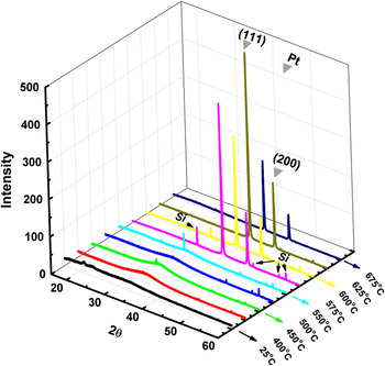
Figure 1 shows the x-ray diffraction (XRD) patterns for platinum films annealed at different temperatures. From as-deposited to samples annealed up to 550 °C, apart from weak diffraction lines originating from the Si substrate, only a broad peak centered around 2θ = 33° was observed. This indicates that the material is amorphous or nanocrystalline. As the annealing temperature was increased to 575 °C, two peaks appeared with high intensity, centered around 39.8° and 46.6°. They correspond to the Pt<111> and the Pt<200> diffraction lines, respectively (CAS number: 7440-06-4). The Pt<111> peak exhibits higher intensity compared with the Pt<200> peak, indicating that preferential growth of the films along the <111> direction. No diffraction peaks from other orientation planes were observed after heat treatment. The appearance of the signal corresponding to the Pt phase upon thermal annealing at 575 °C is in good agreement with the findings of previous work,Reference Saenger, Cabral, Lavoie and Rossnagel15 which showed that <200> metal Pt appeared in the range of 559–626 °C during the heating of platinum oxide films in a pure N2 atmosphere, without the presence of other phases. Also in that study,Reference Saenger, Cabral, Lavoie and Rossnagel15 heating the films in O2 atmosphere delayed the decomposition up to 650 °C with the intermediate formation of small amounts of Pt3O4. The initial morphology and initial bonding state (related to the x-value in PtOx) may also have a strong impact on the decomposition dynamics. In addition, other authors synthesized directly the Pt phase when reducing the oxygen content to around 25% in the oxygen/argon gas flow during the deposition.Reference Saenger, Cabral, Lavoie and Rossnagel15, Reference McBride, Graham, Peters and Weber29
X-ray diffraction pattern of PtOx films annealed at different temperatures.

B. Chemical bonding and electronic structure
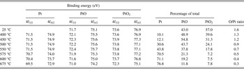
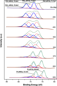
To investigate the evolution of the chemical states of platinum upon thermal decomposition, the surface composition of the films was studied by XPS. The peak shift caused by the sample charging was corrected by taking into account the shift in the position of the C 1s peak registered without charging at 284.6 eV. The XPS peaks were deconvoluted to determine the peak areas and the binding energies belonging to the different bonding states of platinum. These energies are reported in Table I. They are in good agreement with those reported in the literature.Reference Hecq, Hecq, Delrue and Robert30, Reference Chen, Sun, Yu, Hsiung, Gan and Kou31
X-ray photoelectron spectroscopy data for the different thermal treatment of the PtOx films.

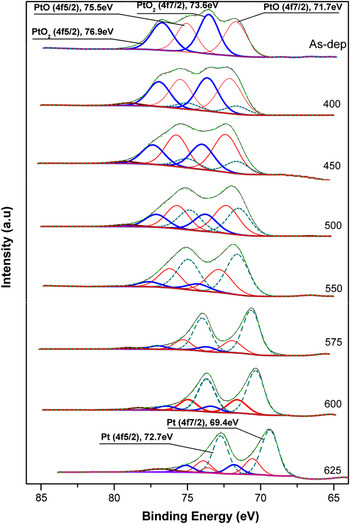
Figure 2 shows the Pt 4f spectra of the as-deposited film and for the films annealed at different temperatures. As can be observed in this figure, several superficial compounds were identified with XPS that could not be identified with XRD. The Pt 4f7/2 and Pt 4f5/2 of the as-deposited film showed that the film is mainly composed of PtO2 and PtO with a composition of 57 and 43 mol%, respectively. For the samples annealed at 400 °C and higher temperature, additional contributions appeared at 71.5 and 74.9 eV, both corresponding to Pt. A low-intensity peak at 79 eV can be attributed to plasmon losses. The contribution of this plasmon peak was less than 2% for all the temperatures. As the annealing temperature was further increased from 400 to 550 °C, the peak intensities of both forms of platinum oxide, PtO2 and PtO, decreased, while the peaks associated to metallic Pt increased. At 575 °C, the intensity of the Pt 4f7/2 (70.7 eV) and Pt 4f5/2 (74.0 eV) peaks ascribed to metallic Pt were higher than those of the oxides present indicating that 70.5 mol% of the total composition are now metallic Pt. These observations demonstrate that the decomposition of the oxide surface already started at 400 °C. The O/Pt atomic ratio is reported in Table I and quantifies this decomposition. Some Pt–O bonds can still be detected after annealing at 625 °C; some of which could be ascribed to the bonding of platinum to the silicon surface oxidized upon the thermal annealing in air. Moreover, all contributions present a shift in the binding energy upon annealing. This could originate from slight changes in the phases’ stoichiometry.
X-ray photoelectron spectroscopy spectra of PtOx thin films after annealing at different temperatures (°C) indicated in the curves.

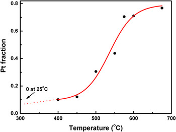
Figure 3 shows the molecular fraction of platinum formed during the decomposition process as measured by XPS. The data follow a sigmoid trend. The decomposition rate was slow below 400 °C and accelerated in the 400–575 °C temperature range. These two stages are in line with the findings of Saenger et al.Reference Saenger, Cabral, Lavoie and Rossnagel15 who observed with XRD a first decomposition stage, with an activation energy of 71 kcal/mol dominating below 400 °C, and a second stage with a lower activation energy of only 25 kcal/mol dominating above this temperature. We additionally observe a third stage above 575 °C for which the process slows down significantly.
Platinum fraction in the remaining material following annealing of the original PtOx films.

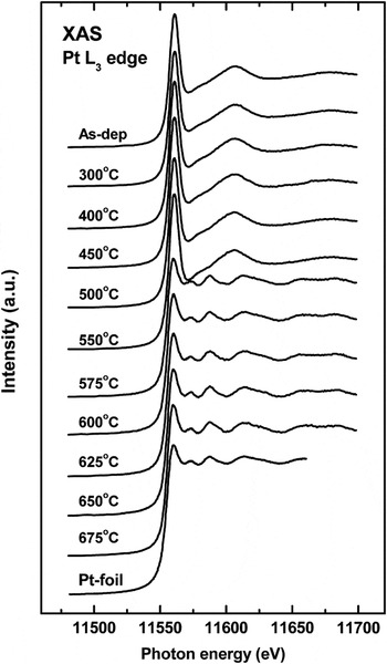
Figure 4 shows the X-ray Absorption Spectra (XAS) at the Pt-L3 absorption edge of PtOx films deposited on a silicon substrate and subsequently annealed at different temperatures. The top spectrum, labeled “As-dep,” corresponds to a PtOx film without any thermal treatment. The bottom spectrum corresponds to a Pt-foil used as a reference. The first peak after the absorption threshold, at 11550 eV, can be assigned to electronic transitions from the 2p core level to the 5d empty states just above the Fermi energy. It is commonly called the “white line.” The intensity of the white line is a measure of the density of empty 5d states. The spectrum of the as-deposited film has the typical lineshape of PtO2.Reference Nagai, Hirabayashi, Dohmae, Takagi, Minami, Shinjoh and Matsumoto32, Reference Hlil, Baudoing-Savois, Moraweck and Renouprez33 The intensity of the white line in PtO2 is higher than in metallic Pt because there is a transfer of four 5d electrons from platinum to oxygen, leaving a higher density of 5d empty states in Pt. The region above the white line is governed by multiple scattering processes and, therefore, it is very sensitive to the chemistry and symmetry of the corresponding compounds.
Pt-L3 XAS spectra recorded after heat treatment of the PtOx samples at temperatures indicated on the curves.

According to Fig. 4, the electronic structure of the films is dominated by the character of oxidized platinum up to a temperature of 550 °C. However, upon annealing between 550 and 575 °C, the XANES of the oxide film approaches that of the Pt foil, which highlights the predominance of metallic Pt in the coatings. Annealing at higher temperatures does not reveal any further significant structural and chemical evolution of the material. The dynamics of the decomposition of PtOx thin films can be followed by XPS and XANES providing a much deeper understanding than that gained by XRD. Aside from the evolution of the relative amounts of PtO2, PtO, and Pt phases, the decomposition process naturally releases oxygen species according to the following reactions:


It leads to the evolution of the mean molecular volume of the coating. Moreover, the annealing activates atomic diffusion. Hence, the film morphology is expected to undergo significant modifications during decomposition.
C. Fractal properties
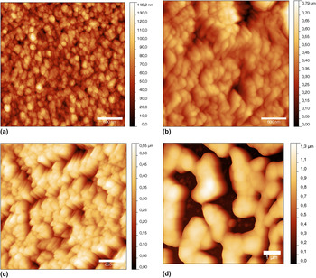
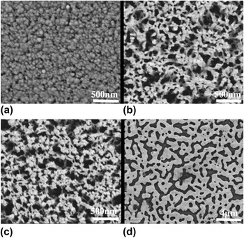
AFM images of PtOx thin films after annealing at different temperatures are shown in Fig. 5. As can be seen, the grains tend to agglomerate at high temperature. This is confirmed by SEM observations (Fig. 6). The topped-dome surface topology as measured by AFM of the samples annealed at 575 and 600 °C [Figs. 5(b) and 5(c)] suggests columnar texture, which is not observed by SEM. Therefore, this topology could be due to a combination of the tip shape and surface of the film as recorded by the AFM tip when the actual microstructure of the film is sharper than the AFM tip, resulting in an image distorted by tip artifactsReference Westra, Mitchell and Thomson34, Reference Grütter, Zimmermann-Edling and Brodbeck35 with strong tip convolution effects. This effect is more evident at the imaged slopes of the granular structures.
Atomic force microscopy (AFM) images of PtOx thin films annealed at different temperatures: (a) 25 °C, (b) 575 °C, (c) 600 °C, and (d) 675 °C.

Scanning electron microscopy (SEM) images of PtOx thin films annealed at different temperatures: (a) 25 °C, (b) 575 °C, (c) 600 °C, and (d) 675 °C.

The SEM images [Figs. 6(b) and 6(c)] reveal a porous morphology due to the decomposition of the platinum oxide phases (PtO2 and PtO) and reduction of the average molecular volume of the films as stated above.
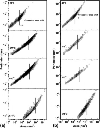
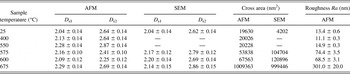
Figure 7 displays the perimeter as a function of the area of the surface features for all the samples measured by SEM and AFM. The calculated values of the surface fractal dimension D s of PtOx thin films annealed at different temperatures are shown in Table II. In general, we can distinguish two regions, each with a characteristic fractal dimension D s1 and D s2. This change of regime (slope) takes place at a given crossover area value.
Perimeter versus area logarithmic plots for samples obtained after annealing at different temperatures and analyzed by (a) SEM and (b) AFM. The change in the slope is the crossover area indicating where the slope changes.

Surface fractal dimension D s and cross area calculated from the analysis of atomic force microscopy (AFM) and scanning electron microscopy (SEM) images.

The average roughness Ra values shown in Table II were calculated from 3 μm × 3 μm AFM pictures. For temperature below 575 °C, the samples roughness tends to be less than 14.9 ± 0.3 nm, and for temperature above 575 °C, sample roughness (Ra) increases with annealing temperature until reaching 301 ± 20 nm.
To correctly analyze fractal dimension, we have to consider the above-mentioned tip-induced artifacts on the AFM images of samples annealed at 575 and 600 °C. Below 575 °C, the AFM data are reliable as judged by the agreement between the AFM and SEM D s values for the room temperature sample. From 575 °C, it is better to use the data obtained from SEM images as the AFM results can be distorted due to strong tip convolution effects (thus, AFM D s values tend to be smaller than the SEM-derived values for this temperature range, particularly the D s2 values). Once these considerations are taken into account, the fractal dimensions obtained are very similar and only slightly evolve with the annealing temperature. The values of D s1 are in the 2.05–2.3 range, i.e., close to 2 indicating that the features exhibiting small area are rather Euclidean without jagged perimeters. In contrast, the values of D s2, which seem to initially increase with the annealing temperature, approach 2.9, which is the percolation limit,Reference Stauffer and Aharony36 indicating highly disordered, fractal surfaces. It is also observed that the crossover area shifts to higher D s values and the features of smaller area disappear as the annealing temperature is increased. This can be put in relation to the agglomeration of grains that form the film. This decrease in surface area of exchange could be at the origin of the slowing down of the decomposition process above 575 °C. Moreover, as the decomposition starts at the film/air interface, it is possible that oxidized volumes are progressively surrounded by a metallic environment (equivalent of a capping layer) as shown for palladium oxides.Reference Horwat, Zakharov, Endrino, Soldera, Anders, Migot, Karoum, Vernoux and Pierson18 Such an environment would limit the oxygen uptake and lower the decomposition rate.
IV. CONCLUSIONS
The thermal decomposition of sputter-deposited platinum oxide PtOx was studied with different techniques. The as-deposited films were XRD-amorphous or nanocrystalline according to the XRD analysis. Pt 4f XPS investigations indicated that the films were actually composed of PtO2 and PtO phases. The decomposition started at the surface when the temperature reached 400 °C in ambient air, and it followed a sigmoid trend with further increasing temperature. The evolution of XANES at the Pt-L3 edge was consistent with the XPS results, but showed that the electronic structure was dominated by oxidized platinum until the temperature reached 575 °C. The AP method applied to SEM and AFM images showed that the film morphology was a combination of large area aggregates and small area granular structures with fractal and Euclidian characteristics, respectively. This analysis also measured the agglomeration of grains in the film with increasing annealing temperature as observed by SEM.
ACKNOWLEDGMENTS
This work was partially supported by the Spanish Ministerio de Educación y Ciencia through project Consolider Ingenio CSD2008-00023 and ENE2010-21198-C04-04. J. Andersson was supported by the Wenner-Gren Foundations and the SSF program MS2E. We gratefully acknowledge beamtime at the SGM beamline (CLS, Saskatoon) and the KMC-2 beamline (BESSY, Berlin). The research performed at the Canadian Light Source is supported by NSERC, NRC, CIHR, and the University of Saskatchewan. The synchrotron work at BESSY-II was supported by the EC “Research Infrastructure Action” under the FP6 “Structuring the European Research Area Programme” through the “Integrated Infrastructure Initiative Integrating Activity on Synchrotron and Free Electron Laser Science” (Contract no. R II 3-CT-2004-506008). Research at Berkeley Lab was supported by the U.S. Department of Energy under Contract no. DE-AC02-05CH11231.