

Introduction
The precise determination of exercise intensity has great importance to some related areas, such as physical conditioning, training and sports performance, making the control of the effort application in a systematized way possible. As a consequence, the objectives' intendeds are reached.
In that sense, several protocols of effort intensity evaluation have been developed in recent decades. Among them, we can mention the ventilatory threshold suggested by Wasserman and McIlroyReference Wasserman and McIlroy1; the use of blood lactate concentration (LAC) to identify the anaerobic threshold (AT), proposed by Kinderman et al. Reference Kindermann, Simon and Keul2; the AT obtained by fixed LAC at 4.0 mmol l− 1 (onset of blood lactate accumulation, OBLA), initially investigated by Mader et al. Reference Mader, Liesen, Heck, Philippi, Rost and Schürich3; the protocol of individual AT presented by Tegtbur et al. Reference Tegtbur, Busse and Braumann4; the double efforts for the critical power identification from null variation of the heart rate and lactataemia among those effortsReference Chassain5; and the non-invasive critical power model proposed by Monod and ScherrerReference Monod and Scherer6.
Currently, the highest workload that can be maintained over time with blood lactate stabilization has been considered the gold standard for aerobic/anaerobic transition determination and is denominated maximal lactate steady state (MLSS)Reference Billat, Siverent, Py, Korallsztein and Mercier7, Reference Beneke8. Daily continuous loads of 25–30 min with blood LACs obtained at each 5 min, for example, are the best way to determinate the MLSSReference Mader and Heck9, Reference Harnish, Swensen and Pate10.
Down the years, one of the resources used most frequently in exercise physiology and correlated areas is animal experimentation, in particular because it enables invasive and controlled manipulations, promoting cellular and molecular analysis including both healthy animals and, with pathology, sedentary or physically trained animals. Besides, animals are frequently used in studies involving different physiological conditions. Among commonly investigated pathologies and physiological conditions including acute or chronic physical stress (training), hypertension, diabetes mellitus, obesity, pregnancy and ageing can be cited among others.
Physical ergometers commonly used for animals include the treadmill and swimming. Mammals that are preferentially used in research are rodents of different species (mice and rats). These animals are chosen due to easy manipulation and good response to exercise, comparable in the results obtained with human beings in compatible effort intensities.
Over the past decade our laboratory has been developing several models of physiological evaluation in swimming and running ratsReference Gobatto, Kokubun, Sibuya and Mello11–Reference Manchado, Gobatto, Contarteze, Papoti and Mello16, applying protocols that were initially proposed for human beings.
The MLSS has been considered a gold standard for aerobic evaluation in human beings and, for this reason, it has been widely studied in our laboratory, using rats as the experimental model. In this way, several protocols were established, including invasive and non-invasive, exhaustive or non-exhaustive tests, validated by MLSS. These procedures may support the understanding and application of lactataemic responses in swimmingReference Gobatto, Mello, Sibuya, Azevedo, Santos and Kokubun12, Reference Gobatto, Manchado, Voltarelli, Contarteze and Mello17–Reference Araujo, Papoti, Manchado, Mello and Gobatto19 or runningReference Manchado, Gobatto, Contarteze, Papoti and Mello15, Reference Contarteze, Manchado, Gobatto and Mello20 rats.
Billat et al. Reference Billat, Mouisel, Roblot and Melki21 demonstrated the possibility of AT determination in treadmill-running mice, using incremental velocity protocol and the AT intensity corresponding to the blood lactate inflection point. Recently, Ferreira et al. Reference Ferreira, Rolim, Bartholomeu, Gobatto, Kokubun and Brum22 have determined the MLSS in running mice.
In this way the present study aims, based on the MLSS protocol, to determine the swimming intensity and maximal blood LAC in sedentary mice, recently adapted to water. Our hypothesis is that, as happens in human beings and rats, there is an intensity of maximal swimming in which there is maximal stabilization of blood LACs.
Materials and methods
Animals
Twenty-five untrained mice, 60 days old, weighing 46–53 g at the beginning and 58–66 g at the end of the experiment, were used for this study. During the experimental period, the animals received ad libitum water and commercial chow (Labina-Purina). The mice were housed in collective cages (ten animals per cage), in a room with a light cycle from 06.00 to 18.00 hours, at 25°C. All experiments involving the animals were conducted in conformance with the policy statement of the American College of Sports Medicine on research with experimental animals.
Experimental protocol
Adaptation to water
All mice (n = 25) were adapted to the water before the start of the experiment in a standardized procedure. The adaptation occurred over 15 uninterrupted days, when the mice were placed in a cylindrical tank of 60 cm diameter and 60 cm depth. It was progressively filled with water at 31 ± 1°C. This deep tank was chosen so that the mice did not touch bottom. The mice were initially inserted in shallow water for 5 days, starting with 5 min, up to 25 min (increments of 5 min per day). Progressively, both the water level and the swimming duration were increased until the 10th day. After that, the mice started the swimming attained with dorsal bags, but without loads. Over the last 2 days, the animals were submitted to swimming exercise for 10 min, supporting a load equivalent to 2% of their body weight (bw). This adaptation to the water procedure has been tested in rodents in previous studies carried out in our laboratory, and it was verified that it did not promote the metabolic alterations expected with physical trainingReference Gobatto, Mello, Sibuya, Azevedo, Santos and Kokubun12, Reference Manchado, Gobatto, Contarteze, Papoti and Mello16.
MLSS determination
After the water adaptation period, 25 mice were submitted to 25 min of swimming performed continuously with loads equivalent to 3.0, 4.0, 5.0, 6.0 and 7.0% of the bw tied to the back. This procedure occurred on alternate days and the five loads used were randomly distributed among the mice. The MLSS was measured as the highest intensity at which the increase on the blood LAC was equal to or below 1 mmol l− 1 between the 10th and 25th minute of exerciseReference Billat, Siverent, Py, Korallsztein and Mercier7, Reference Gobatto, Mello, Sibuya, Azevedo, Santos and Kokubun12, Reference Manchado, Gobatto, Contarteze, Papoti and Mello15, Reference Araujo, Papoti, Manchado, Mello and Gobatto19.
Blood samples and analysis
Blood samples (25 μl) were collected from a cut at the tail tip every 5 min during the exercise tests and deposited in Eppendorf tubes (1.5 ml capacity) containing 50 μl sodium fluoride (1%). These samples were collected over 30 s with animals outside the water. After the pause, the mice went back to exercising. To avoid blood lactate dilution with residual water on the tail of the animal, the mice were quickly dried with a towel, immediately before blood collection. The LACs were determined by a lactate analyser (YSI model 1500 SPORT).
Statistical analyses
The results are presented as mean ± SEM. The statistical procedure consisted of two-way ANOVA. When necessary, the Newman–Keuls post hoc comparison test was used to identify differences among the blood LACs at the various times and intensity of exercise. In all cases, the statistical significance was prefixed at P < 0.05.
Results
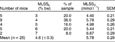
Table 1 shows the mean values of blood LACs for every load. These results by ANOVA analysis show the effect of load over blood LACs obtained during continuous exercise (without rest values). Therefore, the LACs were higher at 7% bw overload than at all inferior intensities (P < 0.05). Moreover, the LACs for 3% bw were lower than other for overloads (P < 0.05). There were no significant differences between 4, 5 and 6% bw. ANOVA did not show any significant effect over time and time–overload interaction. Since the MLSSL is an individual parameter, defined as the highest swimming intensity at which blood lactate elevation did not exceed 1 mmol l− 1 between the 10th and 25th minute of exercise, Table 2 shows the number of animals (and percentage of total sample) and their respective overloads (% bw) when success was obtained in the MLSSL determinations. It will be noted that most of the animals (36%) attained MLSSL at 4% bw. This table also presents the mean of blood lactate stabilization for each intensity. The mean of MLSSL was 4.6 ( ± 0.3)% in a MLSSC of 5.78 ( ± 0.29) mmol l− 1.
Blood lactate concentration (mmol l−1) in the maximal lactate steady-state protocol in swimming mice

Results are expressed as mean ± SEM, with the number of mice in parenthesis.
a Difference (P < 0.05) in relation to 4, 5, 6 and 7% bw, independent of the time effect.
b Difference (P < 0.05) in relation to 3, 4, 5 and 6% bw, independent of the time effect.
Number of animals, MLSS load (MLSSL – % body weight), respective animal sample percentage, mean and SEM of blood lactate stabilization (MLSSC – mmol l−1) for each different exercise intensity

mean ± SEM.
Discussion
The precise determination of physical exercise intensity is important for better a prescription of training, not only in human beings but also in animals, since experimental models using other mammalian species could simulate situations widely observed in humans, aiding in the solution of these problems in acute or chronic exercise.
Measurements of blood lactate during exercise provide necessary information concerning the energy necessary for effort execution, especially in human beings, but recently have also proved to be accurate in rats. In sport training and therapeutic exercise procedures, this parameter is an important tool for exercise intensity prescription and evaluation of physiological adaptation and its development.
MLSS can be used to detect the highest exercise intensity supported without continuous increase in blood lactateReference Heck, Mader, Hess, Mücke, Müller and Hollmann23, Reference Beneke24, representing the highest exercise intensity where there is maximum equilibrium between production and removal of lactateReference Billat, Siverent, Py, Korallsztein and Mercier7.
In spite of MLSS being considered the gold standard for aerobic determination, both in humans and rats, the importance of the present study is that the MLSS of swimming mice with additional weight for the final 15 to 25 min of exercise was determined from our protocol proposed, since there are not yet any conclusive studies, and a very low number of investigations related to AT with these animals, especially in swimming exercise.
Based on the results of the present study, it will be noted that the mean swimming intensity value was 4.6% of bw. Nevertheless, there were marked individual differences among the mice in relation to the load observed at MLSS. Similar results were also obtained in studies with rats submitted to evaluation during swimming, where Voltarelli et al. Reference Voltarelli, Gobatto and Mello14 and Manchado et al. Reference Manchado, Gobatto, Contarteze, Papoti and Mello16 found AT or MLSS at 5% of the bw, while Gobatto et al. Reference Gobatto, Mello, Sibuya, Azevedo, Santos and Kokubun12 verified a stabilization load of 6% in sedentary Wistar rats.
These small differences could be related to the specificity of the swimming pool used. Therefore, Manchado et al. Reference Manchado, Gobatto, Contarteze, Papoti and Mello16 used a deep tank (1.0 m), while Gobatto et al. Reference Gobatto, Mello, Sibuya, Azevedo, Santos and Kokubun12 applied their procedures to a shallower swimming pool (0.6 m), which might have favoured the exercise activity of the rats exploring the bottom of the tank, something that is impossible in a deep pool.
In the treadmill ergometer for rats, there were also different intensities at the aerobic/anaerobic transition, when determined by different protocols. In procedures of MLSS determination, Manchado et al. Reference Manchado, Gobatto, Contarteze, Papoti and Mello15 found a velocity of 20 m min− 1, different from that observed by Pillis et al. Reference Pilis, Zarzeczny, Langfort, Kaciuba-Uscilko, Nazar and Wojtyna25 (25 m min− 1) using a protocol with incremental intensities.
In the present study, even though MLSS has been determined in all loads used (% bw), a large number of mice were not able to support an exercise level above 5% bw up to 25 min, corroborating the findings observed by ManchadoReference Manchado, Gobatto, Voltarelli and Mello18 in the same evaluation protocol with rats.
In relation to blood LAC at MLSS, important individual differences were also found in the present study. Most of the samples (34.62%) showed a mean of 5.78 ( ± 0.29) mmol l− 1 at MLSS. However, the individual variation in blood lactate stabilization ranged from 4.40 to 6.67 mmol l− 1.
Different maximal lactate stabilization concentrations (MLSSC) can also be observed when different ergometers are used in studies involving humans and animals. Manchado et al. Reference Manchado, Gobatto, Contarteze, Papoti and Mello16 found an MLSSC in swimming rats of 5.20 ( ± 0.22) mmol l− 1, whereas in the same study evaluating running rats on the treadmill, the MLSSC obtained was 3.87 ( ± 0.33) mmol l− 1.
In studies with running mice, Ferreira et al. Reference Ferreira, Rolim, Bartholomeu, Gobatto, Kokubun and Brum22 determined the concentration and the intensity of the exercise at MLSS. Later, a training protocol was applied to the mice based on the intensity at MLSS. That velocity (MLSSV) was found in 15.1 ( ± 0.7) m min− 1, corresponding to 60 ( ± 2)% of the maximum speed reached during a test with incremental exercise protocol. The training in that intensity was accomplished during 8 weeks, with a duration of 1 hour per day, 5 days a week. At the end of this period, an enhanced performance was observed (28%). The MLSSV was significantly larger in the trained mice in relation to the sedentary group (19.0 ± 0.5 vs. 14.2 ± 0.5 m min− 1; P = 0.05); however, the MLSSC presented no changes (3.0 mmol l− 1).
In the light of our results, a difference between the lactate stabilization concentrations at MLSS between swimming and running mice can be observed, evidencing the ergometer dependence of this protocol once again, similar to the one observed by Beneke et al. Reference Beneke8 and Manchado et al. Reference Manchado, Gobatto, Contarteze, Papoti and Mello16 in humans and rats, respectively.
In agreement with the present results, we can conclude that the protocol of MLSS could be used for intensity determination of metabolic transition of swimming mice, and that these animals presented MLSSL at 4.6% bw and MLSSC at 5.78 mmol l− 1. However, there are great inter-individual variations of these parameters in mice.
Future studies will be executed in an attempt to supply answers to physical and physiological responses from swimming mice training in a continuous or intermittent exercise based to MLSS intensity. We have no doubts that this is the best way to investigate optimal training intensity prescription, in order to understand physiological benefits of chronic exercise, at least in that animal species.
Acknowledgements
This study was supported by Fundação de Amparo à Pesquisa do Estado de São Paulo (FAPESP-Proc.04/07 070-5, 06/58 411-2, 06/61 189-0), Conselho Nacional de Desenvolvimento Científico e Tecnológico (CNPq – Proc. 301601/2006-2) and CAPES.