Physiology of the healthy ageing kidney
Biological systems work due to the co-ordinated actions of their multiple constitutive systems or microsystems. This co-ordination of systems or complexity makes the organism flexible and adaptable to environmental changes. The process of senescence weakens these microsystems and their co-ordination, resulting in frailty. Therefore, even though healthy elderly people can handle water and electrolyte balance under basal conditions, they easily lose the ability to adapt to challenging situations such as hot weather or sepsis. This explains why older people are equally at risk from opposite electrolyte disorders, such as hyponatraemia or hypernatraemia, depending on the clinical circumstances.
In renal physiology, other common findings present in the majority of healthy aged individuals are the ‘physiological nephrogeriatric giants’. This is a concept, forged by geriatric nephrology, which summarizes the structural and physiological changes that accompany the normal renal ageing process.Reference Musso1 It should always be kept in mind, as it plays an important role in the development of disease and treatment in this late stage of the life cycle.
The physiological nephrogeriatric giants are:
1. Renal vascular dysautonomy: this term refers to a reduction of the autonomic renal vascular reflex that protects the kidney from hypotensive and hypertensive states.
2. Senile hypofiltration: there is a progressive reduction of the glomerular filtration accompanying the ageing process, starting around the age of 30 years and continuing to decline at a rate of approximately 1 ml per year. This diminution in the glomerular filtration rate (GFR) is physiological and it must never be considered as pathological renal insufficiency.
3. Tubular dysfunction: the reduction of the tubular maximal capacity to reabsorb or excrete some substances such as glucose or phosphate or the tendency to reduce both secretion of K+ and reabsorption of Na+.
4. Tubular frailty: in the elderly person, renal tubular cells are more vulnerable to hypoxic or nephrotoxic injury and take longer to recover from acute tubular necrosis.
5. Medullary hypotonicity: in older people, the renal medulla has a lower tonicity compared with younger healthy individuals. This phenomenon produces a reduction in the effect of antidiuretic hormone (ADH) and, as a consequence, a reduction in water reabsorption. The above, combined with a decreased sensation of thirst, may lead to severe dehydration.
Three of these giants (senile hypofiltration, tubular dysfunction, and medullary hypotonicity) modulate the maintenance of water and electrolyte balance in the healthy aged.
Handling of sodium and water
Proximal tubule
This segment is heterogeneous with respect to function and structure, exhibiting different characteristics along its length. Over 60–80% of the filtered sodium and water, bicarbonate and other electrolytes, glucose, uric acid, phosphate, amino acids and small molecular weight proteins are reabsorbed in this segment.
Sodium (Na+). The first movement in the transport of Na+ from the tubular lumen (luminal or apical) to peritubular vessels is a passive movement of Na+ from the lumen to the interior (cytosol) of the tubular cell. Once in the cell, part of it is transported to the paracellular and basal space. Because the interior of the cell is electronegative and Na+ concentration is lower in the cytosol than outside the cell, additional force is needed to transport Na+ from the cell to the paracellular and peritubular vessels. The energy for this transport is provided by a sodium ATPase-dependent pump (Na+–K+-ATPase), located in the basolateral membrane. This pump, which extrudes Na+ from the cell, generates an electrochemical gradient that promotes the passive entry of Na+ from the tubular fluid to the interior of the cell. This movement of Na+ shares some cotransporters that reabsorb glucose, peptides and amino acids.
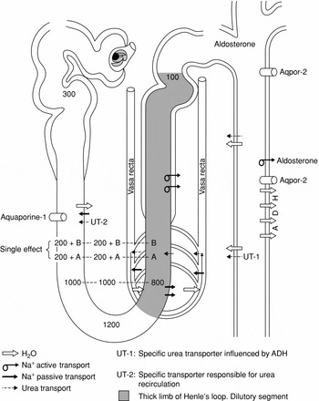
Water. Approximately 60–80% of water filtered by the glomerulus is reabsorbed passively in the proximal tubule, following the gradient of sodium towards the paracellular space. As water accumulates in the basolateral space, the increase of hydrostatic pressure releases water to the peritubular space. The back flow of water and Na+ from the basolateral space to the tubular lumen depends on the degree of extracellular volume expansion. Water and electrolytes enter the peritubular vessels because of the oncotic pressure of the intravascular proteins. Transport in the proximal tubule is iso-osmotic, which means that there is no concentration or dilution of the tubular fluid in this segment, i.e. the tubule reabsorbs 300 mOsm of solutes with 1 l of water (Figure 1).
Representation of the nephron. The shaded area corresponds to the ascending limb of the loop of Henle, in which competence to reabsorb sodium diminishes with age.

Proximal tubular function in the healthy aged. No differences have been detected in proximal tubule function when young individuals (less than 60 years) are compared with elderly healthy aged individuals (older than 60 years) in clearance studies.Reference DeSanto, Anastasio, Coppola, Barba, Jadanza and Capasso2, Reference Durakovic3, Reference Macias Nunez, Garcia Iglesias, Bondia Roman, Rodriguez Commes, Corbacho Becerra and Tabernero Romo4 Moreover, the handling of lithium, a well-known marker of general function of the proximal tubule,Reference DeSanto, Anastasio, Coppola, Barba, Jadanza and Capasso2 does not differ between young and old. Nevertheless, although the renal reabsorption threshold for most compounds handled by the proximal tubule is no different, the maximal tubular reabsorption capacity (Tm) is diminished in aged people.
Thin descending limb of Henle's loop
The characteristic of membrane permeability differs from the previous segments of the proximal tubule (Figure 1). The descending limb is highly permeable to water due to the presence of the channel water aquaporine, but quite impermeable to sodium, potassium and chloride. Although there is some passage of these electrolytes in both directions (from the lumen to the peritubular space and vice versa) it is of limited quantitative importance. This segment is also permeable to urea (from the interstitium to the tubular lumen) by the action of the urea transporter UT2. As tubular fluid flows downward in the descending limb, the osmolarity of the fluid equilibrates with the osmolarity of the interstitium, which increases from 300 to 1200 mOsm/l. Due to this movement of water across the membrane of the cells of the descending limb, the tubular fluid becomes more and more concentrated, reaching 1200 mOsm/l at the tip of the descending limb (i.e. in the bend of the loop).
Thin ascending limb of Henle's loop
The thin ascending limb is impermeable to water and highly permeable to Na+ and urea due to the presence of UT2 channels. In the thin ascending limb, Na+ moves passively from the lumen to the interstitium by means of a concentration gradient, resulting in net Na+ reabsorption.
Thick ascending limb of Henle's loop
This segment is impermeable to water but permeable to Na+, and sustains transport of Na+ from the lumen to the interstitium, thus resulting in diluted urine emerging into the early portion of the distal nephron. In the presence of ADH, it is also responsible for the reabsorption of water from the urine as it traverses the collecting duct and determines the excretion of concentrated urine (low volume, high osmolality). As tubular fluid ascends in the thick ascending limb, electrolytes pass to the interstitium, whilst water remains in the lumen. In essence, the ascending limb and the early part of the distal convoluted tubule are the sites in the nephron where solutes are reabsorbed without water in a process leading to ‘free water formation’ and thus have been named the diluting segment.Reference Macias Nunez, Garcia Iglesias, Bondia Roman, Rodriguez Commes, Corbacho Becerra and Tabernero Romo4
Changes in the healthy aged and clinical implications. The thick ascending limb of the loop of Henle loses some of its capacity to reabsorb Na+, leading to a higher delivery of Na+ to the most distal segments of the nephron. This has been shown to be statistically greater in the old than in the healthy young. This reduction in Na+ reabsorption is found in the healthy aged in situations of normal salt diet or salt restriction, resulting from the normal tubular ageing process.Reference Musso, Lopez Novoa and Macías Núñez5, Reference Musso, Macías and Musso6
Loss of sodium reabsorptive capacity in the thick portion of Henle's loop leads to Na+ and water spillage, making the aged very sensitive to easily becoming dehydrated and desalinated. It is common to find elderly patients in daily practice who exhibit copious natriuresis leading to Na+ depletion and/or hyponatraemia.
Particularly vulnerable to these Na+ abnormalities are aged subjects on a low salt diet plus diuretics. Failure of awareness of this effect of ageing on renal function and failure of timely replenishment of Na+ and water deficits may lead to acute renal failure.Reference Refoyo and Macías-Núñez7, Reference Macias-Nunez, Lopez-Novoa and Martinez-Maldonado8 It is inappropriate to routinely restrict salt intake in the aged unless they suffer from illnesses managed in part by salt restriction, such as liver, heart and renal diseases, in which oedematous syndromes (other than malnutrition) and/or hypertension are manifest. Even under those circumstances it is advisable to periodically monitor plasma electrolytes, particularly if diuretics are a mainstay of the therapeutic regime. Both hypo- and hypernatraemia are dangerous situations in elderly people and should be avoided at all costs.
Elderly people, with or without hypertension, take longer periods of time to eliminate similar amounts of salt overload due to a lower GFR, but if salt is restricted they become easily salt depleted because of the already mentioned limitation of the thick ascending limb of the hoop of Henle to retain salt. Having in mind these differences in sodium handling by the normal aged kidney, it is important to be cautious in describing hypertension in the aged as ‘salt dependent’ or ‘salt sensitive.’
Because of all the above, it is advisable to start fluid replacement with saline isotonic solutions instead of the widespread tendency to start with solutions lacking electrolytes (i.e. glucose or laevulose solutions or Na+ hypo-osmolar solutions) before blood electrolytes determination. Once these results are available, the replacement fluid should be adapted to the concentration of electrolytes in the blood.
Distal convoluted tubule
The early part of the distal convoluted tubule exhibits similar properties to those of the thick ascending limb of Henle's loop and therefore tubular fluid continues to be diluted. The late part is less permeable to Na+. In this segment Na+ is actively reabsorbed against an electrochemical and concentration gradient by action of the Na+–K+-ATPase located at the basolateral membrane of the tubular cell and promoted by aldosterone. In any circumstances, the total capacity for Na+ reabsorption in this segment is limited.
Collecting duct
Sodium. The amount of Na+ reabsorbed in the collecting duct is approximately 3% of the amount filtered. Yet, despite the apparently low Na+ capacity of the collecting duct, its normal function is of paramount importance for overall Na+ balance and for extracellular fluid volume to remain within normal limits.Reference Macias-Nunez, Lopez-Novoa and Martinez-Maldonado8, Reference Macias Nunez, Garcia Iglesias, Tabernero Romo, Rodriquez Commes, Corbacho Becerra and Sanchez Tomero9 Under the influence of aldosterone the collecting duct is also responsible for the final control of urinary Na+ excretion. The synthesis of aldosterone by the adrenal cortex is controlled by angiotensin II, plasma concentrations of Na+ and K+ and ACTH. The level of plasma Na+ is the least potent stimulus on aldosterone synthesis. In contrast, aldosterone-secreting cells in the adrenal cortex are very sensitive to the concentration of K+ in the extracellular fluid. The increase in K+ intake increases extracellular K+, which directly stimulates the production of aldosterone in the adrenal cortex. The increase in plasma aldosterone concentration stimulates Na+ reabsorption and K+ secretion in the cortical collecting duct. This affects final control of Na+ reabsorption and excretion and helps eliminate K+ excesses and prevents hyperkalaemia.
The collecting duct is unable to reabsorb greater quantities of Na+ delivered to it from previous tubular segments, such as the ascending limb of the loop of Henle. Thus decreases in Na+ reabsorption in prior segments will lead to natriuresis and could enhance K+ secretion into the tubular urine and cause hypokalaemia and volume depletion.
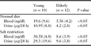
Aldosterone in the healthy aged and clinical implications. Aged people on unrestricted diets of salt and water have lower aldosterone plasma levels and respond more slowly to salt restriction than healthy young people (Table 1).Reference Refoyo and Macías-Núñez7, Reference Macias Nunez, Garcia Iglesias, Tabernero Romo, Rodriquez Commes, Corbacho Becerra and Sanchez Tomero9, Reference Weidmann, De Myttenaere-Bursztein, Maxwell and de Lima10, Reference Ceruso, Squadrito, Quartarone and Parisi11
Blood and urine aldosterone levels (mean and SD) in healthy young and elderly people under normal dietary conditions and after salt restrictionReference Macias Nuñez, Bondía Roman and Rodriguez Commes45

The most common and life-threatening clinical consequence of this is the development of hyperkalaemia in circumstances where an older patient is treated with aldosterone blocking agents such as spironolactone, or angiotensin converting enzyme inhibitors (ACE inhibitors) and angiotensin receptor blocking (AII RB) agents, which diminish aldosterone secretion by blocking AII production or its actions, respectively. Elderly patients with cardiac failure or other co-morbidities treated with anti-aldosterone agents should be periodically investigated for blood potassium, and at least every 3 months if simultaneously treated with drugs interfering with the renin–angiotensin system such as ACE inhibitors or angiotensin II receptor blockers.Reference Macias Nunez, Garcia Iglesias, Tabernero Romo, Rodriquez Commes, Corbacho Becerra and Sanchez Tomero9, Reference Weidmann, De Myttenaere-Bursztein, Maxwell and de Lima10
Water. The final adjustment of urinary osmolarity mediated by ADH takes place in the final part of the distal convoluted tubule (connecting segment) and the collecting duct. Water is passively reabsorbed under the influence of ADH and the transepithelial osmotic gradient. ADH is secreted by the pituitary hypophysis in response to changes in plasma osmolarity. High plasma osmolarity strongly stimulates ADH secretion, whereas low plasma osmolarity attenuates it. The urinary elimination of water is controlled by the circulating levels of ADH. The distal tubule is practically impermeable to water. The permeability of the collecting tubule is low in the absence of ADH and high when ADH is present. ADH makes the collecting tubule permeable to water by insertion of water-permeable channels (aquaporin 2) (Figure 1). The reabsorption of sodium chloride in the absence of ADH is responsible for a hypo-osmotic tubular fluid with respect to plasma and, consequently, the elimination of a great quantity of water in the urine and a urinary osmolarity lower than plasma. In the presence of ADH, this segment of the tubule becomes permeable, allowing the passage of water from the tubular fluid to the inner medulla and renal papilla. As the osmolarity in the renal interstitium and papilla is much higher than that of the tubular fluid, a considerable amount of water passes from the tubular fluid to the interstitium. As a result, the elimination of water diminishes in the urine, but the amount of solutes in the tubular fluid remains unaltered. Consequently the urine becomes concentrated. Because of this the osmolarity of the urine is higher than that observed in the plasma.
Handling of urea
Urea is reabsorbed in the proximal tubule straight and convoluted segments, before the thin descending limb of the loop of Henle. The thick ascending limb of the loop, the distal tubule and the cortical segment of the collecting ducts are impermeable to urea. By contrast, the medullar segment of the collecting duct is permeable. For this reason the intratubular concentration of urea increases along the distal tubule and the medullar collecting duct. When tubular fluid reaches the medullar segment of the collecting duct, the concentration of urea in the tubular fluid is very high. This gradient of concentration is the reason why urea passively diffuses from the tubular fluid to the medullar interstitium. This passive movement is possible by the insertion of two specific transporters of urea (UT1 and UT2) (Figure 1). Both have been identified and cloned.Reference Elias and Schwartz12 UT1, modulated by aldosterone, is responsible for urea transport in the medullar segment of the collecting tubules. The expression of ADH induces the insertion of more UT1. UT2 is not influenced by ADH but it is responsible for urea recirculation because it regulates the entry of urea accumulated in the interstitium to the descending and ascending limbs of the loop of Henle. The high concentration of urea in the interstitium, or trapping of urea, is possible due to medullar recirculation and the countercurrent exchange mechanism.
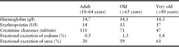
Renal handling of urea by the healthy ageing kidney and clinical implications. The healthy aged exhibit a higher fractional excretion of urea than young controls (Table 2). This phenomenon is not clearly understood, but may result from diminished function of urea transporters (less urea tubular reabsorption). Moreover, the elderly eat less protein, which contributes to lower blood and kidney urea concentration.Reference Musso, Macias Nunez and Oreopoulos13
Key parameters of renal function at different agesReference Macias Nuñez, Lopez Novoa, Macias Nuñez, Cameron and Oreopoulos43

It is possible that the impairment in urine concentration and dilution observed in the healthy aged may be partly the result of the increased urea excretion, rather than its accumulation in the renal interstitium. Low interstitial urea would reduce the steep interstitial osmolar gradient necessary for urinary concentration cannot be maintained.
Handling of potassium
Almost all of the filtered K+ is reabsorbed in the proximal tubule and the loop of Henle, so that less than 10% of the filtered load is delivered to the early distal tubule (CK+/Cin < 0.1). Proximal K+ transport appears to passively follow that of Na+ and water, whereas reabsorption in the thick ascending limb of the loop of Henle is mediated by the Na+–K+–2Cl− carrier in the luminal membrane.
In comparison to these reabsorptive processes, K+ is secreted by the connecting segment, the principal cells in the cortical and outer medullary collecting duct, and the papillary (or inner medullary) collecting duct. Secretion in these segments can be modified according to physiological needs and is generally responsible for most urinary K+ excretion. Distal K+ secretion is regulated by aldosterone, which is in turn regulated by extracellular volume through angiotensin II levels and by plasma K+ concentration.
Distal secretion can be partially counteracted by K+ reabsorption by the intercalated cells in the cortical and outer medullary collecting tubules. This process may be mediated by an active H+–K+-ATPase pump in the luminal membrane, which results in both H+ secretion and K+ reabsorption. The activity of this pump is increased with K+ depletion and is reduced with K+ loading. The former adaptation is probably responsible for the observation that net K+ reabsorption, not secretion, appropriately occurs in the distal nephron with K+ depletion. Selective inhibition of the H+–K+-ATPase pump in the setting of K+ depletion abolishes distal K+ reabsorption.
The K+ reabsorbed in the thick ascending limb initially enters the medullary interstitium. Some of this K+ is then secreted into either the S3 segment of the late proximal tubule or the thin descending limb of the loop of Henle; this extra K+ can be reabsorbed again when it enters the outer medulla. Thus K+ is recycled within the medulla, resulting in the attainment of a relatively high concentration in the interstitium. The physiological function of this phenomenon is uncertain. It is possible, for example, that K+ accumulation in the interstitium promotes K+ excretion by minimizing the degree of passive back leak out of the collecting tubular lumen (where the highest urine K+ concentrations are attained). The high interstitial K+ concentration may also contribute to K+ excretion by a second mechanism, by diminishing the gradient for passive K+ reabsorption via the Na+–K+–2Cl− carrier in the loop of Henle. This process is similar to NH4+ recycling between the loop of Henle and the medullary collecting tubule that promotes net NH4+ excretion.
Potassium management of the healthy aged and clinical implications. Measurements of total exchangeable body potassium by various isotopic dilution methods agree that total body potassium is 15% lower in elderly than young people.Reference Lye14 The reasons for these differences are not altogether clear, but one major factor is that muscle mass, a major site for potassium storage, is diminished in the aged. Despite the spontaneous ingestion of lower amounts of potassium by the elderly (<60 mmol/24 h) because of a diet low in meat, fruit and vegetables, plasma potassium concentrations in aged people do not differ from those of the younger population, although potassium urinary output is lower in the old than in the healthy young (Table 3).Reference Biswas and Mulkerrin15
Renal handling of potassium (mean and SD) in young and elderly subjectsReference Macias Nuñez, Bondía Roman and Rodriguez Commes45

The tendency to retain potassium associated with age predisposes the elderly person to hyperkalaemia.Reference Biswas and Mulkerrin15–Reference Musso, Miguel, Algranati and Farias Edos17 Moreover, elderly patients are more likely to be taking medications that could interfere with potassium secretion, such as non-steroidal anti-inflammatory drugs, angiotensin-converting enzyme inhibitors, and potassium-sparing diuretics. However, when other drugs such as diuretics (especially furosemide and thiazide) are taken, elderly people develop hypokalaemia much more rapidly and frequently than the young.Reference Zuccala, Pedone, Cocchi, Pahor, Carosella and Carbonin18
Handling of chloride
Chloride is reabsorbed with sodium throughout the nephron. As a result, the rate of excretion of these ions is usually similar and measurement of the urine Cl− excretion generally adds little to the information obtained from the more routinely measured urine Na+ excretion. However, as many as 30% of hypovolaemic patients have more than a 15 mEq/l difference between the urine Na+ and Cl− concentrations. This is due to the excretion of Na+ with another anion (such as HCO3− or carbenicillin) or to the excretion of Cl− with another cation (such as NH4+ in metabolic acidosis). Thus it may be helpful to measure the urine Cl− concentration in a patient who seems to be volume depleted, although Cl− urinary output is lower than that of Na+.
Studies specifically addressing the segmental tubular handling of Cl− in healthy aged people are lacking. We have found that Cl−, in basal conditions and after volume expansion, parallels Na+.Reference Macias Nunez, Garcia Iglesias, Bondia Roman, Rodriguez Commes, Corbacho Becerra and Tabernero Romo4
Handling of phosphorus
Approximately 1% of total body phosphorus is in the extracellular space. Homeostasis of phosphorus is maintained by the transport of phosphate (Pi) across renal and intestinal epithelia.
The majority of body phosphorus comes from the digestive tract where it is absorbed in the small intestine, more specifically in the duodenum and jejunum and enhanced by vitamin D.
Approximately 80% of renal phosphate reabsorption takes place in the proximal tubule by means of a sodium-dependent phosphate transporter.Reference Forster, Hernando, Biber and Murer19 Transport of Pi across the apical membrane is mediated by SLC34 of solute carriers, NaPi-IIa (SLC34A1) and PI-IIc (SCL34A3). Both are specifically expressed in the brush-border membrane of the proximal tubular cells. This mechanism is stimulated by serum phosphate depletion. Severe phosphate depletion can reduce the electrolyte reabsorption capability of the proximal and distal tubules.Reference Forster, Hernando, Biber and Murer19, Reference Goldfarb, Westby, Goldberg and Agus20
About 5–10% of filtered phosphorus is reabsorbed by the distal tubules. Phosphorus excretion is stimulated by high natriuresis, metabolic acidosis, and parathyroid hormone.Reference Sorensen21 Moreover, there are factors that can stimulate phosphorus movement towards the intracellular compartment such as glucose infusion, insulin and hyperventilation.
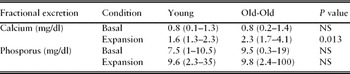
Phosphorus in the healthy aged and clinical implications. There is no significant difference in either the serum phosphorus level or its fractional excretion between the young and the healthy aged. Furthermore, when healthy old people undergo volume expansion, they do not show a significant increase in their fractional excretion of phosphorus compared with their basal level (see Table 4).
Fractional excretion of calcium and phosphorus (mean and range) in young and old subjectsReference Musso, Alvarez Gregori, Macias Nuñez, Macias Nuñez, Cameron and Oreopoulos46

Most of the major clinical manifestations of hyperphosphataemia are derived from the hypocalcaemia that frequently accompanies it.
Hypophosphataemia is defined as a serum phosphate level below 2.5 mg/dl in adults. However, symptoms of hypophosphataemia usually appear when the serum phosphate level is below 1 mg/dl.Reference Forster, Hernando, Biber and Murer19, Reference Knochel22 Malnutrition is one of the main causes of hypophosphataemia in the elderly population.
Increased renal excretion of phosphorus is generally the result of an excessive PTH level. Both primary and secondary hyperparathyroidism may lead to hyperphosphaturia.
Other causes of urinary phosphate loss are polyuric acute renal failure, post-obstructive polyuria and some drugs often wrongly managed in the elderly population such as proximal diuretics, corticosteroids, and cytotoxic drugs.
Hypophosphataemia is a common problem in alcoholic patients (because of proximal tubule dysfunction)Reference Shields23, Reference Suki24 and in those suffering from diabetes mellitus due to their poor phosphorus intake, vitamin D deficiency and heavy use of phosphate-binding antacids. It may be also observed in leukaemia sepsis, hepatic disease and stroke.
Urinary concentration and dilution
The generation and maintenance of the hyperosmolar interstitium is equally necessary for both the concentration and the dilution of urine. The former will occur in the presence of hypertonic interstitium, ADH and maximal increase in permeability of the collecting duct, while the latter will occur with a hypertonic interstitium in the absence of ADH and ADH-mediated collecting duct water permeability. In both processes, normal function of the thick ascending limb of the loop of Henle is mandatory in order to establish the interstitial to lumen gradient. Transport of electrolytes from the lumen to the interstitium makes it hypertonic. The special loop configuration of the vasa recta, that run countercurrent to the flow of tubular fluid is able to sustain the hyperosmolar profile of the interstitium.
Concentration ability in the healthy aged. Aged people have a reduced renal capacity to maximally concentrate the urineReference Dontas, Marketos and Papanayiotou25 and lose 5% of this capacity with every 10 years of age.Reference Lewis and Alving26 From a maximum urinary specific gravity of 1.030 at 40 years of age, they fall to 1.023 at 89 years of age. Rowe et al.Reference Rowe, Shock and DeFronzo27 found a mean maximum osmolality of 1109 mOsm/kg in individuals aged 20–39 years, with a minimum urine flow rate of 0.49 ± 0.03 ml/min; 1051 mOsm/kg in 40- to 59-year-olds; and only 882 mOsm/kg in those aged 60–79 years. In this group, the minimum urinary flow rate of 1.03 ml/min was more than double that of the young. Other groups have recorded even healthy individuals aged 60–80 years have a greater excretion of water, sodium and potassium during the night than younger individuals.Reference Takazakura, Sawabu, Handa, Takada, Shinoda and Takeuchi28
The origin of the decreased concentration ability is multifactorial. It has been related to the decrement in GFR that occurs with age in most studies,Reference Davies and Shock29–Reference Lindeman and Goldman32 although some data do not reveal a close relation between the reduction in the GFR and the capacity to concentrate urine in elderly people. The relative increase in medullary blood flow is unlikely to contribute to the impairment of renal concentration capacity.Reference Takazakura, Sawabu, Handa, Takada, Shinoda and Takeuchi28, Reference Kirkland, Lye, Levy and Banerjee33 As already discussed, inappropriately low ADH does not seem to be a factor in the genesis of the defect since elderly people secrete as much hormone as the young. The defect in sodium chloride reabsorption in the ascending limb of the loop of Henle, which is the basic event for the operation of the countercurrent concentration mechanism, together with a higher urinary urea output (that means a lower amount of urea reabsorbed by the tubule and shifted to the interstitium), could be the most relevant factors for the decrease in the capacity to concentrate urine seen in aged individuals.
Impaired urine concentration may play some role in the frequency of nocturia in the elderly, although this alteration in circadian rhythm does not depend upon inability to concentrate the urine alone, but also reflects defects in sodium handling and the decreased aldosterone secretion and sensitivity already discussed.
Dilution ability in the healthy aged. Few data in a few number of papers are available to explain the change in the capacity of the ageing kidney to dilute urine. Nevertheless, it has been found to be decreased.Reference Lewis and Alving26, Reference Kirkland, Lye, Levy and Banerjee33 The minimum urine concentration of only 92 mOsm/kg in elderly individuals is almost half of that (52 mOsm/kg) achieved in the young. Maximum free water clearance (CH2O) was also reduced in elderly individuals from 16.2 to 5.9 ml/min. This is, probably, mostly dependent on the reduced GFR, but when CH2O was factored by GFR to normalize it as a percentage of filtration rate, it was still somewhat reduced in the older participants (9.1 versus 10.2%). Again, the functional impairment of the diluting segment (diminished sodium reabsorption) of the thick ascending limb described above seems to account for the remainder of the diminution in the capacity to dilute urine observed in aged people.
Renal acidification
The kidney is a key organ for the maintenance of acid–base equilibrium. This is broadly achieved by two processes: reabsorption of filtered bicarbonate (HCO3−) and excretion of fixed acids mediated by the same basic process: renal H+ secretion.Reference Rodriguez Soriano34
The reabsorption of HCO3− is mainly accomplished in the proximal tubule, where 70–90% of it is reabsorbed, whereas excretion of fixed acid, achieved through the acidification of urinary buffers and the excretion of ammonium ions, mainly occurs in the distal tubule. In the thick ascending limb of the loop of Henle about 15% of the filtered HCO3− is reabsorbed by a mechanism equal to that present in the proximal tubular cells, i.e. a Na+–H+ apical exchanger and a basolateral Na+–HCO3− cotransporter.
Between 50 and 80% of the NH3 /NH4+ secreted by the proximal tubule and delivered to the loop of Henle is reabsorbed in the thick ascending limb of the loop by ionic transport.
The acidification of the urine is accomplished in the distal nephron (mainly cortical and medullary collecting ducts) by three related processes:
1. Reabsorption of the small amount of bicarbonate that escapes proximal reabsorption (5–20%).
2. Conversion (titration) of the divalent basic phosphate HPO4= to the monovalent acid form HPO4− (titratable acid).
3. Accumulation of ammonia (NH3) intraluminally, which reacts with H+ to form the non-diffusible ammonium ion (NH4+).
All these processes rely on the active secretion of H+ ions by an active transport, H+-ATPase, although it should be remembered that there are further factors influencing the acidification in the distal nephron, including aldosterone, potassium stores, sodium intake, extracellular fluid volume and acid–base status.Reference Adrogué, Madias, Davison, Cameron and Grünfeld35
Acidification by the normal ageing kidney. So far there are only five studies that have evaluated the response of the normal ageing kidney to a challenge with acid overload.Reference Adler, Lindeman, Yiengst, Beard and Shock36–Reference Macías Núnez, García Iglesias and Tabernero40
The kidney of healthy aged people is able to lower the urinary pH in response to acid overload to a value similar to that reached by healthy young adult subjects in response to the same stimulus.Reference Schuck and Nadvornikova39 Furthermore, in one studyReference Adler, Lindeman, Yiengst, Beard and Shock36 it was reported that the fall in urinary pH was more intense in the aged, i.e. 4.85 ± 0.23 versus 4.96 ± 0.52 in the young subjects. In a different study the urinary pH reached in response to acid overload varied from 4.5 ± 0.1 in the young to 4.93 ± 0.07 in the aged.Reference Agarwal and Cabebe37
Titratable acid elimination behaves similarly in the young and in the aged,Reference Adler, Lindeman, Yiengst, Beard and Shock36, Reference Agarwal and Cabebe37 but in one study it was reported that it is higher in subjects above 60 years compared with the young.Reference Schuck and Nadvornikova39
There is almost a consensus that NH4+ elimination is lower in the aged than in young subjects, except in one studyReference Macías Núnez, García Iglesias and Tabernero40 in which differences between young and aged people were not found. In this study, it was proven that the aged need more time to reach peak acid excretion (between 6 and 8 h) and that the same dose of ammonium chloride induces a greater decline in blood bicarbonate in the aged than in young individuals.
There are several factors that can influence NH4+ metabolism, such as blood electrolyte status previous to the acid challenge, nutrition, subject position (recumbent, sitting, standing or walking), urine collection time, degree of blood acidification following the acute acid overload and health assessment. Some of these factors have not been evaluated and the results from the different studies are difficult to compare.
In an attempt to find an explanation for the discrepancy regarding the dysfunction in NH4+ excretion in aged people found in the above studies, a few possible mechanisms have been postulated. The absorption of NH4+ in the thick ascending limb of Henle's loop takes place by the substitution of K+ for NH4+ in the cotransporter Na+–K+–2Cl−. There is a certain degree of incompetence in the thick ascending limb of the loop of Henle to retain Na+. It is known that the cotransporter Na+–K+–2Cl− plays a first-order role in the absorption of sodium in this segment. So, one possibility is that the Na+–K+–2Cl− cotransporter is less efficient in the aged than in the young in reabsorbing both Na+ and NH4+. Although the clinical significance of the altered acidification in the loop of Henle is poorly understood, in some patients renal tubular acidosis may originate, at least in part, from an abnormal function of the loop of Henle. Another possible explanation is that standing from a supine position is associated with a significant decrease in creatinine clearance.Reference Adachi, Kawamura, Owada and Hiramori41 As all data are corrected for GFR, it may introduce some error in the final calculation. An important difference among the studies is the observation period between acid administration and collection time. In some papers the time varied from subject to subject in the same study.Reference Adler, Lindeman, Yiengst, Beard and Shock36, Reference Agarwal and Cabebe37 In others these data are not given.Reference Shock and Yiengst38 In one study the collection period was 5 hReference Schuck and Nadvornikova39 and in our study the collection time was extended to 8 h.Reference Macías Núnez, García Iglesias and Tabernero40 Another finding is that the healthy aged person has a lower plasma concentration rate of aldosterone,Reference Macías, Cameron, Davison, Cameron and Grunfeld42, Reference Macias Nuñez, Lopez Novoa, Macias Nuñez, Cameron and Oreopoulos43 which influences urinary acidification, as already mentioned.
In the only study that has specifically examined the renal tubular handling of bicarbonate following an acute acid overload, it was found that age had no effect.Reference Schuck and Nadvornikova39, Reference Macías Núnez, García Iglesias and Tabernero40
In summary, from the few studies available it can be concluded that:
(a) The basal conditions and techniques used differ from study to study, resulting in difficulties in comparing results.
(b) Urinary pH and titratable acid behave similarly in the aged and in the young.
(c) The renal threshold for bicarbonate is comparable in the aged and in the young.
(d) Plasma pH and PCO2 take longer be to restored after an acid load in the aged compared with the young.
(e) Ammonium ion excretion is less efficient or, at least, takes longer to reach its peak in response to an acute acid overload in the aged than in the young.
Clinical implications. The elderly are prone to metabolic acidosis. This situation can be produced by several circumstances such as accumulation of fixed acids (e.g. lactic acid) or from loss of bicarbonate (e.g. through diarrhoea, pancreatic or small bowel fistulas, or renal tubular damage). Any significant period of circulatory failure results in the accumulation of lactic acid, reflecting anaerobic metabolism. In the geriatric patient, the impaired GFR leads to less direct filtration of lactic acids. In addition, the reduced tubular function and lower aldosterone levels lead to less regeneration of bicarbonate. Thus the elderly patient is more at risk of developing disturbances of acid–base balance and, compared with the younger patient, has decreased mechanisms of compensation.
The older population is also less well able to tolerate metabolic alkalosis and may experience the development of rapid dangerous hypokalaemia as the body preferentially conserves sodium (under the influence of aldosterone) and hydrogen (which is titrated by the excess bicarbonate to carbon dioxide and excreted by the lungs).Reference Kirkland, Lye, Levy and Banerjee33, Reference Rodriguez-Puyol44 The resultant hypokalaemic alkalosis is a risk for cardiac dysrhythmias in the elderly patient, especially in those with coronary artery disease, those taking digitalis, or those who may be taking long-term potassium-losing diuretics.
In surgical procedures Ringer solution is commonly administered as the maintenance intravenous fluid intraoperatively. This lactate is metabolized to bicarbonate. Because of the impaired renal excretion of alkali in elderly patients, bicarbonate may accumulate to an unacceptable level. In this case, it is preferable that a balanced salt solution such as normal saline be used. Potassium chloride can be added to this solution as needed and the plasma level followed by intraoperative monitoring of potassium levels.
Conclusion
The healthy ageing kidney has some functions comparable to that in the healthy young individual. Proximal functions do not differ, in the absence of restriction or overload, from those of the younger adult. Some other functions, such as creatinine clearance, are lower in the healthy aged than in the young. Other functions, such as the renal management of urea or the handling of Na+ by the thick ascending limb of the loop of Henle and the segments where aldosterone works, are less efficient in the aged than in the young.
As a rule, under normal circumstances the aged kidney is able to maintain plasma electrolyte concentrations.Reference Refoyo and Macías-Núñez7 However, as with other organs, the functional characteristic of the aged kidney is a slow response or sometimes a complete inability to adapt to situations of overload or restriction.
Conflicts of interest
The authors declare no conflicts of interest.