Introduction
Attempts have focused on the prediction of the outcomes of the assisted reproductive technique (ART) as well as improving the success rates in assisted reproduction treatment (Ceviren et al., Reference Ceviren, Ozcelik, Urfan, Donmez and Isikoglu2014). The evaluation of oocyte and embryo competences to determine the best embryo(s) for transfer is a critical step in IVF treatment. In general, the ART centers perform multiple embryo transfer to enhance the implantation and pregnancy rates, which may result in a significantly higher risk of multiple gestations. Multiple gestations related to an increased risk of preterm birth and complications, such as infant death and cerebral palsy (Uyar et al., Reference Uyar, Torrealday and Seli2013), as well as maternal complications (Molina et al., Reference Molina, Martínez, Pertusa, Balasch, Iniesta and Pellicer2014). However, single-embryo transfer (SET) is now the preferable practice in IVF treatment to lower the risk for damaging outcomes associated with multiple gestations (Kirkegaard et al., Reference Kirkegaard, Agerholm and Ingerslev2012).
Oocyte morphology is one of the factors that may affect the success rate of intra-cytoplasmic sperm injection (ICSI) (Halvaei et al., Reference Halvaei, Khalili, Razi and Nottola2012). Previous studies showed that 80% of metaphase II (MII) oocytes retrieved from stimulated cycles have one or more abnormal morphological characteristics (Ashrafi et al., Reference Ashrafi, Karimian, Eftekhari Yazdi, Hasani, Bahmanabadi and Akhond2015). The morphological parameters of oocytes were divided on the basis of the presence of extra- and intra-cytoplasmic abnormalities. Extra-cytoplasmic abnormalities include a dark or non-Hemogen zona pellucida (ZP), a wide perivitelline space (PVS), PVS debris and a fragmented polar body (PB). Intra-cytoplasmic abnormalities include dark cytoplasm, ooplasm granulation, irregular shape, refractile bodies (RF), bull eye (central aggregation of organelles and vesicles within ooplasm), smooth endoplasmic reticulum (SER) and vacuoles (Fancsovits et al., Reference Fancsovits, Tóthné, Murber, Rigó and Urbancsek2012). Oocyte morphology and its influence on embryo competence is still a controversial subject in the literature (Loutradis et al., Reference Loutradis, Drakakis, Kallianidis, Milingos, Dendrinos, Michalas and Stylianos1999; Esfandiari et al., Reference Esfandiari, Burjaq, Gotlieb and Casper2006; Nichi et al., Reference Nichi, Figueira, Braga, Setti, Iaconelli and Borges2011; Rienzi et al., Reference Rienzi, Vajta and Ubaldi2011; Kaser et al., Reference Kaser, Reichman, Ginsburg, Politch and Racowsky2012; Braga et al., Reference Braga, Setti, Rita de Cássia, Machado, Iaconelli and Borges2013; Sauerbrun-Cutler et al., Reference Sauerbrun-Cutler, Breborowic, Gonzales, Stein, Lederman and Keltz2015). Hence, the assessment of oocyte morphology remains one of the most significant purposes of ART advancement (Uyar et al., Reference Uyar, Torrealday and Seli2013).
The selection of the most suitable embryo(s) for transfer is an important challenge in the ART programme. The failure to properly recognize embryos with the highest implantation potential can lead to failed IVF outcomes. Despite hopeful metabolic profiling, O2 respiration, aneuploidy screening and gene expression survey, the grading systems based on morphology still remain the preferred way of identifying embryonic development (Chamayou et al., Reference Chamayou, Patrizio, Storaci, Tomaselli, Alecci, Ragolia, Crescenzo and Guglielmino2013). However, embryo selection based on primary subjective and static morphology methods provides limited data as punctual, periodic observations (Chawla et al., Reference Chawla, Fakih, Shunnar, Bayram, Hellani, Perumal, Divakaranm and Budak2015). So, more dynamic methods are needed to select the embryos with the highest implantation potential for transfer. The morphokinetic assessment of embryos at early cleavage stages improved the prediction of implantation potential. Morphokinetic information also aids the embryologists to discriminate impalpable timing diversities between developmental stages in embryos with same morphological appearance (Liu et al., Reference Liu, Chapple, Roberts and Matson2014).
In recent years, continuous monitoring of embryo development throughout the culture period has been possible using TLM. This technology is considered as a secure tool in ART laboratories. It has no adverse effects on the embryo development, nevertheless maintaining stable culture conditions (Motato et al., Reference Motato, de los Santos, Escriba, Ruiz, Remohí and Meseguer2016). However, the high expenses of this technology do not let their fulfillments in many ART laboratories (Molina et al., Reference Molina, Martínez, Pertusa, Balasch, Iniesta and Pellicer2014). The purpose of this prospective study was to assess the relationship between oocyte morphology and morphokinetic variables and cleavage patterns of embryos in clinical setting. To the best of our knowledge, this preliminary study is the first to investigate the possible impact of oocyte morphology on the early embryo development as reflected by TLM in ICSI treatment cycles.
Materials and methods
Study design
In total, 478 MII oocytes were retrieved from 52 women (mean age 30.0 ± 3.42 years) undergoing ICSI cycles between June 2015 to June 2016. Cycles with severe male factors, frozen and surgically retrieved spermatozoa were eliminated from study due to poor recovery of motile spermatozoa. Study inclusion criteria were as follows: patients with fresh autologous oocytes and at least one normal fertilization. The ethics committee of our institution approved this study.
Ovarian stimulation and oocyte preparation
Ovarian stimulation was achieved by long pituitary down-regulation using a combination of gonadotrophin-releasing hormone (GnRH) antagonist and follicle-stimulating hormone (FSH) (Gonal-F; Serono, Geneva, Switzerland). Oocyte collection was done 36 h after human chorionic gonadotrophin (hCG; 10 000 IU) injection. Retrieved cumulus–oocyte complexes (COCs) were incubated in culture medium (G-IVF; VitroLife, Kungsbacka, Sweden) covered with mineral oil (Ovoil; VitroLife) at 37°C and 6% CO2 for 2–3 h. After mechanic and enzymatic removal of cumulus cells (CCs) with 80 IU/ml hyaluronidase (Sigma Co, USA), MII oocytes were injected using the husband's prepared sperm.
Evaluation of oocyte morphology
Under an inverted microscope (TE300; Nikon), the morphology of oocytes was assessed after the denudation of CCs. The oocytes morphologic features were classified according to the presence of intra- and extra-cytoplasmic abnormalities. Intra-cytoplasmic abnormalities included dark oocyte, ooplasm granulation, presence of vacuoles, RF, irregular shape, bull eye and SER. Extra-cytoplasmic abnormalities included wide PVS, PVS debris, and fragmented PB (Omidi et al., Reference Omidi, Khalili, Ashourzadeh and Rahimipour2014).
Embryo culture
After ICSI, each oocyte was individually placed in a pre-equilibrated culture slide (Primo Vision dish, VitroLife, Sweden). The Primo Vision dish with a central depression containing nine straight-sided circular wells filled with 40 μl of G-1+ medium, and covered with 3 ml of mineral oil. The culture slides were pre-equilibrated (37°C and 6% CO2) using fresh G-1+ medium and mineral oil. The culture slide was placed in a time-lapse microscope at 37°C, 5 % O2 and 6 % CO2 immediately after ICSI for 3 days.
Time-lapse imaging system
A Primo Vision camera was set to take a picture of the embryos every 10 min and the total scan of embryos was taken every 20 min. On day 3, embryos were morphologically assessed for timing of cell divisions and development. The following early kinetic markers were evaluated: absolute time point including time to second PB extrusion (ESPB), pronuclei (PN) appearance (PNA), PN fading (PNF), time to 2-cells (t2), 3c (t3), 4c (t4), 5c (t5), 6c (t6), 7c (t7), 8c (t8) and relative timing parameters S1, CC2, CC3, S2 and S3. Durations of the second cycle (cc2; t3-t2) and third cycle cells (cc3: t5-t3), period time of complete first synchronous division (s1:t2-tPNf), the time to complete second synchronous divisions (s2:t4-t3) were also monitored.
In addition, cleavage patterns including uneven blastomeres embryos were evaluated. Also, three cleavage anomalies specifically monitored were reverse cleavage (RC), where a blastomere was reabsorbed after cleavage. RC was defined as either: (1) two daughter blastomeres recombining after complete separation following cleavage division (type I); or (2) failed cytokinesis, the incomplete separation of blastomeres that later recombine fully (type II) (Liu et al., Reference Liu, Chapple, Roberts and Matson2014). Direct cleavage (DC), in which a single blastomere divided directly from one to three cells in less than 5 h (Rubio et al., Reference Rubio, Kuhlmann, Agerholm, Kirk, Herrero, Escriba, Bellver and Meseguer2012). Arbitrary cleavage, including abnormal syngamy, (embryos with disordered PN movement in the cytoplasm, accompanied by delayed dispersion of the nuclear envelopes (AS), abnormal first cytokinesis (embryos exhibiting oolemma ruffling, and possible pseudofurrows, before first cytokinesis (A1cyt), abnormal cleavage more than two cells during a single cell division event (AC)), and chaotic cleavage (disordered cleavage behaviour by the 4-cell stage (CC)) (Wirka et al., Reference Wirka, Chen, Conaghan, Ivani, Gvakharia, Behr, Suraj and Tan2014).
Data analysis
Results are expressed as mean ± standard deviation (SD) for normal numeric variables, median ± interquartile range and percentage for categorical variables. The means of samples with normal distribution and of sufficient size were compared by independent samples test parametric test. In case of non-normal distribution, the medians were compared by Mann–Whitney test non-parametric test. Categorical variables were compared using chi-squared test. A P-value < 0.05 was considered as significant. Statistical analysis was performed using the Statistical Package for the Social Sciences 20 (SPSS Inc., Chicago, IL, USA).
Results
In total, 577 oocytes were retrieved, of which 19 (3.30%) were discarded after denudation because of an abnormal appearance (e.g. extensive vacuolization or degeneration). Of the remaining, 478 (85.67%) MII oocytes were injected and 368 (76.98%) of them fertilized normally. Some of the zygotes were excluded from analysis due to arresting in the early development stages or excessive fragmentation.
Incidence of the oocyte morphologic features
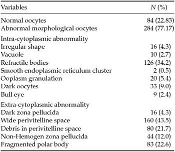
On assessment of normal fertilized oocytes, 284 (77.17%) had at least one abnormal morphological characteristic. From 284 abnormal MII oocytes, intra-cytoplasmic abnormalities were seen in 43 (11.7%) of the oocytes. Moreover, extra-cytoplasmic abnormalities were observed in 109 (29.6%), while combined intra- and extra-cytoplasmic abnormalities were responsible for the remaining (35.9%) oocytes (Table 1).
Evaluation of the morphological parameters in normally fertilized oocytes.

Values in parenthesis are (%).
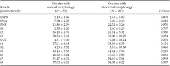
Correlation of oocyte morphology with embryo morphokinetic variables (absolute and relative time)
As shown in Table 2, TLM demonstrated insignificant relationship between oocytes morphology and early embryo developmental events (timing of PNA, PNF, t2, S1, t3, CC2, t4, S2, CC3, t6, t7, t8 and S3) (P < 0.05), except for ESPB time. There was a significant difference between normal and abnormal oocyte morphology (P < 0.05) (Table 2).
Correlation of oocyte morphology with embryo morphokinetic variables (absolute and relative time)

Note: Results are expressed as mean ± standard deviation (SD) for normal numeric variables, median ± interquartile range and percentage for categorical variables. Second PB extrusion (ESPB), pronuclei (PN) appearance (PNA), PN fading (PNF), t2 = first cleavage (2-cell stage); t3 = second cleavage (3-cell stage); t4 = 4-cell stage; t5 = 5-cell stage; t6 = 6-cell stage; t7 = 7-cell stage t8 = 8-cell stage. The time to complete first synchronous divisions s1 (t2-tPNf), durations of the second cycle (cc2; t3-t2), the time to complete second synchronous divisions (s2:t4-t3), durations of the third cycle (cc3:t5-t3) and the time to complete third synchronous divisions (s3:t8-t5). NS: not significant.
Correlation of oocyte morphology with uneven blastomeres embryos
The data showed that oocytes with abnormal morphology was correlated with a higher rate of uneven blastomeres embryos compared with normal morphology oocytes (6.5% vs. 93.5%; P = 0.037) (Table 3).
Correlation between oocyte morphology and rate of uneven blastomeres embryos

CI, confidence interval.
Correlation of oocyte morphology with reverse cleavage embryos
The findings demonstrated that there was significant correlation between oocytes morphology and rate of RC embryos. Normal morphology oocytes had lower rate of RC embryos compared with abnormal morphology oocytes (7.0% vs. 93.0%) (Table 4).
Correlation between oocyte morphology and rate of reverse cleavage embryos

CI, confidence interval.
Correlation of oocyte morphology with direct cleavage embryos
Also, there was significant relationship between oocyte morphology and rate of RC embryos. Embryos derived from abnormal oocyte morphology showed higher rates of direct cleavage compared with embryos derived from normal oocyte morphology (5.0% vs. 95.6%) (Table 5).
Correlation between oocyte morphology and rate of direct cleavage embryos

CI, confidence interval.
Correlation of oocyte morphology with arbitrary cleavage embryos
TLM showed that there was abnormal oocytes morphology significantly had a higher rate of arbitrary cleavage embryos compared with normal morphology oocytes (6% vs. 94%) (Table 6).
Correlation between oocyte morphology and rate of arbitrary cleavage embryos

CI, confidence interval.
Discussion
Since the advent of ICSI, it has become possible to predict embryo viability in relation to oocyte morphological features. Also, TLM is an emerging tool which can assess the parameters that can foretell the developmental potential of cleaving embryos under stable development conditions (Faramarzi et al., Reference Faramarzi, Khalili and Soleimani2015). TLM reveals abnormalities in embryo division correlated to reduce implantation potential that cannot be seen by conventional morphological assessment (Liu et al., Reference Liu, Chapple, Roberts and Matson2014). With the use of continuous TLM, the present study aimed to assess oocyte dysmorphisms and their influence on the embryo morphokinetic parameters and cleavage patterns in the ICSI programme. To our knowledge, this preliminary study is the first to report the impact of oocyte morphology on early embryo development as reflected by time-lapse analysis. We found that 77.17% of the MII oocytes had at least one abnormal morphological characteristic. According to another study, at least 20% of MII oocytes from stimulated cycles had abnormal morphology and the remaining 80% exhibited one or more abnormal morphological characteristics (Ashrafi et al., Reference Ashrafi, Karimian, Eftekhari Yazdi, Hasani, Bahmanabadi and Akhond2015). Intrinsic factors, such as genetic defects and age, as well as extrinsic factors including controlled ovarian hyperstimulation (COH) protocols and the ovarian response to COH may result in oocyte morphological variations (Qassem et al., Reference Qassem, Falah, Aghaways and Salih2015).
The relation between oocyte morphological abnormalities and ICSI outcomes have been discussed previously (Kahraman et al., Reference Kahraman, Yakın, Dönmez, Şamlı and Bahce2000; Plachot et al., Reference Plachot, Selva, Wolf, Bastit and de Mouzon2002; Khalili et al., Reference Khalili, Mojibian and Sultan2005; Yakin et al., Reference Yakin, Balaban, Isiklar and Urman2007; Rienzi et al., Reference Rienzi, Ubaldi, Iacobelli, Minasi, Romano, Ferrero, Sapienza, Baroni, Litwicka and Greco2008; Yu et al., Reference Yu, Ahh, Lee, Jee and Kim2015). Controversies still exist on the effects of oocyte morphology on the embryo quality in ART programme (Kahraman et al., Reference Kahraman, Yakın, Dönmez, Şamlı and Bahce2000; Fancsovits et al., Reference Fancsovits, Murber, Gilan, Rigo and Urbancsek2011; Setti et al., Reference Setti, Figueira, Braga, Colturato, Iaconelli and Borges2011; Ashrafi et al., Reference Ashrafi, Karimian, Eftekhari Yazdi, Hasani, Bahmanabadi and Akhond2015, Yu et al., Reference Yu, Ahh, Lee, Jee and Kim2015). In this study, we did not detect any effect of oocyte morphology on the morphokinetic time points of derived embryos, except for ESPB time. However, deleterious abnormal oocyte morphology influenced embryo cleavage patterns. Although, Lemmen and colleagues found that early cleavage timing was positively associated with embryo developmental potential in clinical practice (Lemmen et al., Reference Lemmen, Agerholm and Ziebe2008). During ICSI, the sperm entry time point for each oocyte may be imprecise as seen, for example, where all embryos cultured on the same slide share one single starting time point. Furthermore, mature oocytes were shown to extrude the second PB at various time intervals, indicating that MII oocytes may have different degrees of cytoplasmic maturity at the point of sperm injection (Liu et al., Reference Liu, Chapple, Feenan, Roberts and Matson2016). In the other words, we assumed that since the injecting time of the oocytes is not the same, the ESPB is variable in different generated embryos.
In this work, oocyte dysmorphism significantly affected cleavage patterns, as well as increasing uneven blastomeres, RC and arbitrary cleavage in derived embryos. Significantly lower fertilization and cleavage rates were reported in dysmorphic oocytes, also reduced rates of top- or good-quality embryos formation, compared with normal morphology oocytes (Yu et al., Reference Yu, Ahh, Lee, Jee and Kim2015). Recently, Ashrafi et al. (Reference Ashrafi, Karimian, Eftekhari Yazdi, Hasani, Bahmanabadi and Akhond2015) demonstrated that MII oocyte morphology had minor effects on fertilization rate, pronuclear morphology and embryo quality in women with regular menstrual cycles and normal ovarian response (Ashrafi et al., Reference Ashrafi, Karimian, Eftekhari Yazdi, Hasani, Bahmanabadi and Akhond2015). Whereas, Khalili and associates (Reference Khalili, Mojibian and Sultan2005) mentioned that normal oocyte morphology had the most significant role in producing high-grade embryos (Khalili et al., Reference Khalili, Mojibian and Sultan2005). Higher rate of aneuploidy found in the embryos derived from abnormal morphologic oocytes and lower intra-cytoplasmic pH and adenosine triphosphate content are postulated mechanisms in order to explain the adverse effect of such oocytes on clinical outcomes (Ceviren et al., Reference Ceviren, Ozcelik, Urfan, Donmez and Isikoglu2014). Embryos developed from centrally located granular ooplasm presented chromosomal abnormality in 52.2% of the biopsied blastomeres (Kahraman et al., Reference Kahraman, Yakın, Dönmez, Şamlı and Bahce2000).
Some previous studies indicated that cleavage pattern, such as embryo with uneven blastomere size at 2-cell stage, direct cleavage, RC (types I and II) and arbitrary cleavage (abnormal first cytokinesis, abnormal cleavage and chaotic cleavage) resulted in lower embryo development and implantation potentials (Meseguer et al., Reference Meseguer, Herrero, Tejera, Hilligsoe, Ramsing and Remoh2011, Rubio et al., Reference Rubio, Kuhlmann, Agerholm, Kirk, Herrero, Escriba, Bellver and Meseguer2012, Liu et al., Reference Liu, Chapple, Roberts and Matson2014, Wirka et al., Reference Wirka, Chen, Conaghan, Ivani, Gvakharia, Behr, Suraj and Tan2014). The molecular mechanisms that cause abnormal cleavage are not obvious. Yet, it seems rational to suppose that mitotic errors may play a main role in this phenomenon (Almagor et al., Reference Almagor, Or, Fieldust and Shoham2015). Liu and colleagues also showed that RC related factors are not limited to those affecting the oocyte, but also include sperm parameters (Liu et al., Reference Liu, Chapple, Roberts and Matson2014), which is not repaired by abnormal morphologic oocytes.
All earlier studies could be limited by the fact that they were performed on the basis of a static conventional observation. However, the present study was focused on oocyte morphology and its relationship with morphokinetic parameters of ICSI generated embryos and atypical cleavage with the aid of TLM. A limitation of this study was the lack of information on implantation and pregnancy outcomes. Therefore, future studies are warranted to validate findings in the present study. In conclusion, the cleavage patterns of embryos were affected by abnormal oocyte morphology. The use of TLM revealed that abnormalities of embryo division, such as uneven blastomere, RC, direct cleavage and arbitrary cleavage increased by abnormal oocyte morphology during ICSI. Therefore, evaluation of oocyte morphology followed by TLM of generated embryos in an ICSI setting is vital.