Introduction
Adult digeneans belonging in the Callodistomidae infect the gallbladder of some freshwater fishes in Africa and America (Bray, Reference Bray, Gibson, Jones and Bray2002). In Africa, the family is represented by Callodistomum Odhner, 1902 and Cholepotes Odhner, 1910. In America, Teratotrema dubium Travassos, Artigas & Pereira, 1928 is monotypic and infects Pseudocurimata plumbea (Eigenmann & Eigenmann, 1889), and Prosthenhystera Travassos, 1922 represents the only other genus, in which three species are currently valid in North, Middle and South America (Tkach & Curran, Reference Tkach and Curran2015). Prosthenhystera caballeroi Jiménez-Guzmán 1973 infects Astyanax mexicanus De Filippi, 1853 in Mexico (Jiménez-Guzmán, Reference Jiménez-Guzmán1973; Pérez-Ponce de Leon & Choudhury, Reference Pérez-Ponce de Leon and Choudhury2005), and Prosthenhystera oonastica Tkach & Curran, 2015 infects three species of catfishes (Siluriformes: Ictaluridae) in the US; Prosthenhystera obesa (Diesing, 1850) Travassos, 1922, the only species that has been reported from South America, infects a wide variety of Characiformes, Perciformes and Siluriformes (Diesing, Reference Curran, Tkach and Overstreet1850; Kohn et al., Reference Kohn, Fernandes and Cohen2007). Additionally, Martínez-Aquino et al. (Reference Martínez-Aquino, Ceccarelli and Perez-Ponce de Leon2013) discovered an additional species with a distinct DNA linage from Panamá parasitizing a characid fish.
In the present study, we investigated the identity of a digenean conforming to the generic diagnosis for Prosthenhystera that we found infecting the gallbladder of a characid fish (Bryconamericus ikaa Casciotta, Almirón, & Azpelicueta, 2004) from Yacuí stream (Lower Iguazú River) in Argentina. Tkach & Curran (Reference Tkach and Curran2015) suggested that since P. obesa is thought to infect such a wide array of hosts over such a large region, that molecular analysis may be useful for prospecting for cryptic species of Prosthenhystera sp. in South America.
Morphological examination combined with comparative analysis using partial 28S ribosomal DNA (rDNA) and internal transcribed spacer (ITS) sequences indicated that the species found in B. ikaa was distinct from all known congeners, and it is herein described as a new species. Also, we report cytogenetic analysis for the new species, which represents the first such data for a species in the Callodistomidae.
Materials and methods
Specimens of B. ikaa were collected from the Yacuí stream (Lower Iguazú River) in Iguazú National Park, Argentina, upstream from the waterfalls (25°40′50.2″S, –54°10′05.8″W) during autumn and spring of 2016 using gill nets. The fish were transported alive in plastic bags with water from the sample site to the field laboratory at the Centro de Investigaciones Ecológicas Subtropicales (CIES), where they were necropsied. Digeneans were removed from the gallbladders of fish and heat-killed with hot water and fixed in 10% formalin. Live specimens for the molecular study were preserved using cold 96% ethanol and stored for later DNA extraction.
Whole-mount specimens were made using specimens preserved in 10% formalin solution and stained according to standard parasitological techniques using hydrochloric carmine (Pritchard & Kruse, Reference Petkevičiute, Stunženas and Stanevičiute1982). Measurements and digital images of specimens were taken using an Olympus Bx51 (Tokyo, Japan)microscope equipped with an AmScope MU 1000 10 MP digital camera(Irvine, USA). The structures were measured using ImageJ software (Schneider et al., Reference Schneider, Rasband and Eliceiri2012). Drawings were made with the aid of a drawing tube. The mean followed by minimum and maximum values in parentheses are given in micrometres (μm). The type-material is deposited in the Invertebrate Collection of the Museo de La Plata, La Plata, Argentina.
Total genomic DNA was extracted from two specimens using a Wizard® Genomic DNA Purification Kit (Promega, Madison, Wisconsin, USA) according to the manufacturer's protocol. The partial fragment of the 28S rDNA gene was amplified from individual worms. Amplification was conducted using polymerase chain reaction (PCR) on an Eppendorf Mastercycler thermal cycler(Hamburg, Germany) using forward primer LSU-5 (5′-TAG GTC GAC CCG CTG AAY TTA AGC A-3′) and 1500R (5′-GCT ATC CTG AGG GAA ACT TCG-3′) (Tkach et al., Reference Tkach, Littlewood, Olson, Kinsella and Swiderski2003). The ITS1-5.8S-ITS2 gene fragment was amplified from individual worms using the forward primer MITF1 (5′-CGT AAC AAG-GTT TCC GTA G-3′) (Tkach & Curran, Reference Tkach and Curran2015). The PCR reactions were performed according to protocols described by Tkach et al. (Reference Tkach, Littlewood, Olson, Kinsella and Swiderski2003). Both strands of the PCR products were sequenced using an ABI 3730XLs sequencer (Macrogen Inc., Korea). The sequences were assembled using the platform Geneious Pro version 5.1.7 (Drummond et al., Reference Drummond, Ashton, Buxton, Cheung, Cooper, Duran and Wilson2016) and compared with sequences from Olson et al. (Reference Olson, Cribb, Tkach, Bray and Littlewood2003) of P. obesa (AY222206); Rosas-Valdez et al. (Reference Rosas-Valdez, Choudhury and Perez-Ponce de Leon2011) and Tkach & Curran (Reference Tkach and Curran2015) of P. caballeroi (KM871183–KM871186, HQ325029); Curran et al. (Reference Diesing2006) and Tkach & Curran (Reference Tkach and Curran2015) of P. oonastica (EF032690, KM871180–KM871182); and Martínez-Aquino et al. (Reference Martínez-Aquino, Ceccarelli and Perez-Ponce de Leon2013) of Prosthenhystera sp. (KC899973–KC899974). Sequences were aligned using the online version of MAFFT version 7 (Katoh & Standley, Reference Katoh and Standley2013). Then, to exclude ambiguously aligned regions of the rDNA, we used the online program Gblocks version 0.91 (Castresana, Reference Castresana2000; Talavera & Castresana, Reference Talavera and Castresana2007) with relaxed parameters. Dicrocoelium dendriticum (Rudolphi, 1819) Odhner, 1910 (AF151939) was used as the outgroup for the phylogenetic analyses. The best partitioning scheme and substitution model for the DNA partition was chosen under the Bayesian Information Criterion (Schwarz, Reference Schwarz1978) using the ‘greedy’ search strategy in Partition Finder version 1.1.1 (Lanfear et al., Reference Lanfear, Calcott, Ho and Guindon2012, Reference Lanfear, Calcott, Kainer, Mayer and Stamatakis2014). The appropriate nucleotide substitution model implemented for the matrix resulting after the Gblock program was HKY + G for the 28s rDNA matrix, and K80+G for the ITS1-5.8S-ITS2 matrix.
Bayesian inference (BI) through MrBayes version 3.2.p1 (Ronquist et al., Reference Ronquist, Teslenkovan and van der Mark2012) was used for the phylogenetic reconstructions. Phylogenetic trees, based on the 28S rDNA gene and ITS sequences, were reconstructed for each gene using two parallel analyses of Metropolis-coupled Markov chain Monte Carlo for 20 million of generations each, to estimate the posterior probability (PP) distribution. Topologies were sampled every 1000 generations once the average standard deviation of split frequencies was less than 0.01, as suggested by MrBayes 3.2. The robustness of the clades was assessed using Bayesian PP, where PP > 0.95 was considered strongly supported. A majority consensus tree with clade lengths was reconstructed for each run after discarding the first 25% of sampled trees.
Additionally, the proportion (p) of absolute nucleotide sites (p-distance) (Nei & Kumar, Reference Nei and Kumar2000) was obtained to compare the genetic distance between lineages and with the outgroup. The P-value matrix was obtained using MEGA version 6.0 (Tamura et al., Reference Tamura, Stecher, Peterson, Filipski and Kumar2013) with the bootstrap method (1000 replicates) and with a nucleotide substitution (transition + transversions) uniform rate. Newly generated sequences from the new species were submitted to the National Center for Biotechnology Information GenBank database (http://www.ncbi.nlm.nih.gov) under GenBank accession numbers MF664223 and MF664224 for 28S rDNA, and MN521830 and MN521831 for ITS1-5.8S-ITS2.
For the cytogenetic analysis, one specimen was fixed in Carnoy solution (three parts ethyl alcohol 100% to one part glacial acetic acid). The preparation was made by crushing and dilation of the specimen over a slide in acetic orcein, followed by a 5 min period prior to adding a coverslip.
Results
Prosthenhystera gattii n. sp.
ZooBank registration: urn:lsid:zoobank.org:pub:3C687CF4-7DE7-4772-BAE8-C54293B107B4.
Description
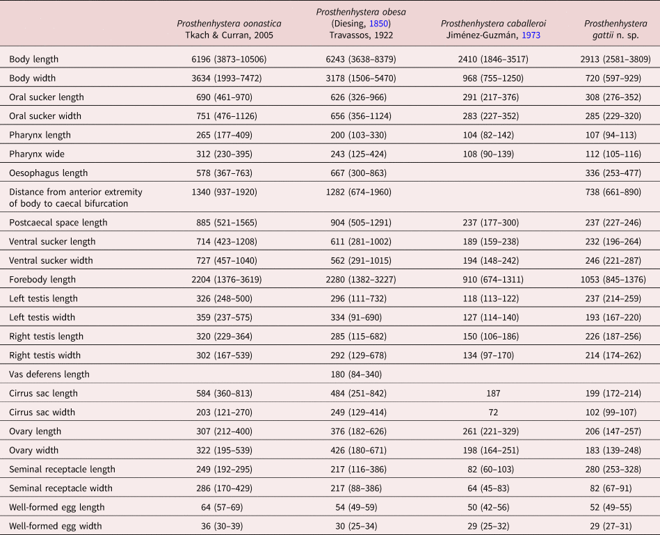
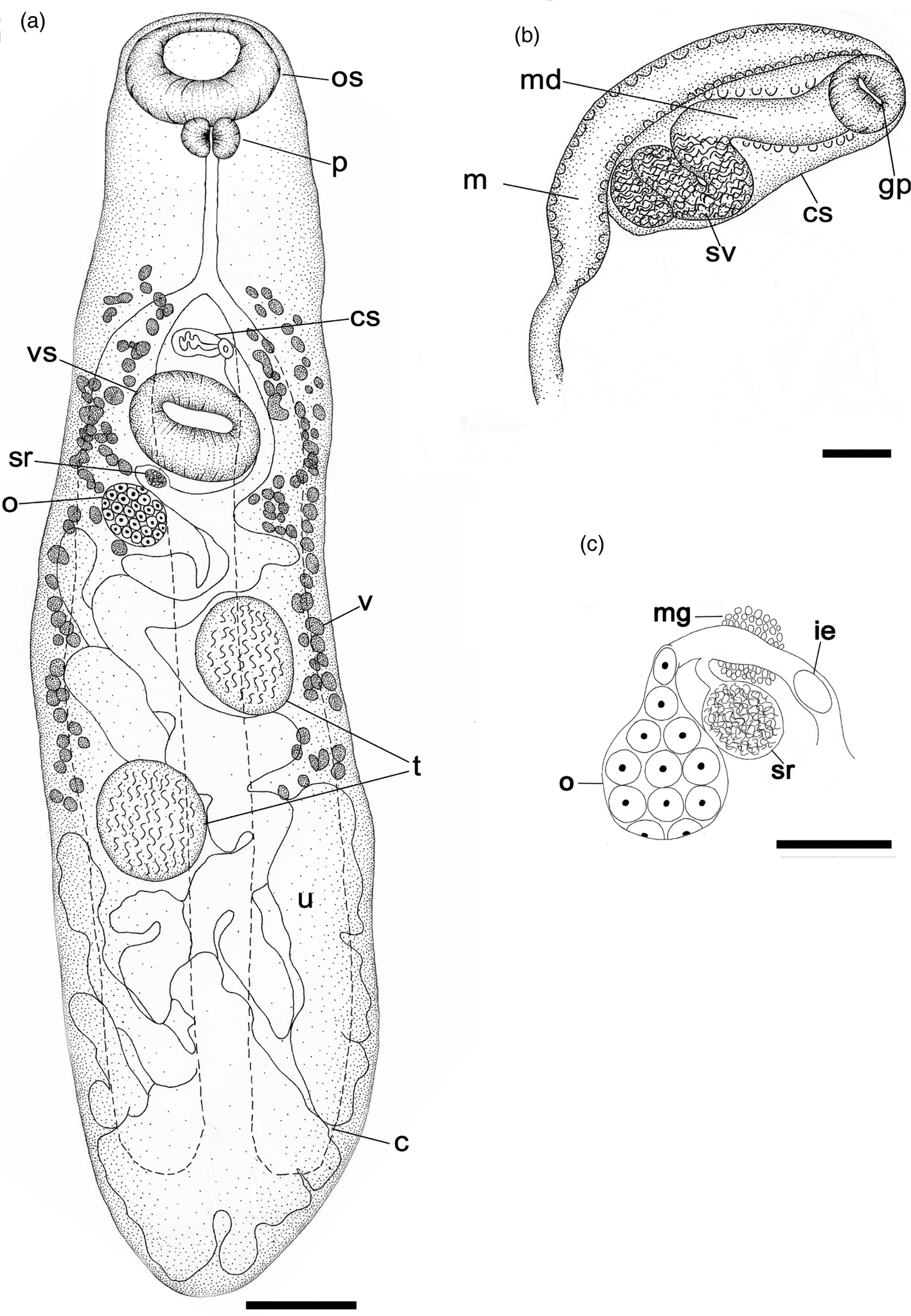
The measurements based on seven specimens are presented in table 1. Body elongated (fig. 1), ratio of body length to width 3.36–5:1. Tegument spineless. Remnants of cercarial eyespots present in anterior body region. Oral sucker subterminal, posterior margin ranging from rounded to nearly straight. Prepharynx absent. Pharynx muscular, fusiform. Ratio of body length to pharynx length 21–36:1. Oesophagus three times longer than pharynx. Ventral sucker nearly round in outline. Ratio of oral to ventral sucker width 1:0.57–0.83. Forebody represents 28–40% of body length. Intestinal bifurcation at midpoint between pharynx and ventral sucker. Caeca wide; terminating blindly near body posterior extremity. Postcaecal space represents 6–9% of body length (fig. 1a).
Comparative measurements for the four species of Prosthenhystera (in micrometres).

(a) Prosthenhystera gattii n. sp. ventral view; (b) terminal genitalia; (c) proximal end of the female genitalia. Abbreviations: c, caecum; cs, cirrus sac; gp, genital pore; ie, immature egg; m, metraterm; md, male duct; mg, Mehlis glands; o, ovary; os, oral sucker; p, pharynx; sr, seminal receptacle; sv, seminal vesicle; t, testicle; u, uterus; v, vitelline follicle; vs, ventral sucker. Scale bars: (a) 200 μm; (b) 40 μm; (c) 100 μm.

Testes 2, subspherical, oblique in anterior region of hindbody, intercaecal or overlapping caeca in ventral hindbody. Vas deferens not observed. Cirrus sac claviform, rounded at both ends, containing spiralling internal seminal vesicle at proximal end and straight male duct surrounded by prostatic cells distally; duct opening into dorsal aspect of small genital atrium; genital atrium submedian. Genital pore on ventral surface posterior to intestinal bifurcation (fig. 1b).
Ovary subspherical, submedian, smaller than testes, situated in hindbody anterior quarter. Seminal receptacle oval, adjacent to ovary and close to ventral sucker. Mehlis’ gland surrounding the middle part of the ootype. Uterus thin-walled, looping throughout hindbody, with mature eggs at distal end. Metraterm extending into forebody, ventral to and longer than cirrus sac, communicating with posteroventral aspect of genital atrium. Vitellarium comprising two groups of relatively large follicles surrounding caeca; anteriorly reaching level of intestinal bifurcation; extending posteriorly to anterior level of posterior testis. Measured eggs from distal half of uterus; with well-developed miracidia possessing two prominent fused eyespots.
Excretory vesicle I-shaped, extends to posterior level of ventral sucker. Excretory pore opens slightly subterminal on ventral surface (not drawn).
Taxonomic summary
Type host. Bryconamericus ikaa Casciotta, Almirón & Azpelicueta, 2004.
Site of infection. Gallbladder.
Type locality and collection date. Yacuí Stream, March and September of 2016.
Prevalence of infestation. Nine of 48 fish examined (19%).
Mean intensity. One per infected host.
Mean abundance. 0.15 individuals per host examined.
Material. The holotype (7407 MLP-He) and the paratype (7408 MLP-He) are deposited in the Museo de Ciencias Naturales, La Plata, Buenos Aires, Argentina.
Etymology. The species epithet ‘gattii’ is in honour of Fabian Gatti, a Forestry Engineer who works in Iguazu National Park and joined us on several field trips, during which he provided us with invaluable support and guidance during collections.
Remarks
The digeneans we collected from the gallbladder of B. ikaa conformed morphologically to the diagnosis for Prosthenhystera provided by Bray (Reference Bray, Gibson, Jones and Bray2002).
Prosthenhystera gattii n. sp. is most similar to P. caballeroi, but the testes size, cirrus sac size, seminal receptacle size and the ratio of body length to width are all greater in P. gattii n. sp. Additionally, the vitellarium of P. gattii n. sp. is more extensive than in P. caballeroi. The posterior extent of the vitellarium is in the testicular region in both species; however, the anterior extent is at a level anterior to the intestinal bifurcation in P. gattii n. sp., while it is restricted to a level posterior to the intestinal bifurcation in P. caballeroi. Additionally, the uterus is confined to the hindbody in P. gattii n. sp., whereas it extends to the level of the pharynx in P. caballeroi.
Prosthenhystera gattii n. sp. shares similar testis size, seminal receptacle size and egg size with both P. obesa and P. oonastica, but differs from both by having smaller dimensions for the body, oral sucker, pharynx and ovary. Additionally, the oesophagus length, postcaecal space length, ventral sucker length, forebody length and cirrus sac length are all shorter in P. gattii n. sp. than in P. obesa and P. oonastica. In P. gattii n. sp., the uterus is confined to the hindbody, but in P. oonastica it reaches to the caecal bifurcation with a proximal region extending into the left side of the body then extending into the right hindbody, and, according to Kohn et al. (Reference Kohn, Fernandes and Baptista-Farias1997), the uterus in P. obesa fills the hindbody and extends to the oral sucker.
Molecular analyses
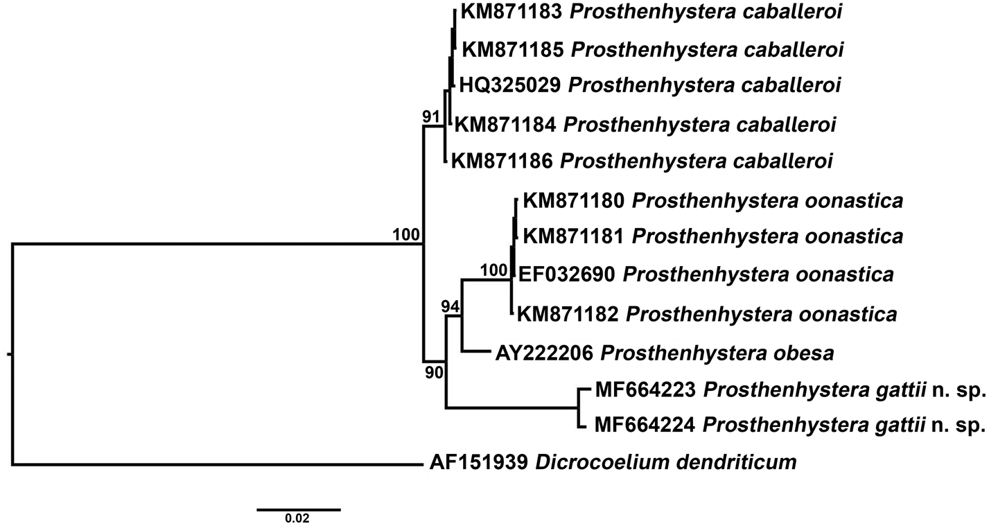
The partial sequences of the 28S rDNA gene from two sequenced individuals were 1201 bp long. The alignment containing the new sequences and the sequences obtained from GenBank was 1276 bp long. The BI analysis of the dataset containing the four sequences of P. oonastica, the AY222206 P. obesa sequence, the five sequences of P. caballeroi, the two sequences of P. gattii n. sp. and rooted by the dicrocoelid, D. dendriticum, produced a phylogram (fig. 2) with strong nodal support that indicates that P. gattii n. sp. is more closely related to the branch formed by P. obesa + P. oonastica than it is to P. caballeroi. The P-value matrix obtained using MEGA version 6.0 shows 4% of genetic distance between P. gattii n. sp. against all the species analysed (table 2).
Phylogram resulting from Bayesian inference (20,000,000 generations) of partial 28S rDNA gene sequences of Prosthenhystera gattii n. sp. rooted by Dicrocoelium dendriticum (Dicrocoeliidae). Branch support values indicate posterior probabilities. Scale bar shows number of substitutions per site.

Distance matrix on 28S rDNA of uncorrected p-distances among Prosthenhystera spp. and the outgroup (Dicrocoelium dendriticum).

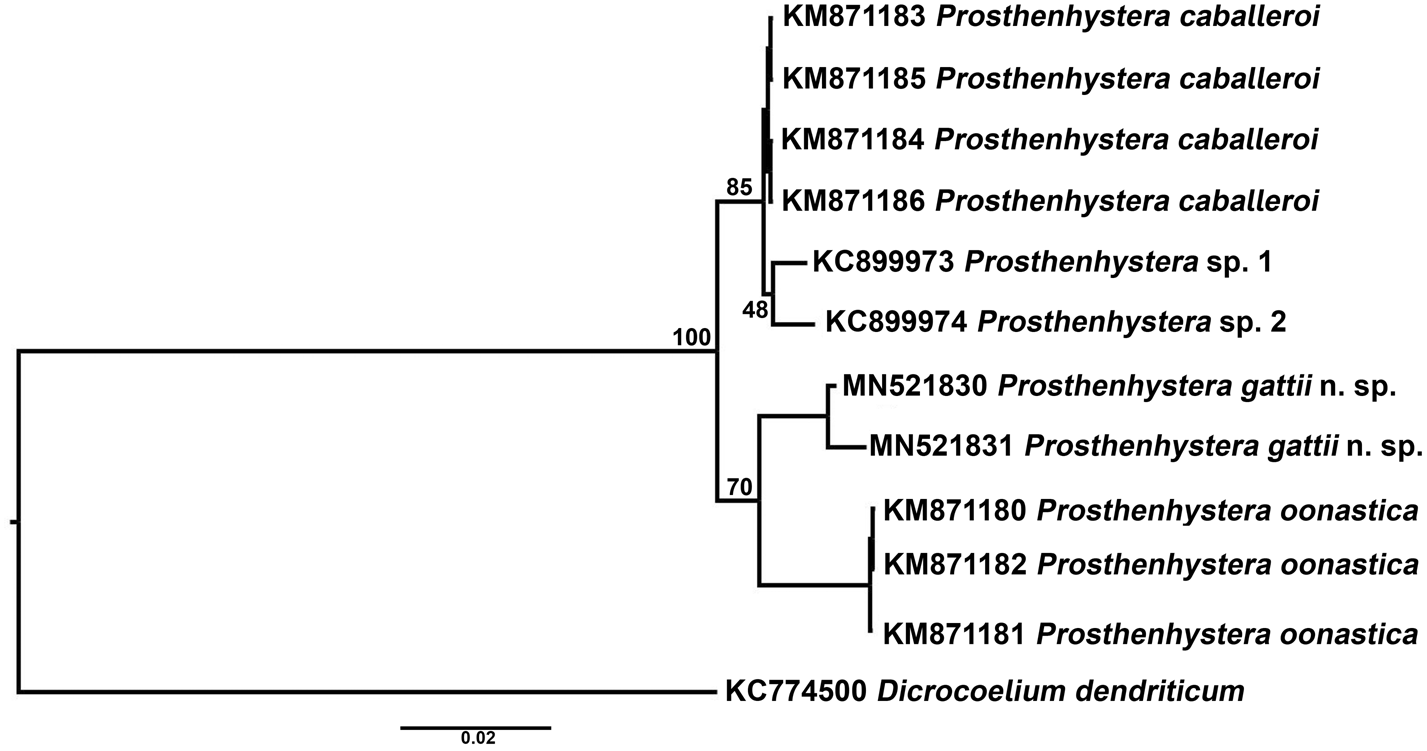
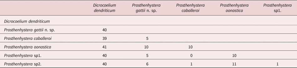
The two fragments of the partial ITS rDNA gene from the new species measured between 802 and 810 bp. After aligning the new sequences with the sequences obtained from GenBank, a matrix of 815 bp was constructed. The BI of the dataset containing the three sequences of P. oonastica, the four sequences of P. caballeroi, two sequences of Prosthenhystera sp., the two sequences of P. gattii n. sp. and rooted by the dicrocoelid, D. dendriticum, produced a phylogram (fig. 3) that indicates P. gattii n. sp. is related to P. oonastica. The P-value matrix obtained using MEGA version 6.0 show 5–6% of genetic distance between P. gattii n. sp. against P. caballeroi and Prosthenhystera sp., and 10% with P. oonastica (table 3).
Phylogram resulting from Bayesian inference (20,000,000 generations) of partial ITS1-5.8S-ITS2 gene sequences of Prosthenhystera gattii n. sp. rooted by Dicrocoelium dendriticum (Dicrocoeliidae). Branch support values indicate posterior probabilities.

Distance matrix on ITS1-5.8S-ITS2 of uncorrected p-distances among Prosthenhystera spp. and the outgroup (Dicrocoelium dendriticum).

Cytogenetic analysis
The division rate is very low, with the total number of cells in division not exceeding 20 in the different stages (Montes et al., Reference Montes, Barneche, Croci, Rodriguez Gil, Curran, Ferrari, Casciotta and Martorelli2020a). We observed ten metacentric chromosomes in mitotic metaphase, five bigger than the other five. The ten chromosomes in anaphase are displaced in each pole with a ‘V’ shape (Montes et al., Reference Montes, Barneche, Croci, Rodriguez Gil, Curran, Ferrari, Casciotta and Martorelli2020b).
Discussion
The two gene regions produced phylograms with identical topologies estimating the relationships among the Prosthenhystera species. The 28S rDNA fragment alone supported a closer relationship between P. obesa and P. oonastica with P. gattii n. sp., whereas the ITS1-5.8S-ITS2 region suggested a close relationship between P. gattii n. sp. and P. oonastica, with very low nodal support (70%), which was confirmed with a high genetic distance between P. oonastica and P. gattii n. sp. (10%). Considering only the genetic distance, we could say P. gattii n. sp. is more closely related with P. caballeroi and the Prosthenhystera sp. reported by Martínez-Aquino et al. (Reference Martínez-Aquino, Ceccarelli and Perez-Ponce de Leon2013). Kohn et al. (Reference Kohn, Fernandes and Baptista-Farias1997) conducted an exhaustive survey for parasites in the gallbladder of several fish species from Brazil and concluded that P. obesa exhibits a large variation in body shape and size, whether infecting the same or different host species, and from different localities. The present discovery and the observations of Tkach & Curran (Reference Tkach and Curran2015) highlight the need for further revision of the Callodistomidae involving molecular tools to uncover cryptic species.
Presently, no sequence data representing the African genera Callodistomum and Cholepotes or the other American genus Teratotrema are available. The disjunct distribution between the African genera and American genera is intriguing since no marine forms are known from the family. Clearly, the evolutionary lineage of the family needs examining with molecular tools to determine whether this distribution is legitimate or if convergence is present. Such morphological convergence might be due to influence by evolutionary forces related to site specificity in the gallbladder of hosts. Alternatively, the morphological similarities might be manifested by sharing an ancient genetic lineage that predates the separation of America from Africa and the formation of the formidable barrier to gene flow that is the Atlantic Ocean. In either case, when molecular data finally become available from African species and such questions can be addressed, solving the conundrum of whether the family represents a natural lineage or a convergent unrelated lineage should excite all biologists. So, in the absence of life history data, the revision of the family must rely heavily on molecular analyses of South American and African forms to effectively elucidate the relationships among this group's species, which exhibit minimal interspecific morphological variation yet infect a broad range of hosts on two isolated continents.
The present study provided the first cytological data for the Callodistomidae. The karyotype was not analysed in detail by the low number of division cells found. The number of chromosomes of closely related Gordoderidae (Tkach & Curran, Reference Tkach and Curran2015) vary between 12 and 18 (Britt, Reference Britt1917; Walton, Reference Walton1959; Petkevičiute et al., Reference Pritchard and Kruse2004; Sofi et al., Reference Sofi, Ahmad, Sheikh, Mohi, Din and Fazili2015), against the ten chromosomes present in the new Callodistomidae species. Prosthenhystera gattii n. sp. has all metacentric chromosomes and the diploid chromosomes number is lower than in any previously studied species from the Gorgoderidae. In that family, the species studied until now have acrocentric, submetacentric and metacentric chromosomes. The present results provide new data and complement the description made by Petkevičiute & Stanevičiute (Reference Petkevičiute and Stanevičiute2008) about the karyotype homogeneity respect the number, morphology and number of chromosomes in these two close families.
Acknowledgements
Our thanks to the Administration de Parques Nacionales for allowing us to work inside the National Park Iguazú; to the Facultad de Ciencias Naturales y Museo (UNLP), Consejo Nacional de Investigaciones Cientificas y Tecnicas (CONICET) and Centro de Estudios Parasitologicos y Vectores (CEPAVE) for providing the infrastructure necessary to carry out this work; to Adriana Almiron, Liliana Ciotek and Pablo Giorgis for their assistance with gill netting; to the people of the CIES, Nahuel, Facundo Luque, Leonardo Quiroga, Modesta Rodriguez; to the Delegacion Regional Noreste for their assistance, especially Andres Bosso, Dalma Raymundi and Veronica Bernava; to Guillermo Gil, coordinator of the CIES for providing our accommodation; and to Marcia Montes for making the description drawings.
Financial support
This work was supported by the Consejo Nacional de Ciencia y Tecnologia (CCT-CONICET-La Plata) (S.R.M., grant number PIP-0015); Centro de Estudios Parasitologicos y Vectores (grant number PUE 3334/16); Universidad de La Plata for (J.D. and S.R.M., grant number N859); and Fondo para la Investigación Científica y Tecnológica (FONCyT) (M.M.M., grant number BID PICT 2016-4153).
Conflicts of interest
None.
Ethical standards
The authors assert that all procedures contributing to this work comply with the ethical standards of the relevant national and institutional guides on the care and use of laboratory animals.