Introduction
Conduct disorder (CD) is characterized by a repetitive pattern of aggressive and antisocial behavior (American Psychiatric Association, 2000). It results in substantial personal and financial costs for the affected individuals, their families, and society in general (Erskine et al., Reference Erskine, Ferrari, Polanczyk, Moffitt, Murray, Vos and Scott2014; Rivenbark et al., Reference Rivenbark, Odgers, Caspi, Harrington, Hogan, Houts and Moffitt2018), and is the most common reason for referral to Child and Adolescent Mental Health Services in the UK (National Institute for Health and Care Excellence, 2013). CD is also linked to negative adult outcomes, such as mental and physical health problems (Copeland, Wolke, Shanahan, & Costello, Reference Copeland, Wolke, Shanahan and Costello2015; Odgers et al., Reference Odgers, Caspi, Broadbent, Dickson, Hancox, Harrington and Moffitt2007) and personality disorders (Burt, Donnellan, Iacono, & McGue, Reference Burt, Donnellan, Iacono and McGue2011; Robins, Reference Robins1978). It is therefore important to understand its etiology, which may help in developing effective treatments and prevention programs.
There is increasing evidence that CD may have a neurobiological basis, with many studies reporting differences in brain function or structure in children or adolescents with CD or conduct problems compared with typically developing controls (Alegria, Radua, & Rubia, Reference Alegria, Radua and Rubia2016; Fairchild et al., Reference Fairchild, Hawes, Frick, Copeland, Odgers, Franke and De Brito2019; Rogers & De Brito, Reference Rogers and De Brito2016). This research has been extremely valuable in identifying the neuroanatomical and functional correlates of CD and callous-unemotional (CU) traits. However, the cross-sectional nature of these studies means it is unclear whether structural or functional abnormalities in regions such as the orbitofrontal cortex or insula precede the disorder or reflect a secondary effect of having CD or lifestyle factors associated with the condition (e.g. substance abuse or sustaining head injuries when fighting). It is possible that these brain abnormalities are caused by the same etiological factors (genetic or environmental risk factors) that led the individual to develop the disorder (Bidwell, Willcutt, Defries, & Pennington, Reference Bidwell, Willcutt, Defries and Pennington2007). Relevant to this point, twin studies have shown that brain volume, and particularly volume of the frontal lobe, is highly heritable (Jansen, Mous, White, Posthuma, & Polderman, Reference Jansen, Mous, White, Posthuma and Polderman2015). It has also been shown that CD and related phenotypes such as criminal behavior and antisocial personality disorder cluster within families. For example, Blazei, Iacono, and McGue (Reference Blazei, Iacono and McGue2008) found a strong resemblance between biological fathers and sons in terms of antisocial behavior – particularly if the father resided in the home. Similarly, Christiansen et al. (Reference Christiansen, Chen, Oades, Asherson, Taylor, Lasky-Su and Faraone2008) found that the siblings of those with attention-deficit/hyperactivity disorder (ADHD) and conduct problems were five times more likely to develop ADHD and conduct problems and three times more likely to develop conduct problems than the siblings of children with ADHD alone. Similar patterns of familial aggregation have been reported in criminological studies – for example, the Cambridge Study in Delinquent Development found that just 6% of the families accounted for 50% of all criminal convictions, and conviction rates were two times higher in the sons of fathers with a history of criminal behavior than the sons of fathers without such a history (Farrington, Barnes, & Lambert, Reference Farrington, Barnes and Lambert1996).
Although these studies have provided compelling evidence that antisocial behavior clusters within families, far less is known about the brain mechanisms which explain this family resemblance, even though this is an important issue with implications for the development of intervention and prevention programs. One strategy that can be adopted to study the brain mechanisms that may mediate genetic or environmental risk for CD is to study the first-degree relatives of affected probands who do not show the disorder themselves, but may still carry markers of familial risk. For example, Ersche et al. (Reference Ersche, Jones, Williams, Turton, Robbins and Bullmore2012) employed this strategy to investigate whether brain abnormalities are associated with familial risk for substance dependence or reflect the neurotoxic effects of prolonged drug use. Substance-dependent individuals and their unaffected siblings were found to display common neuroanatomical abnormalities in brain regions involved in inhibitory control, suggesting that they are a risk factor for substance dependence, rather than reflecting the secondary consequences of drug use (i.e. drug-induced damage). A similar study investigating familial risk markers for autism found common reductions in activation in brain regions involved in biological motion perception and social cognition (e.g. the superior temporal sulcus) in children with autism and their unaffected relatives (URs), relative to controls (Kaiser et al., Reference Kaiser, Hudac, Shultz, Lee, Cheung, Berken and Pelphrey2010).
Applying this logic to CD, if similar alterations in brain structure are observed in adolescents with CD and their URs, this would indicate that neuroanatomical abnormalities and CD co-segregate within families (are inherited together) and that such structural changes may mediate the effects of genetic risk for CD. Studying unaffected first-degree relatives, as well as affected probands with CD, would therefore help us to address the question of whether neuroanatomical changes increase risk for developing CD or reflect the secondary consequences of having CD and associated lifestyle factors. A further advantage of studying URs, who ‘beat the odds’ by remaining free from severe antisocial behavior despite being at increased risk, is that protective or compensatory brain changes might be observed in this group which counteract the effects of familial risk. Relevant to this point, Kaiser et al. (Reference Kaiser, Hudac, Shultz, Lee, Cheung, Berken and Pelphrey2010) identified potential compensatory effects in the URs of ASD probands. They showed greater ventromedial prefrontal cortex activity than the autistic or typically developing control groups when viewing point-light displays of biological motion.
Most existing studies investigating brain structure in CD have employed voxel-based morphometry (VBM) methods which test for differences in gray matter volume across the whole brain. However, using this composite, intensity-based measure is problematic because volume in a given region is a function of its cortical thickness and surface area, as well as cortical folding, which show distinct genetic etiologies (Panizzon et al., Reference Panizzon, Fennema-Notestine, Eyler, Jernigan, Prom-Wormley, Neale and Kremen2009), developmental trajectories (Raznahan et al., Reference Raznahan, Shaw, Lalonde, Stockman, Wallace, Greenstein and Giedd2011), and underlying cellular mechanisms (Rakic, Reference Rakic2009).
Accordingly, we compared adolescents with CD and their URs using surface-based morphometry (SBM), to examine whether these groups show common or distinct abnormalities in cortical structure compared with typically developing adolescents. On the basis of previous SBM findings (Fairchild et al., Reference Fairchild, Toschi, Hagan, Goodyer, Calder and Passamonti2015; Hyatt, Haney-Caron, & Stevens, Reference Hyatt, Haney-Caron and Stevens2012; Smaragdi et al., Reference Smaragdi, Cornwell, Toschi, Riccelli, Gonzalez-Madruga, Wells and Fairchild2017; Wallace et al., Reference Wallace, White, Robustelli, Sinclair, Hwang, Martin and Blair2014), we predicted that adolescents with CD would show structural alterations in the insula, orbitofrontal cortex, superior temporal gyrus, and inferior parietal cortex compared with typically developing adolescents. We also hypothesized that the URs of CD probands would show similar structural abnormalities, albeit possibly at an intermediate level, as their loading of genetic or environmental risk may be lower than the probands. Consistent with this, a recent study found that ADHD probands and their unaffected siblings both showed lower orbitofrontal cortex volume (Bralten et al., Reference Bralten, Greven, Franke, Mennes, Zwiers, Rommelse and Buitelaar2016). Given prior evidence that ADHD comorbidity may be important in determining the extent of structural changes observed in CD (Fairchild et al., Reference Fairchild, Toschi, Hagan, Goodyer, Calder and Passamonti2015; Smaragdi et al., Reference Smaragdi, Cornwell, Toschi, Riccelli, Gonzalez-Madruga, Wells and Fairchild2017), we controlled for ADHD symptoms in a supplementary analysis. Although exploratory in nature, we also tested for potential ‘compensatory’ or ‘protective’ structural changes in the URs. However, due to the lack of previous studies, we had no a priori predictions regarding the loci and direction of such effects. Lastly, based on evidence of subcortical alterations in youths with CD (Rogers & De Brito, Reference Rogers and De Brito2016; Wallace et al., Reference Wallace, White, Robustelli, Sinclair, Hwang, Martin and Blair2014), we tested for group effects on subcortical volumes. We predicted that the CD group would show lower amygdala volume compared to the controls, and similar reductions might be observed in URs.
Methods
Participants
Healthy control participants (n = 41) were recruited from mainstream schools and colleges, whereas participants with CD (n = 43) were mainly recruited from specialist schools, pupil referral units and Youth Offending Services in the Hampshire area. The URs (n = 24) were recruited directly from the families of the CD participants, as well as the aforementioned recruitment sources. Participants were aged between 12 and 18 years. All parents/carers completed a Family History Screen, consisting of three questions assessing current and lifetime psychopathology, behavioral problems, and criminal convictions, in the participants' first-degree relatives. This screen was designed to identify siblings of adolescents with CD who did not meet the diagnostic criteria for CD themselves. In addition, it enabled us to identify the unaffected offspring of parents who had previously displayed CD and ensure that the controls had no family history of CD.
Diagnostic and questionnaire measures
The Kiddie-Schedule of Affective Disorders and Schizophrenia-Present and Lifetime version (K-SADS-PL; Kaufman et al., Reference Kaufman, Birmaher, Brent, Rao, Flynn, Moreci and Ryan1997), a semi-structured diagnostic interview based on DSM-IV-TR criteria (APA, 2000), was used to screen for CD and other common psychiatric disorders. The participants and their parents/carers were interviewed separately to ensure confidentiality, and the information from each interview was combined such that a symptom was considered present if it was endorsed by either informant (Kaufman et al., Reference Kaufman, Birmaher, Brent, Rao, Flynn, Moreci and Ryan1997). Even if the initial screen items for CD and ADHD were not endorsed, the full supplements for these disorders were always completed to obtain dimensional information on these disorders for all participants.
The self-report version of the Inventory of Callous-Unemotional traits (ICU; Essau, Sasagawa, & Frick, Reference Essau, Sasagawa and Frick2006) was used to assess CU traits. It contains 24 items scored on a 0–3 scale, from ‘not at all true’ to ‘definitely true’ (Cronbach's Alpha in present sample = 0.82). Factor analysis has revealed that the ICU captures three distinct dimensions of behavior termed callousness, uncaring, and unemotional (Essau et al., Reference Essau, Sasagawa and Frick2006), therefore scores for these subscales are also reported. The self-report Youth Psychopathic traits Inventory (Andershed, Kerr, Stattin, & Levander, Reference Andershed, Kerr, Stattin, Levander, Blaauw and Sheridan2002) was used to measure psychopathic personality traits. It contains 50 items, each scored on a 1–4 point scale, from ‘does not apply at all’ to ‘applies very well’ (Cronbach's Alpha in present sample = 0.93), and as well as total scores, it can be divided into Grandiose-Manipulative, CU, and Impulsive-Irresponsible subscales corresponding to the three-facet model of psychopathy (Andershed et al., Reference Andershed, Kerr, Stattin, Levander, Blaauw and Sheridan2002). Further information about these questionnaires' psychometric properties and their factor structures can be found in Supplementary Materials. The Wechsler Abbreviated Scale of Intelligence was used to estimate full-scale IQ (Wechsler, Reference Wechsler1999). Lastly, the Edinburgh Handedness Inventory (Oldfield, Reference Oldfield1971) was used to assess handedness.
Ethical approval
The study was approved by the University of Southampton Ethics Committee, the University Hospital Southampton NHS Trust, Southampton City Council Children's Services and the Hampshire County Council Research and Evaluation Unit. Participants aged ⩾16 provided written informed consent, whereas parents or carers provided informed consent and participants provided assent if below age 16.
Procedure
Once they had been screened for psychiatric disorders and standard MRI exclusion criteria, such as claustrophobia, participants were invited to the Southampton General Hospital for a magnetic resonance imaging (MRI) scan lasting 35–40 min. The structural (T1-weighted) scan was the first sequence performed during the scanning session, and was repeated as needed until usable data, uncontaminated by movement, had been collected. This was determined by a trained radiographer.
Data acquisition
Structural MRI data were acquired using a 1.5-Tesla Siemens Avanto scanner (Siemens Medical Solutions, Erlangen, Germany). We acquired T1-weighted three-dimensional MPRAGE images (voxel size = 1.2 × 1.2 × 1.2 mm, repetition time = 2400 ms, echo time = 3.62 ms, flip angle = 8°, 160 slices). Total scanning time was 7 min, 41 s. After scanning, the structural images were reviewed by a consultant neuroradiologist to screen for neurological abnormalities. We initially included 108 participants, but five participants (three controls; two CD) were excluded due to having cysts or tumors, leaving 103 participants with usable MRI data.
SBM analyses: cortical volume, thickness, surface area (SA) and local gyrification index (lGI) and subcortical volumes
MRI-based quantification of cortical volume, thickness, SA and folding (quantified using lGI) was performed using FreeSurfer 5.3.0 (http://surfer.nmr.mgh.harvard.edu). This method has been described in detail (Fischl, Reference Fischl2012). Briefly, the procedure involves segmentation of white matter, tessellation of the gray-white matter junction to construct representations of the gray/white matter boundary and cortical surface. Each participant's cortex was then visually inspected and, if necessary, manually edited by one of the authors (N.T.), blind to group status. This involved: (i) realignment of each subject's image to the Montreal Neurological Institute template; (ii) setting intensity normalization control points where brain matter was erroneously skull-stripped; and (iii) adjustment of the watershed parameters of the skull strip. From this reconstruction, vertex-wise estimates of both cortical thickness and cortical area were obtained. lGI, which measures the amount of cortical folding within v. outside the sulcus, was calculated using the method outlined by Schaer et al. (Reference Schaer, Cuadra, Schmansky, Fischl, Thiran and Eliez2012). In order to map the participants' brains to a common space, reconstructed surfaces were registered to an average cortical surface atlas using a nonlinear procedure that optimally aligns sulcal and gyral features across individuals (Fischl, Sereno, & Dale, Reference Fischl, Sereno and Dale1999). Finally, we estimated amygdala, hippocampus, caudate, pallidum, putamen, thalamus, and nucleus accumbens volumes using FreeSurfer's automatic segmentation pipeline (Fischl et al., Reference Fischl, Salat, Busa, Albert, Dieterich, Haselgrove and Dale2002).
Statistical analyses
We tested for group differences in demographic and clinical variables using analyses of variance, with independent t tests used to follow up significant F tests; chi-square tests were used for group comparisons of nominal variables (e.g. sex).
For each hemisphere, group differences in cortical volume, thickness, surface area and lGI at each vertex were tested using a general linear model (GLM) with age, sex, IQ and total intracranial volume (TIV) orthogonalized to sex included as covariates of no interest. We also repeated the analyses including lifetime ADHD symptoms as a further covariate (these results are reported in online Supplementary Table 1, available on-line). Given previous evidence suggesting that childhood-onset (CO-CD) and adolescence-onset (AO-CD) variants of CD may differ quantitatively in brain structure or function (Fairchild et al., Reference Fairchild, Hagan, Walsh, Passamonti, Calder and Goodyer2013), we initially ran analyses comparing these subgroups (i.e. CO-CD>AO-CD, AO-CD>CO-CD). As there were no significant differences between subgroups, they were treated as a combined group in the comparisons with URs and HCs.
After applying a vertex-wise/cluster-forming threshold of p = 0.05, the level of statistical significance was subject to a further cluster-wise p (CWP) value correction procedure for multiple comparisons based on a Monte Carlo z-field simulation (Hagler, Saygin, & Sereno, Reference Hagler, Saygin and Sereno2006). Clusters are only reported if they met a whole-brain corrected threshold of CWP ⩽0.05.
Lastly, we tested for group differences in subcortical volumes using one-way analyses of covariance with age, sex, IQ and TIV orthogonalized to sex included as covariates of no interest, whilst applying a false-discovery-rate (FDR) correction for multiple comparisons at q = 0.05. Significant group effects were followed up with pairwise t tests. These analyses were repeated including lifetime ADHD symptoms as a covariate.
Results
Sample characteristics
Demographic and clinical characteristics of the sample are reported in Table 1. The groups differed in age (p = 0.01), with the URs being around a year younger than the other groups. The groups also differed in estimated IQ (p < 0.001), with the CD group having the lowest average IQ and the control group having the highest. However, the groups were matched in terms of sex and handedness. As expected, the CD group reported higher rates of CD symptoms, ADHD symptoms, CU traits, and psychopathic traits than the other groups (ps < 0.001). Critically, the HCs and URs did not differ on any clinical or personality variable, confirming the ‘unaffected’ nature of the latter group.
Demographic and clinical characteristics of the sample

Note. Means are presented with standard deviations in parentheses. Group differences were assessed with one-way ANOVA F-tests and pairwise Bonferroni-adjusted t tests (continuous variables) and Chi-squared tests (categorical variables).
HC = healthy controls; UR = unaffected relatives; CD = conduct disorder; M = male; F = female; R = right-handed; L = left-handed; A = ambidextrous; IQ = intelligence quotient; ADHD = attention-deficit/hyperactivity disorder; ICU = Inventory of Callous-Unemotional traits (self-report version); YPI = Youth Psychopathic traits Inventory. Participants who developed symptoms of CD before age 10 were classified as having ‘childhood-onset’ CD, whereas those who only displayed symptoms of CD after age 10 were classified as having ‘adolescence-onset’ CD.
a n missing = 1 (CD group).
b n missing = 1 (HC group).
c n missing = 2 (HC group).
d n missing = 3 (2 HCs, 1 CD).
SBM results: potential markers of familial risk for antisocial behavior
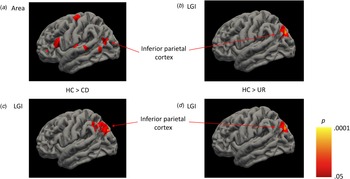
Relative to controls, the CD group showed lower left inferior parietal cortex surface area, whereas the URs showed lower cortical folding in this region (Fig. 1; Table 2). When controlling for comorbid ADHD symptoms, both the CD and UR groups showed lower left inferior parietal cortical folding (online Supplementary Table 1).
Neuroanatomical markers of familial risk for CD that were observed in both the CD probands and the URs compared with controls. (a) Left inferior parietal cortical surface area was lower in participants with CD compared with healthy controls (HC). (b) Left inferior parietal cortical folding was lower in URs than healthy controls. (c) Left inferior parietal cortical folding was reduced in participants with CD compared with healthy controls when adjusting for comorbid ADHD symptoms. (d) Left inferior parietal cortical folding was lower in URs than healthy controls when adjusting for comorbid ADHD symptoms.

Cortical volume, thickness, surface area and gyrification differences between the conduct disorder, unaffected relative and healthy control groups, when not including lifetime ADHD symptoms as a covariate

ADHD, attention-deficit/hyperactivity disorder; CD, conduct disorder; CWP, cluster-wise-p value; HC, healthy control; L, left; NVtxs, number of vertices; Max, maximum -log10(p value) in the cluster; R, right; UR, unaffected relatives.
Note. Only significant pairwise comparisons between the groups are reported.
Effects related to CD but not observed in URs – non-familial risk
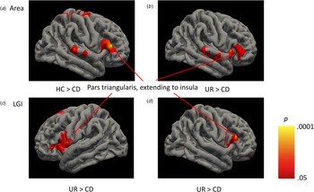
In contrast, the CD group showed lower volume in left insula and right pars triangularis extending to right insula compared with controls, and lower surface area in left insula and right pars triangularis/insula compared with both the controls and URs (Fig. 2; Table 2).
Cortical structure alterations observed in the Conduct Disorder group compared to the healthy controls and URs, reflecting non-familial risk. (a) Right pars triangularis surface area (extending to insula) was lower in participants with CD compared with healthy controls (HC). (b) Right pars triangularis surface area (extending to insula) was lower in participants with CD compared with the URs. (c) Left pars triangularis cortical folding (extending to insula) was lower in participants with CD compared with the URs. (d) Right pars triangularis cortical folding (extending to insula) was lower in participants with CD compared with the URs.

Further structural differences between the CD and control groups that were not observed in the URs included lower bilateral pericalcarine, left pars opercularis and right precentral gyrus surface area in the CD group (Table 2). In addition, the CD group showed greater cortical thickness in left superior frontal gyrus and superior temporal cortex and right frontal pole compared with the URs (Table 2).
Potential compensatory effects in the URs
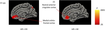
There were also several effects unique to the URs – they showed increased folding in rostral anterior cingulate cortex compared with controls and increased medial orbitofrontal cortex folding compared with the CD group (Fig. 3; Table 2). The URs also showed greater folding in lingual gyrus and inferior temporal cortex compared with controls, and greater folding in bilateral pars triangularis/insula and left superior frontal gyrus compared with the CD group.
Potential protective or compensatory structural changes observed in the URs compared with the healthy control and CD groups. (a) Medial orbitofrontal cortical folding was higher in the URs compared with the CD group. (b). Rostral anterior cingulate cortical folding was higher in the URs compared with the healthy controls (HC).

Impact of adjusting for comorbid ADHD
When including ADHD symptoms as a covariate, the similarities between the CD and UR groups were amplified – as mentioned above, both groups showed lower left inferior parietal folding compared with controls (online Supplementary Table 1). There were also differences in cortical thickness which appeared specific to the CD group – they showed greater medial orbitofrontal cortical thickness compared to controls and greater superior frontal cortical thickness compared to URs (online Supplementary Table 1). However, some of the CD-control differences were rendered non-significant, such as differences in left insula and right pars opercularis volume (HC >CD). In addition, the group differences in surface area (e.g. lower insula and pars triangularis surface area in CD) were not significant when adjusting for ADHD symptoms. As might be expected given the low level of ADHD symptoms in URs, differences between this group and the controls remained significant when adjusting for ADHD – including increased rostral anterior cingulate folding.
Subcortical volumes
There were no group differences in amygdala, hippocampus, thalamus, caudate, pallidum, putamen or nucleus accumbens volumes when controlling for multiple comparisons. There was, however, a trend toward a group effect in left thalamus (p = 0.088, uncorrected), which became nominally significant when IQ was not included as a covariate. Post-hoc tests revealed lower left thalamus volume in CD v. HC participants (p = 0.028, Hedges's g = −0.50), but no other pairwise differences. See Supplementary Materials for more information.
Discussion
In the present study, we used SBM and subcortical segmentation methods to investigate whether probands with CD and their URs show similar or distinct changes in brain structure compared with typically developing adolescents, and identify potential protective or compensatory alterations in the URs. The observation of common structural alterations in CD probands and URs would indicate that CD and related neuroanatomical changes co-segregate within families, suggesting the latter may partly mediate the effects of genetic or environmental risk for CD. We also assessed four different measures of cortical structure – cortical volume, thickness, surface area, and folding – and subcortical volumes to provide greater specificity regarding the structural changes associated with CD and its familial risk.
Our first key finding was that similar alterations in left inferior parietal cortical structure were observed in individuals with CD and their URs. The CD participants showed lower surface area in left inferior parietal cortex, whereas the URs showed lower folding in this region, compared with controls. When controlling for comorbid ADHD, both groups showed lower left inferior parietal folding compared with controls. These findings provide the first available evidence that reductions in inferior parietal surface area and folding might constitute a neuroanatomical endophenotype for CD which is present in CD probands and their URs. This suggests that alterations in inferior parietal cortical structure may partly mediate the effects of familial risk for CD. Previous SBM studies have reported structural abnormalities in the inferior parietal cortex, or adjacent areas such as supramarginal gyrus, in CD (Fahim et al., Reference Fahim, He, Yoon, Chen, Evans and Perusse2011; Jiang et al., Reference Jiang, Guo, Zhang, Gao, Wang, Situ and Huang2015; Smaragdi et al., Reference Smaragdi, Cornwell, Toschi, Riccelli, Gonzalez-Madruga, Wells and Fairchild2017; Wallace et al., Reference Wallace, White, Robustelli, Sinclair, Hwang, Martin and Blair2014). The left inferior parietal cortex is implicated in language comprehension, theory of mind, action observation (Molenberghs, Cunnington, & Mattingley, Reference Molenberghs, Cunnington and Mattingley2012), and perhaps most intriguingly, facial emotion recognition (Zhang, Song, Liu, & Liu, Reference Zhang, Song, Liu and Liu2016). The latter function appears relevant to our earlier finding that CD probands and their URs show similar deficits in facial emotion recognition (Sully, Sonuga-Barke, & Fairchild, Reference Sully, Sonuga-Barke and Fairchild2015).
Our second key finding was that the volume of the insula and surrounding frontal lobe structures such as the pars opercularis was lower in CD compared with control participants. Critically, these changes in volume appeared to be driven by reductions in insula and pars opercularis surface area, rather than cortical thickness, in the CD group. The CD group also showed lower insula and pars triangularis surface area compared to URs. To our knowledge, although several VBM studies have reported lower insula gray matter volume in CD (Fairchild et al., Reference Fairchild, Passamonti, Hurford, Hagan, von dem Hagen, van Goozen and Calder2011, Reference Fairchild, Hagan, Walsh, Passamonti, Calder and Goodyer2013; Sterzer, Stadler, Poustka, & Kleinschmidt, Reference Sterzer, Stadler, Poustka and Kleinschmidt2007) and the insula was identified in a recent meta-analysis of VBM studies (Rogers & De Brito, Reference Rogers and De Brito2016), this is the first study to examine the underlying basis of such volumetric changes. The finding that reductions in surface area, rather than cortical thickness, drive reductions in insula volume observed in CD is consistent with data from normative studies showing that surface area is more strongly related to volume than cortical thickness (Im et al., Reference Im, Lee, Lyttelton, Kim, Evans and Kim2008). The anterior insula is considered to play a critical role in processing emotional (especially negative) stimuli (Calder, Keane, Manes, Antoun, & Young, Reference Calder, Keane, Manes, Antoun and Young2000), empathy (Singer et al., Reference Singer, Seymour, O'Doherty, Kaube, Dolan and Frith2004), and awareness of one's own physiological and emotional states (Craig, Reference Craig2009). Consequently, structural deficits in the insula might explain why adolescents with CD show deficits in empathy (Martin-Key, Brown, & Fairchild, Reference Martin-Key, Brown and Fairchild2017) and learning from punishment (Kohls et al., Reference Kohls, Baumann, Gundlach, Scharke, Bernhard, Martinelli and Konrad2020), and reduced sensitivity to losses when making decisions (Fairchild et al., Reference Fairchild, van Goozen, Stollery, Aitken, Savage, Moore and Goodyer2009). On the other hand, the fact that reductions in insula volume and surface area were not observed in the URs challenges the idea that they mediate the effects of familial risk for CD. Of interest, we found that adolescents with CD, but not their URs, showed heightened risk-taking in a gambling task (Sully, Sonuga-Barke, Savage, & Fairchild, Reference Sully, Sonuga-Barke, Savage and Fairchild2016). It should also be noted that these insula volume and surface area differences were rendered non-significant when adjusting for comorbid ADHD symptoms, suggesting that they are not related to CD specifically or are more pronounced in participants with comorbid CD + ADHD.
We also found greater medial orbitofrontal cortical thickness in the CD group compared with controls, although only when adjusting for comorbid ADHD, whereas cortical thickness in the superior frontal gyrus and frontal pole was increased in the CD group compared with the URs. The medial orbitofrontal cortex is involved in representing the reward value of stimuli (Liu, Hairston, Schrier, & Fan, Reference Liu, Hairston, Schrier and Fan2011) and social cognitive processes (Molenberghs, Johnson, Henry, & Mattingley, Reference Molenberghs, Johnson, Henry and Mattingley2016). The frontal pole is implicated in executive functions – especially tasks in which multiple cognitive processes must be monitored simultaneously (Mansouri, Koechlin, Rosa, & Buckley, Reference Mansouri, Koechlin, Rosa and Buckley2017). Previous studies have reported lower cortical thickness (Fahim et al., Reference Fahim, He, Yoon, Chen, Evans and Perusse2011; Jiang et al., Reference Jiang, Guo, Zhang, Gao, Wang, Situ and Huang2015; Smaragdi et al., Reference Smaragdi, Cornwell, Toschi, Riccelli, Gonzalez-Madruga, Wells and Fairchild2017), lower surface area (Fairchild et al., Reference Fairchild, Toschi, Hagan, Goodyer, Calder and Passamonti2015; Sarkar et al., Reference Sarkar, Daly, Feng, Ecker, Craig, Harding and Murphy2015), and atypical folding in the orbitofrontal cortex in CD (Hyatt et al., Reference Hyatt, Haney-Caron and Stevens2012; Wallace et al., Reference Wallace, White, Robustelli, Sinclair, Hwang, Martin and Blair2014). fMRI studies have also observed atypical medial orbitofrontal cortex activation in adolescents with CD during facial emotion processing (Fairchild et al., Reference Fairchild, Hagan, Passamonti, Walsh, Goodyer and Calder2014; Passamonti et al., Reference Passamonti, Fairchild, Goodyer, Hurford, Hagan, Rowe and Calder2010).
Although we did not observe reduced superior temporal gyrus cortical thickness in the CD group, contrary to the findings of our previous study (Fairchild et al., Reference Fairchild, Toschi, Hagan, Goodyer, Calder and Passamonti2015) and earlier results obtained in younger children or individuals with non-comorbid CD (Fahim et al., Reference Fahim, He, Yoon, Chen, Evans and Perusse2011; Hyatt et al., Reference Hyatt, Haney-Caron and Stevens2012; Wallace et al., Reference Wallace, White, Robustelli, Sinclair, Hwang, Martin and Blair2014), left superior temporal gyrus folding was reduced in CD participants compared with controls. This is in line with previous VBM studies reporting lower superior temporal gray matter volume in adolescents with CD (Rogers & De Brito, Reference Rogers and De Brito2016), and adults with antisocial personality disorder and psychopathy (de Oliveira-Souza et al., Reference de Oliveira-Souza, Hare, Bramati, Garrido, Azevedo Ignacio, Tovar-Moll and Moll2008; Muller et al., Reference Muller, Ganssbauer, Sommer, Dohnel, Weber, Schmidt-Wilcke and Hajak2008). As with the insula results, these findings further implicate the superior temporal gyrus in the pathophysiology of CD but challenge the idea that structural changes in this region fall on the causal pathway between familial (genetic or environmental) risk and CD. The superior temporal gyrus is implicated in social cognition, including facial emotion processing, as well as auditory and vocal perception and language comprehension (Redcay, Reference Redcay2008). Of note, a range of social cognitive deficits have been reported in CD, such as impairments in facial and vocal emotion recognition (Blair, Budhani, Colledge, & Scott, Reference Blair, Budhani, Colledge and Scott2005; Fairchild, van Goozen, Calder, Stollery, & Goodyer, Reference Fairchild, van Goozen, Calder, Stollery and Goodyer2009), empathic accuracy and affective empathy (Martin-Key et al., Reference Martin-Key, Brown and Fairchild2017, Reference Martin-Key, Allison and Fairchild2020; Schwenck et al., Reference Schwenck, Mergenthaler, Keller, Zech, Salehi, Taurines and Freitag2012), and social competence in real-life situations (Oliver, Barker, Mandy, Skuse, & Maughan, Reference Oliver, Barker, Mandy, Skuse and Maughan2011).
Strengths and limitations
To our knowledge, this is the first neuroimaging study to investigate familial risk markers for antisocial behavior by assessing cortical structure in CD probands and their URs. The use of SBM methods enabled us to disaggregate the cortical properties that give rise to volume and demonstrate that reductions in insula and pars opercularis/triangularis volume were driven by changes in surface area. We also assessed the volume of key subcortical structures such as the amygdala. Another strength of the study was that the URs were truly free of CD – most had no CD symptoms – rather than being elevated in CD symptoms, but not quite meeting the diagnostic criteria. The URs were also relatively free of psychopathology in general and did not differ from controls in personality traits linked to CD, such as CU traits. Lastly, our sample was well-characterized from a psychiatric perspective, with data collected from multiple informants (in most cases, participants and parents/carers). The participants were also screened carefully for comorbid disorders such as ADHD, and we systematically examined the impact of ADHD comorbidity.
In terms of limitations, it was not optimal that the groups contained a mixture of males and females (although males were over-represented), as the relationship between CD and brain structure may partly differ by sex (Fairchild et al., Reference Fairchild, Hagan, Walsh, Passamonti, Calder and Goodyer2013; Smaragdi et al., Reference Smaragdi, Cornwell, Toschi, Riccelli, Gonzalez-Madruga, Wells and Fairchild2017). This was almost unavoidable, given the scale of the study and the fact that URs were more likely to be female, although the groups did not differ in sex. Critically, we controlled for sex, age, IQ and total intracranial volume, in our analyses. However, future studies should recruit enough males and females in each group to examine whether similar findings are obtained when analyzing data from males and females separately. Stronger familial effects may be observed in the unaffected siblings of female, v. male, probands, as girls might require a higher loading of genetic risk to develop CD (Meier, Slutske, Heath, & Martin, Reference Meier, Slutske, Heath and Martin2011). It should be noted that some of the group differences, particularly those obtained for volume and surface area, were rendered non-significant when controlling for ADHD (e.g. insula), whereas others were only significant when adjusting for ADHD (e.g. differences in inferior parietal folding in CD). This is consistent with previous SBM studies which found that controlling for ADHD symptoms attenuated some of the group differences in cortical structure – particularly for surface area (Smaragdi et al., Reference Smaragdi, Cornwell, Toschi, Riccelli, Gonzalez-Madruga, Wells and Fairchild2017) – whereas other CD-related effects were only present when adjusting for ADHD symptoms (Fairchild et al., Reference Fairchild, Toschi, Hagan, Goodyer, Calder and Passamonti2015). Nevertheless, we note that our unadjusted findings are probably more representative of clinical reality, given that ADHD comorbidity is common in CD (Angold, Costello, & Erkanli, Reference Angold, Costello and Erkanli1999) and there is significant genetic overlap between ADHD and CD/ODD (Tuvblad, Zheng, Raine, & Baker, Reference Tuvblad, Zheng, Raine and Baker2009), so controlling for ADHD symptoms might be ‘overcorrecting’. Finally, the UR group included both the unaffected siblings of the CD probands who were included in the study and the siblings of CD probands who were unwilling or ineligible to participate in the study (e.g. due to being too old or incarcerated). It has been argued that investigating URs who are related to the included CD participants and those that are not offers advantages in terms of identifying markers that are specifically related to psychiatric disorders (Kaiser et al., Reference Kaiser, Hudac, Shultz, Lee, Cheung, Berken and Pelphrey2010), rather than simply identifying heritable aspects of brain structure, but this also restricted the analyses that we could perform. One benefit of recruiting a large number of CD proband-unaffected sibling pairs would be to investigate whether abnormalities in cortical structure (e.g. in inferior parietal cortex) are shared by both family members. Future studies could also examine whether neuroanatomical abnormalities are transmitted inter-generationally from parents to children and whether this predicts risk for CD (Thissen et al., Reference Thissen, Rommelse, Hoekstra, Hartman, Heslenfeld, Luman and Buitelaar2014), consistent with the hypothesis that brain structure abnormalities mediate familial risk for CD.
Conclusions
In the first study to investigate whether neuroanatomical abnormalities associated with CD co-segregate within families, we found evidence that reductions in inferior parietal cortex surface area and folding may be familial risk markers for CD, as these were present in both affected probands and their unaffected relatives. These alterations in inferior parietal cortical structure merit further investigation as candidate endophenotypes for CD. Conversely, we identified neuroanatomical abnormalities that were specific to the CD group, such as lower insula and pars opercularis/ triangularis volume and surface area. Although this suggests that alterations in insula structure play an important role in the development of CD, such that they distinguish between affected and unaffected members of the same families, they also challenge the idea that such structural alterations mediate the effects of familial risk for CD. We also observed increased folding in the rostral anterior cingulate, medial orbitofrontal and inferior temporal cortices in the URs compared with the CD and control groups, which may reflect compensatory or protective effects.
Supplementary material
The supplementary material for this article can be found at https://doi.org/10.1017/S0033291721003202.
Financial support
The study was funded by an Institute for Disorders of Impulse and Attention PhD studentship from the University of Southampton to Kate Sully and an Adventure in Research grant from the University of Southampton to Graeme Fairchild. Luca Passamonti was funded by the Medical Research Council (grant number MR/P01271X/1).
Conflict of interest
Edmund Sonuga-Barke has received speaker fees, consultancy or research funding from Medice, Takeda, Neurotech Solutions and QBTech. The other authors have no conflicts of interest to report.