Scimitar syndrome is usually defined as partially anomalous pulmonary venous connections characterised by drainage of all or part of the right lung into a curved collector vein, resembling a curved Turkish sword, draining into the inferior caval vein above or below the diaphragm.Reference Rose and Vosshenrich 1 , Reference Legras, Guinet, Alifano, Lepilliez and Regnard 2 Hypoplasia of the right lung and aberrant systemic arterial supply to the right lower lung are commonly associated with the defect.Reference Rose and Vosshenrich 1 , Reference Legras, Guinet, Alifano, Lepilliez and Regnard 2
Within the years, however, various definitions have been used to describe this complex syndrome. The term “scimitar syndrome” was first used in a 1960 article by Neill et al,Reference Neill, Ferencz, Sabiston and Sheldon 3 to describe a combination of defects including anomalous venous drainage of the right lung to the right atrium or the inferior caval vein plus right lung hypoplasia and systemic arterial collateral vessels. Later, in 2003, Ben Felson and colleagues redefined this combination of anomalies as “congenital pulmonary venolobar syndrome”.Reference Konen, Raviv-Zilka and Cohen 4 Finally, in 2006, Freedom et alReference Freedom, Yoo, Goo, Mikailan and Andresom 5 included the syndrome in the broad complex of “broncho-pulmonary foregut malformation”. Difficulties in the definition seem to reflect the wide spectrum of pulmonary developmental anomalies, and anatomical variants of scimitar anomaly that may be encountered.Reference Rose and Vosshenrich 1 – Reference Yoo, Al-Otay and Babyn 8 We describe a rare variant that not only shares some features with variants already describedReference Rose and Vosshenrich 1 – Reference Yoo, Al-Otay and Babyn 8 but also presents some original peculiarities.
Case report
A 33-year-old man, who underwent echocardiography as a part of diagnostic workup for retinal thrombosis, was found to have dilatation of the right ventricle and the presence of a tiny patent foramen ovale with a minimal left-to-right shunt.
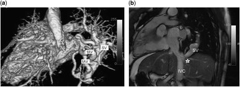
A subsequent MRI, ordered because of a suspicion for partially anomalous pulmonary venous connections, revealed the presence of scimitar syndrome, with the anomalous vessel collecting all of the right pulmonary veins. There were also two large and tortuous anomalous vessels decompressing the scimitar vessel into the left atrium just proximal to its connection to the inferior caval vein. The right ventricular dilatation was also confirmed and a low pulmonary-to-systemic flow ratio (Qp/Qs) of 1.4 was calculated by aortic and pulmonary flow analysis (Fig 1).
Volume rendered, 3D reconstruction of gadolinium-enhanced magnetic resonance angiography ( a ) showing the scimitar vessel (SV) and the junction with the inferior caval vein (IVC) from behind. Axial reformat of balanced steady-state free precession (SSFP) 3D volume images ( b ) showing the SV and the tortuous anomalous vessels (tAV) departing from SV.

Cardiac catheterisation was then performed to clarify the amount of the left-to-right shunt towards the inferior caval vein through the scimitar vein with respect to the flow reaching the left atrium. The scimitar was stenotic at its junction with the inferior caval vein (pressure gradient 5 mmHg).
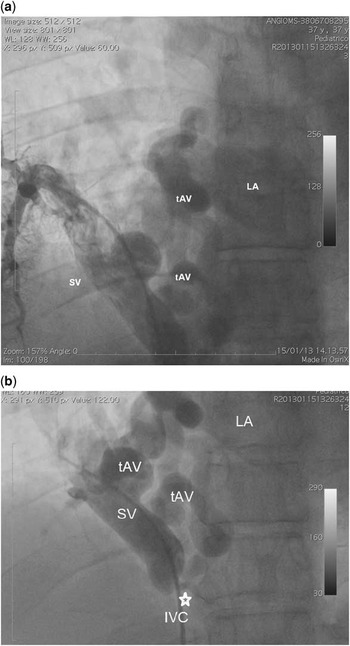
This stenosis created an obstruction to flow to the inferior caval vein with decompression to the left atrium by the way of the two tortuous vessels departing from the scimitar vein. As a result, the modest left-to-right shunt was also confirmed by angiography (Qp/Qs 1.5).
The patient was also found to have a pulmonary sequestration with two relatively small anomalous feeding vessels from the descending aorta to the right inferior lobe (Fig 2).
Anterior–posterior angiographic projections ( a and b ) allowing for a comprehensive visualisation of the scimitar variant. The scimitar vessel (SV) draining most of the right lung, its stenotic junction (arrow) with the inferior caval vein (IVC), and the tortuous anomalous vessels (tAV) departing from the SV and draining the majority of its flow to the left atrium (LA).

On the basis of the decompression of the scimitar vein into the left atrium, with a modest Qp/Qs, it was decided that surgery would not be performed.
Discussion
The term “scimitar anomaly” is commonly used to describe a wide complex of anomalies including drainage of all or part of the right lung to the inferior caval vein, hypoplasia of the right lung, and aberrant systemic arterial supply. In the classical form, a curved collector vein resembling a Turkish sword drains into the inferior caval vein; however, various anatomical variants have been described.Reference Rose and Vosshenrich 1 – Reference Yoo, Al-Otay and Babyn 8 The case we describe not only shares similarities with some of these variants but also show some original features.
In particular, the anomalous tortuous vessels resemble a scimitar variant known as a “meandering” pulmonary vein, where the anomalous pulmonary venous collector is directed to the left atrium instead of the inferior caval vein.Reference Legras, Guinet, Alifano, Lepilliez and Regnard 2 – Reference Yoo, Al-Otay and Babyn 8 This meandering vein may be the result of an abnormal left atrial drainage via the persistent Thebesian veinsReference Pearl 7 or a delay in the development of the common pulmonary vein maintaining the foetal connection between the pulmonary and systemic vascular beds and forming the scimitar vein.Reference Takeda, Imachi, Arimitsu, Minami and Hayakawa 6 In the meandering vein variant, the scimitar collector has an upward direction to the left atrium, whereas in this case it is normally downward oriented to the inferior caval vein and the connection with the left atrium is mediated by accessory vessels.
Connection of the scimitar collector vein both with the inferior caval vein and the left atrium have also been described previously in at least seven cases;Reference Pearl 7 , Reference Yoo, Al-Otay and Babyn 8 however, in those cases the anomalous vessel connected directly with the left atrium and not by the interposition of other anomalous vessels, into which the sequestration also drained.
The strange anatomical pattern we describe could be interpreted as the result of an anomaly at an early stage of the developmental process. The unusual anatomy makes its diagnosis particularly challenging and at times a single examination may provide unclear results. Therefore, the synergy and complementarity of various imaging modalities combined to make the diagnosis apparent.
Acknowledgements
None.
Financial Support
This research received no specific grant from any funding agency, commercial, or not-for-profit sectors.
Conflicts of Interest
None.
Ethical Standards
The authors assert that all procedures contributing to this work comply with the ethical standards of the relevant national guidelines on human experimentation and with the Helsinki Declaration of 1975, as revised in 2008, and has been approved by the institutional committees.