INTRODUCTION
Motor imagery (MI) involves the mental simulation of movement without overt action (Decety, Reference Decety1996; Guillot, Di Rienzo, MacIntyre, Moran, & Collet, Reference Guillot, Di Rienzo, MacIntyre, Moran and Collet2012; Jeannerod, Reference Jeannerod2006). Although the application of MI training has largely been confined to sports settings, there is growing interest in the potential benefit it may offer as a neurorehabilitative tool (Malouin & Richards, Reference Malouin and Richards2013; Sharma, Pomeroy, & Baron, Reference Sharma, Pomeroy and Baron2006; Williams, Pearce, Loporto, Morris, & Holmes, Reference Williams, Pearce, Loporto, Morris and Holmes2012). The guiding theory is that MI may preserve or foster neural function in motor-related circuitry without necessitating overt action (Lacourse, Orr, Cramer, & Cohen, Reference Lacourse, Orr, Cramer and Cohen2005; Sharma et al., Reference Sharma, Pomeroy and Baron2006; Williams et al., Reference Williams, Pearce, Loporto, Morris and Holmes2012). However, the nature of neurological impairment itself not only influences MI performance, but the neural systems that support it (Di Rienzo, Collet, Hoyek, & Guillot, Reference Di Rienzo, Collet, Hoyek and Guillot2014). Clarifying the neural basis of MI (and indeed, MI difficulties) is therefore paramount to appropriately planning MI therapy parameters. Given this, in light of limitations in our understanding of the neural mechanisms underpinning MI the mixed findings concerning the efficacy of MI intervention in rehabilitative contexts are perhaps unsurprising (Blefari, Sulzer, Hepp-Reymond, Kollias, & Gassert, Reference Blefari, Sulzer, Hepp-Reymond, Kollias and Gassert2015). This study attempts to elucidate the involvement of the PMC during MI using transcranial magnetic stimulation (TMS) and introduces a novel method that permits participants to respond to stimuli without a gross motor response.
The PMC is known to be the terminal processing site for the selection and initiation of voluntary motor commands before descending pathways transmit them to the spinal cord, en route to relevant effectors (Rathelot & Strick, Reference Rathelot and Strick2009; Stinear, Coxon, & Byblow, Reference Stinear, Coxon and Byblow2009). However, it is less clear whether involvement of the PMC extends to mentally simulated movement. In order to clarify the role of the PMC during MI, a growing number of studies have applied TMS to the hand area of the PMC (hPMC) prior to, or during, one of the most widely adopted and well-validated implicit measures of MI, the hand laterality task (HLT) (Parsons, Reference Parsons1994; Parsons & Fox, Reference Parsons and Fox1998). Here, participants make laterality judgments about hand stimuli presented at varying angles and in different postural orientations. The HLT holds several key advantages over alternative measures of mental movement as an index of MI. First, implicit measures of MI such as the HLT arguably provide a more objective performance measure than explicit measures of MI which rely on conscious introspection of mentally simulated movement (Zapparoli et al., Reference Zapparoli, Invernizzi, Gandola, Berlingeri, De Santis, Zerbi and Paulesu2014). Also, there is some evidence that explicit imagery instructions may affect participants differently (Williams, Thomas, Maruff, Butson, & Wilson, Reference Williams, Thomas, Maruff, Butson and Wilson2006). Furthermore, unlike many other measures of MI, implicit or otherwise, the objective behavioral performance elicited by the HLT can then be used to both infer an underlying performance strategy (i.e., MI or otherwise- see below) and performance quality. Still, evidence concerning involvement of the hPMC in mental hand movements using TMS remains equivocal, with some work suggesting involvement (Date, Kurumadani, Watanabe, & Sunagawa, Reference Date, Kurumadani, Watanabe and Sunagawa2015; Ganis, Keenan, Kosslyn, & Pascual-Leone, Reference Ganis, Keenan, Kosslyn and Pascual-Leone2000; Pelgrims, Michaux, Olivier, & Andres, Reference Pelgrims, Michaux, Olivier and Andres2011; Tomasino, Borroni, Isaja, & Ida Rumiati, Reference Tomasino, Borroni, Isaja and Ida Rumiati2005) and others not (e.g., Bode, Koeneke, & Jäncke, Reference Bode, Koeneke and Jäncke2007; Sauner, Bestmann, Siebner, & Rothwell, Reference Sauner, Bestmann, Siebner and Rothwell2006).
The suggestion that HLT performance should activate the hPMC is largely predicated on the assumption that participants adopt a specific MI strategy (Bode et al., Reference Bode, Koeneke and Jäncke2007; Date et al., Reference Date, Kurumadani, Watanabe and Sunagawa2015; Hanakawa, Reference Hanakawa2015; Kosslyn, Ganis, & Thompson, Reference Kosslyn, Ganis and Thompson2001). That is, hPMC modulation would be conditional on participants performing the task from a first-person perspective (i.e., mentally moving one’s own body), a view typically supported by psychophysical and self-report data (Butson, Hyde, Steenbergen, & Williams, Reference Butson, Hyde, Steenbergen and Williams2014; Fuelscher, Williams, Wilmut, Enticott, & Hyde, Reference Fuelscher, Williams, Wilmut, Enticott and Hyde2016; Ionta, Perruchoud, Draganski, & Blanke, Reference Ionta, Perruchoud, Draganski and Blanke2012; Kosslyn, Digirolamo, Thompson, & Alpert, Reference Kosslyn, Digirolamo, Thompson and Alpert1998; Parsons & Fox, Reference Parsons and Fox1998; Ter Horst, van Lier, & Steenbergen, Reference Ter Horst, van Lier and Steenbergen2010). Indeed, the implicit use of MI is commonly inferred when behavioral profiles are characterized by traits that are unique to motoric forms of imagery. Given the consistency of the latter, the broad assumption that the HLT implicitly elicits MI at a group level is generally well founded. Still, there is variation in the strategies adopted. For example, participants may use a visual rotation strategy (i.e., non-motoric), whereby they view the hand as an inanimate and self-rotating object (Ionta et al., Reference Ionta, Perruchoud, Draganski and Blanke2012; Kosslyn et al., Reference Kosslyn, Ganis and Thompson2001; Spruijt, van der Kamp, & Steenbergen, Reference Spruijt, van der Kamp and Steenbergen2015). Here, activation of motor systems including the PMC is expected to be attenuated (see Kosslyn et al., Reference Kosslyn, Ganis and Thompson2001). Accordingly, it is only valid to ascribe modulation of PMC activity (or lack thereof) to MI to the extent that we can be confident that participants engaged MI during HLT performance.
There is considerable variation in the methods used by earlier TMS HLT studies to infer a group-level imagery strategy. In some cases, no analysis is presented which would allow one to infer whether MI was adopted (e.g., Date et al., Reference Date, Kurumadani, Watanabe and Sunagawa2015; Ganis et al., Reference Ganis, Keenan, Kosslyn and Pascual-Leone2000; Tomasino et al., Reference Tomasino, Borroni, Isaja and Ida Rumiati2005). Indeed, contemporary researchers identify behavioral phenotypes unique to MI before inferring its use. For the HLT, those rotations that pose greater biomechanical complexity (e.g., laterally rotated hands) generally take longer to imagine than less complex rotations (e.g., medially rotated hands) (Butson et al., Reference Butson, Hyde, Steenbergen and Williams2014; Ionta et al., Reference Ionta, Perruchoud, Draganski and Blanke2012; Spruijt et al., Reference Spruijt, van der Kamp and Steenbergen2015; Ter Horst et al., Reference Ter Horst, van Lier and Steenbergen2010). While two recent studies that implemented TMS during HLT performance have reported on the presence of such biomechanical effects to support the inference that participants engaged in MI, inspection of the reported data for one appeared to show biomechanical effects for hands shown in “back” but not “palm” view (Pelgrims et al., Reference Pelgrims, Michaux, Olivier and Andres2011), yet vice versa was observed in the second instance (Sauner et al., Reference Sauner, Bestmann, Siebner and Rothwell2006). It may be that MI was employed in both studies but not uniformly across stimuli types. Taken together, we must be cautious when inferring the performance strategies of participants in these earlier studies, and hence temper any attributions of hPMC activity to MI.
Finally, while HLT responses have traditionally been manual, those studies employing TMS have adopted alternative response methods to avoid response related activation of the hPMC and/or hand muscles. These have predominantly involved recording verbal (Ganis et al., Reference Ganis, Keenan, Kosslyn and Pascual-Leone2000; Pelgrims et al., Reference Pelgrims, Michaux, Olivier and Andres2011; Tomasino et al., Reference Tomasino, Borroni, Isaja and Ida Rumiati2005) or pedal responses (Date et al., Reference Date, Kurumadani, Watanabe and Sunagawa2015; Sauner et al., Reference Sauner, Bestmann, Siebner and Rothwell2006). In other studies, no behavioral data were collected (Bode et al., Reference Bode, Koeneke and Jäncke2007; Lebon, Byblow, Collet, Guillot, & Stinear, Reference Lebon, Byblow, Collet, Guillot and Stinear2012), limiting the degree to which performance strategies and quality can be inferred and, accordingly, the attribution of corticospinal excitability to mental processes. Alternatively, leg responses allow hand MEPs and behavioral performance to be measured simultaneously, although activation from proximal foot and leg PMC areas may result in “spill-over” activation of the hPMC (Eisenegger, Herwig, & Jäncke, Reference Eisenegger, Herwig and Jäncke2007). Similarly, there is direct evidence that verbal responses may alter hPMC excitability (Meister et al., Reference Meister, Boroojerdi, Foltys, Sparing, Huber and Töpper2003; Sparing et al., Reference Sparing, Meister, Wienemann, Buelte, Staedtgen and Boroojerdi2007; Tokimura, Asakura, Tokimura, Oliviero, & Rothwell, Reference Tokimura, Asakura, Tokimura, Oliviero and Rothwell1996). Accordingly, where leg and verbal responses are adopted it is difficult to disentangle the degree to which hPMC excitability can be attributed to the response method and/or a given cognitive process (MI or otherwise). Similarly, where a “virtual lesion” approach to TMS has been adopted, TMS to the hPMC could reasonably be expected to influence the response method, irrespective of whether or how the PMC is involved in the cognitive processes preceding a response.
The Present Study
We aimed to clarify whether hPMC activity is modulated during mental simulation of hand movement. We applied single-pulse TMS to the left hPMC of healthy young adults, and recorded MEPs from contralateral hand muscles to determine PMC activation, while they performed a highly innovative adaptation of the HLT task. Participants’ visual gaze was tracked using eye-tracking, allowing them to respond visually. Voluntary saccades of this kind are thought to largely bypass the PMC, and are instead subserved by corticostriatal circuitry that is independent of limb movements, especially when visual information is present (Alexander & Crutcher, Reference Alexander and Crutcher1990; Frens & Erkelens, Reference Frens and Erkelens1991). Thus, visual responses allowed us to reduce the possibility of hPMC activation associated with manual and verbal responses, while maintaining the measurement accuracy and reliability of manual responses. A visual response method also ensured that changes in contralateral hand MEPs could not be attributed to the act of responding. Thus, at a neurophysiological level, we could be confident that corticospinal excitability was not unduly influenced by the act of responding.
We predicted that most participants would engage a MI strategy during the HLT. As noted, this is ordinarily inferred when group level analysis suggests the presence of biomechanical constraints on response efficiency. Intriguingly, this form of screening is rarely conducted at an individual level (see Spruijt et al., Reference Spruijt, van der Kamp and Steenbergen2015). Despite the HLT being a well-validated measure of MI, a degree of individual variability in performance strategy nonetheless exists. Where group-wise analysis suggests use of MI, a small proportion of participants will likely have adopted an alternative strategy. This poses a threat to study sensitivity given the modest sample sizes typical of studies examining hPMC activation during HLT performance via TMS. Thus, we only included those participants whose performance efficiency profile on the HLT was consistent with the use of a MI strategy prior to investigating group-level hPMC activity during HLT performance.
We predicted that the hPMC would be modulated during HLT performance in “MI users.” We used single-pulse TMS as a comparative technique with the view that changes in contralateral hand MEPs during the HLT would reflect activation of the left hPMC. Only the left hPMC was subjected to TMS given evidence that the left hemisphere shows predominance during MI tasks when TMS is applied (Fadiga et al., Reference Fadiga, Buccino, Craighero, Fogassi, Gallese and Pavesi1998). Also, where hPMC involvement has been inferred following TMS during HLT performance, consistent effects have been reported for the left hemisphere (Date et al., Reference Date, Kurumadani, Watanabe and Sunagawa2015; Ganis et al., Reference Ganis, Keenan, Kosslyn and Pascual-Leone2000; Pelgrims et al., Reference Pelgrims, Michaux, Olivier and Andres2011; Tomasino et al., Reference Tomasino, Borroni, Isaja and Ida Rumiati2005). Hence, assuming that the left hPMC is activated when participants engage in MI during mental hand rotation, we hypothesized that this would manifest as increased corticospinal excitability during HLT performance relative to baseline when participants engaged in MI, indicated by higher amplitude right hand MEP values. We reasoned that if this activation could be attributed, at least in part, to the use of MI then hPMC activation would be modulated by the biomechanical constraints of movement- again, a unique characteristic of MI. Accordingly, we predicted that hPMC activation during MI would be magnified for biomechanically more awkward rotations (lateral) compared to simpler ones (medial).
METHOD
Participants
The sample comprised 22 healthy adults aged 18 to 36 years. No participants reported a neurological or developmental disorder or presented with significant motor problems, as indicated by motor ability above the 15th percentile on the McCarron Assessment of Neuromuscular Development (MAND) (McCarron, Reference McCarron1997). All participants self-reported being right handed (n=20) or were ambidextrous showing a right hand preference on the MAND (n=2). They were recruited through the university setting and gave written informed consent. All participants were free of TMS contra-indications and none reported negative side-effects during or following TMS. The project received ethical clearance from relevant university Human Research Ethics Committees.
Measures
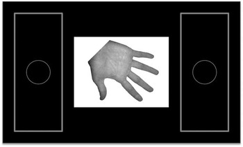
The ability to perform MI was assessed using the HLT (Parsons, Reference Parsons1994), programmed using E-Prime software (Version 2.0, Psychology Software Tools, Pittsburgh, PA). Participants were presented with single hand stimuli that subtended 9.7° × 12.6° of visual angle centered in the middle of a 40-inch LCD monitor. Response boxes were set to the immediate left or right of hand stimuli (see Figure 1 for visual display). Each response box subtended 15.4°×10.3° of visual angle. The response boxes had a small sphere within them indicating their center.
A visual representation of the experimental display.

Participants sat upright with both hands in view, opened and resting palm down on a cushion positioned on their lap. Prior to each trial, a fixation cross appeared at the center of the screen for 1000 ms prior to the hand stimuli. Participants were instructed to fixate on this cross until an image of a hand appeared. They were then asked to determine whether the hand was a left or right hand as quickly and accurately as possible. Participants responded using eye-movements by fixating in the middle of the box positioned either to the left- or right-most side of the screen. A Tobii X60 eye-tracker was used to record participant eye-movements. This apparatus records gaze positions recorded as x,y coordinates on the screen at a sampling rate of 60 Hz.
An algorithm was written that coded eye-movement data in real-time. The algorithm detected when participants fixated at either the left or right response box, operationalized as gaze positions that were in a response box for at least 100 ms. Following this, the trial terminated. Response time was calculated as the difference (in ms) between stimulus onset and fixation onset to the response box. Visual response methods have been successfully adopted for recording accuracy on the HLT in patient groups [e.g., spinal-cord injury (Fiori et al., Reference Fiori, Sedda, Ferre, Toraldo, Querzola, Pasotti and Redaelli2014) and Amyotrophic lateral sclerosis (Fiori et al., Reference Fiori, Sedda, Ferrè, Toraldo, Querzola, Pasotti and Lunetta2013)]. Unlike the present study, however, reaction times were not simultaneously recorded in these earlier studies. Given ceiling effects on accuracy are often reported when healthy young adults perform the HLT (Hyde, Wilmut, Fuelscher, & Williams, Reference Hyde, Wilmut, Fuelscher and Williams2013), our measurement of response times was not only novel but central to valid performance measurement in the principal group of interest here.
The validity and reliability of our response method is supported by the response patterns and mean values reported here which were very similar to those reported in our earlier HLT studies where young adults have responded manually (e.g., Hyde et al., Reference Hyde, Fuelscher, Buckthought, Enticott, Gitay and Williams2014). Indeed, the mean inverse efficiency scores (IES) (see below for details) for palm-view stimuli from this earlier study (M IES =1643) fell within the CI95% of the mean IES of participants here (M IES =1584, CI95%=1100–2067).
Hand stimuli were presented randomly in 45° increments between 0 and 360° and remained on screen for a maximum of 10 s or until a response was recorded. Rotated stimuli were shown in palm-view only (palm facing toward participants). This view was chosen since healthy adults often report ceiling effects on accuracy for the simpler back-view images (Hyde et al., Reference Hyde, Wilmut, Fuelscher and Williams2013). Furthermore, hPMC activation during MI may reasonably be expected to be magnified for more complex movements (Sauner et al., Reference Sauner, Bestmann, Siebner and Rothwell2006), with recent work from the HLT showing that motor-related activation may indeed be greater for posturally more complex movements (Zapparoli et al., Reference Zapparoli, Invernizzi, Gandola, Berlingeri, De Santis, Zerbi and Paulesu2014). Hence inclusion of simpler back-view images had the potential to reduce the measurable motor cortical response. We did however include a stimulus presented in back-view at 0° to ensure that participants were able to make laterality decisions above chance level (60%). Data from the latter were not included in subsequent analyses.
For each stimulus, we recorded response time (RT) to the nearest 1ms, and accuracy. Participants completed as many practice trials as required before they reported being comfortable with the task. This was followed by two blocks of 72 test trials (half left; half right hands), resulting in 12 trials per angle in palm-view and 12 trials at 0° in back-view. Participants did not receive specific instructions cueing MI in light of previous research indicating that the effect of explicit imagery instructions on HLT performance might differ across participants (Williams et al., Reference Williams, Thomas, Maruff, Butson and Wilson2006).
Procedure
Single-pulse TMS (MagStim-200 stimulator, Magstim Company Ltd, UK) was delivered to the left hPMC using a hand held 70-mm figure-eight coil. The coil was oriented tangentially to the scalp with the handle angled backward and 45° away from the midline. As with previous HLT and mental rotation studies where TMS has been applied to the hPMC (Bode et al., Reference Bode, Koeneke and Jäncke2007; Eisenegger et al., Reference Eisenegger, Herwig and Jäncke2007; Sauner et al., Reference Sauner, Bestmann, Siebner and Rothwell2006), electromyogram (EMG) of the right first dorsal interosseous (FDI) was recorded via three self-adhesive surface electrodes: an active electrode was positioned over the muscle belly, a reference electrode over the interphalangeal joint of the right index finger, and the ground electrode was positioned over the ulnar styloid process. PowerLab/4SP (AD instruments, Colorado Springs, CO) was used to amplify and sample the EMG signals, which were collected using LabChart v7. hPMC location was defined as the scalp site that produced the largest motor-evoked potential (MEP) amplitude from the right FDI at rest. Individual resting motor thresholds (RMT) were then defined as the minimum stimulus intensity that yielded an average 1 mV response across 10 trials. Twenty baseline pulses at resting motor threshold were then administered to each participant and the median MEP recorded. No explicit behavioral instructions were provided.
Single-pulse TMS at the 1mV RMT was then delivered randomly during the HLT at latencies of 50 ms, 400 ms, and 650 ms post stimulus presentation. These latencies were empirically driven since single-pulse TMS of the hPMC at 400 ms and 650 ms has previously shown to reduce response efficiency on the HLT (Ganis et al., Reference Ganis, Keenan, Kosslyn and Pascual-Leone2000; Tomasino et al., Reference Tomasino, Borroni, Isaja and Ida Rumiati2005). An earlier stimulation (50 ms) was added since neither of these studies suggesting hPMC involvement in HLT performance included an earlier stimulation reference point, limiting inferences about the time-course of hPMC involvement (Sauner et al., Reference Sauner, Bestmann, Siebner and Rothwell2006).
As per our earlier work (Fitzgibbon, Fitzgerald, & Enticott, Reference Fitzgibbon, Fitzgerald and Enticott2014), to trigger the TMS pulse at each latency (i.e., 50 ms, 400 ms, and 650 ms post stimulus) a light sensor device was fixed to the top left hand corner of the screen to time-lock the TMS pulse with the precise presentation onset of the hand stimulus. The device sent a trigger (5 V TTL pulse via BNC connector) to the TMS stimulator upon light detection. This was achieved by embedding a white box in the top left hand corner of the stimulus display, which appeared for 200 ms at the desired moment post stimulus presentation (50 ms, 400 ms, or 650 ms). This was detected by the light sensor device, which subsequently sent a trigger to the stimulator and a single-TMS pulse was emitted. A second trigger followed that was sent from the stimulator to the EMG device to signal EMG recording.
Design and Analysis
Alpha was set at .05 and adjusted for multiple comparisons using the Benjamini-Hochberg FDR procedure (Benjamini & Hochberg, Reference Benjamini and Hochberg1995). No violations of normality that could reasonably be expected to unduly influence interpretation of the results were observed.
HLT data
For each participant, mean RT and accuracy for each hand at each angle of rotation was calculated. Both correct and incorrect trials were used in the analysis since participants can reasonably be expected to use a comparable mental rotation strategy on either trial type (Fuelscher et al., Reference Fuelscher, Williams, Wilmut, Enticott and Hyde2016; Hyde et al., Reference Hyde, Wilmut, Fuelscher and Williams2013, Reference Hyde, Fuelscher, Buckthought, Enticott, Gitay and Williams2014). Preliminary analysis of the data here supports this view since none of the effects of interest altered upon exclusion of incorrect trials. Performance was averaged across angular rotations of 0°, 45°, 90°, 135°, and 180°. Trials with RTs less than 250 ms were excluded, as were trials that were +/− 3 SD for an individual’s mean RT across all trials. When all exclusions criteria were considered, an average of ≈3% of trials were removed. Mean accuracy was calculated as the proportion of correct responses over all trials. Similarly to our earlier research (Fuelscher, Williams, Enticott, & Hyde, Reference Fuelscher, Williams, Enticott and Hyde2015; Hyde et al., Reference Hyde, Fuelscher, Buckthought, Enticott, Gitay and Williams2014), we calculated mean IES for each participant by dividing mean RT by the proportion of correct responses at each of the stimuli presentation conditions (Townsend & Ashby, Reference Townsend and Ashby1978, Reference Townsend and Ashby1983). For a detailed discussion of the benefits and criterion (which were met here) for adopting IES as the HLT performance measure, see Hyde et al. (Reference Hyde, Fuelscher, Buckthought, Enticott, Gitay and Williams2014).
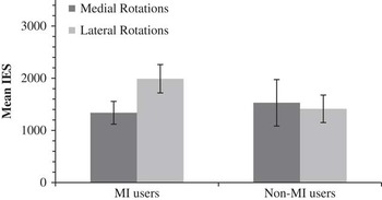
We first differentiated between those participants who were likely (MI users) and unlikely (non-MI users) to have engaged MI during the HLT. We examined the individual performance profile of each participant to verify whether it was consistent with the biomechanical constraints of action, a profile consistent and unique to a MI strategy (Spruijt et al., Reference Spruijt, van der Kamp and Steenbergen2015). Specifically, a MI strategy was inferred when performance efficiency was greater for biomechanically simpler rotations compared to more complex, shown by lower absolute mean IES (indicating greater efficiency) for the medially rotated images compared to laterally rotated (Hyde et al., Reference Hyde, Wilmut, Fuelscher and Williams2013; Spruijt et al., Reference Spruijt, van der Kamp and Steenbergen2015; Ter Horst et al., Reference Ter Horst, van Lier and Steenbergen2010). Because any efficiency measure requires consideration of accuracy (as is the case here), it is not possible to gain an efficiency measure for individual trials since accuracy is binary at this level of analysis (i.e., 0=incorrect, 1=correct). For this reason, parametric comparison of mean IES between medially and laterally rotated images within each participant was not possible. In the absence of an empirically justifiable minimum difference value, MI strategy was inferred when mean efficiency for medially compared to laterally rotated images was greater by any value. We identified 16 participants as probable “MI users” (M Age =24.88; SD=4.69) and 6 as probable “non-MI users” (M Age = 27.82; SD=5.49).
As expected, the sample size of non-MI users precluded meaningful interpretation of statistical analysis, which would offer little insight into HLT performance and hPMC excitability beyond consideration of descriptive statistics alone. Accordingly, this group was not subjected to parametric analysis. Still, in the interest of transparency and as a preliminary descriptive comparison point against MI users, group means for non-MI users are presented throughout. Mean IES values for medially and laterally rotated hands for MI and non-MI users are presented in Figure 2.
Mean IES values (+/− SE) for medially and laterally rotated images for MI and non-MI users.

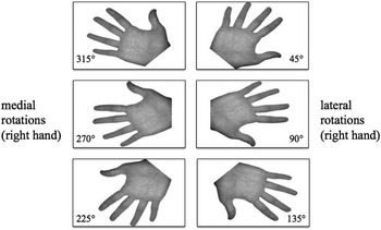
Medial rotation performance was calculated as the average of responses for left hands presented at 45◦, 90◦, and 135◦ and right hands presented at 315◦, 270◦, and 225◦. Lateral rotation performance was calculated as the average responses for left hands presented at 315◦, 270◦, and 225◦ and right hands presented at 45◦, 90◦, and 135◦ (see Figure 3 for a visual representation).
Visual representation of angles included for right hand medial and lateral rotations.

Preliminary analyses included repeated-measures analysis of variance (ANOVA) comparing RTs across TMS latencies of 50 ms, 400 ms, and 650ms post stimulus for all participants (N=22) to test whether TMS latency affected RTs on the HLT. Since no effect was observed, F(2, 42)=0.50, p=.608, ηP 2=.02), we can be confident that HLT performance was not unduly influenced by TMS. Further preliminary analyses included a repeated-measures ANOVA for MI users on IES with angular rotation (i.e., 0°, 45°, 90°, 135°, and 180°) as the within subjects factor (Fuelscher et al., Reference Fuelscher, Williams, Wilmut, Enticott and Hyde2016) to examine whether participants were engaged in a mental rotation strategy on the HLT as expected. In line with seminal work using the HLT (Kosslyn et al., Reference Kosslyn, Digirolamo, Thompson and Alpert1998; Parsons, Reference Parsons1994), general hand rotation performance was analyzed by collapsing medial and lateral rotations to provide mean values for responses from 0° to 180° in palm-view (45° increments; 24 trials per angle).
MEP data
Peak-to-peak MEP amplitude (A
t
) was calculated for each trial in mV and normalized against the median baseline amplitude
 $$\left( {\widetilde{{A_{b} }}} \right)$$
using the following equation:
 $$A_{n} {\equals}{{A_{t} {\minus}\tilde{A}_{b} } \over {s_{b} }}$$
Here, A n represents the normalized amplitude for a given trial and s b represents the standard deviation of the baseline amplitude. All MEPs below 0.15 mV were excluded; as noted, in total ≈3% of all trials were removed as a result of exclusion criteria. Mean normalized MEP amplitude was then calculated at each angle. Performance was averaged across angular rotations of 0°, 45°, 90°, 135°, and 180°. Averages were calculated for TMS latencies of 50ms, 400ms and 650ms respectively, providing eight trials per angle at each of the latencies. Again, MEPs from both correct and incorrect trials were included for the reasons stated above.
To determine whether the hPMC corticospinal excitability was modulated during HLT performance, we ran three single sample t-tests for the MI users comparing normalized MEP amplitude during the HLT at each TMS stimulation latency (50 ms, 400 ms, and 650 ms) to MEP amplitude at baseline. We ran single sample t-tests (using 0 as the test-value), rather than an omnibus ANOVA, as these analyses provided a direct test of our research question whether MEP amplitudes differed during the HLT relative to baseline. Furthermore, to determine whether excitability was influenced by angle of rotation, separate repeated measures ANOVAs (i.e., at each TMS latency) were conducted on MEP values with angular rotation (0°, 45°, 90°, 135°, 180°) as the repeated measures factor.
Finally, to test whether movement complexity (i.e., medial vs. lateral rotations) resulted in changes to corticospinal excitability levels, separate repeated-measures t-tests for TMS latencies of 50 ms, 400 ms, and 650 ms respectively were run for each group on MEP amplitude with direction of rotation (i.e., medial vs. lateral) as the repeated-measures factor. We ran separate t-tests for each latency, rather than an omnibus mixed design ANOVA, as these analyses provided a direct test of our research question and did not result in an unnecessary loss of degrees of freedom that might otherwise have reduced the power of our analysis.
RESULTS
Analysis of HLT Data
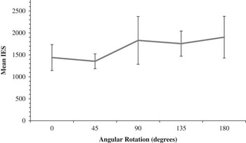
Repeated-measures ANOVA comparing mean efficiency across angular rotation for MI users suggested that efficiency values increased linearly with greater angular rotation F(1, 15)=10.57, p=.005, ηP 2=.41. Mean values are presented in Figure 4.
Mean IES values (+/− SE) as a function of angular rotation for MI users.

Analysis of MEP Data
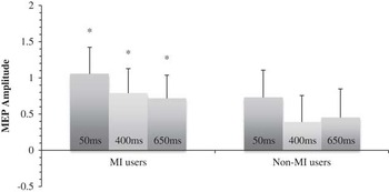
Single-sample t-tests comparing MEP amplitude during the HLT to MEP amplitude at baseline revealed significantly greater MEPs during the HLT for MI users when TMS was delivered at 50 ms, t(15)=2.88, p=.012, d=0.72, at 400 ms, t(15)=2.33, p=.034, d=0.58, and at 650 ms, t(15)=2.24, p=.040, d=0.56. Mean values for MI and non-MI users are presented in Figure 5.
Mean (normalized to baseline) MEP amplitude (+/− SE) during HLT performance following single-pulse TMS at 50ms, 400ms and 650ms post stimulus presentation.Note. * p<.05

Repeated measures ANOVAs comparing MEP amplitude across angular rotation failed to reveal a significant linear trend (or any other form of trend) for angle when TMS was delivered at 50 ms, F(1,15)=0.26, p=.874, ηP 2=.00, 400 ms F(1, 15)=0.34, p=.567, ηP 2=.02 and 650 ms F(1,15)=0.21, p=.654, ηP 2=.01.
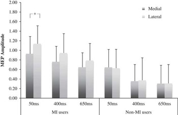
Repeated-measures t-tests comparing MEP amplitude for medially and laterally rotated hand stimuli in MI-users showed that MEP amplitude was greater during lateral rotations than during medial rotations when TMS was delivered at 50ms, t(15)=3.10, p=.007, d=0.78. No significant differences were found when TMS was delivered at 400 ms, t(15)=0.96, p=.352, d=0.26 and at 650 ms t(15)=0.91, p=.379, d=0.24. Mean values for MI and non-MI users are presented in Figure 6.
Comparison of mean (normalized relative to baseline) MEP values (+/- SE) for medial compared to laterally rotated images following single-pulse TMS at 50ms, 400ms and 650 ms post stimulus presentation.

DISCUSSION
This study aimed to investigate the degree to which corticospinal excitability of the hPMC was modulated during the mental simulation of hand movements (i.e., MI) using a novel adaptation of the HLT. Data from MI users showed that hPMC activity was significantly greater during HLT performance relative to baseline, with activation increasing as a function of the biomechanical complexity of movement (i.e., contralateral hand MEPs were larger for lateral compared to medially rotated hands following single-pulse TMS to the hPMC). Since such biomechanical effects are unique to motoric forms of imagery, this pattern of activation supports the view that hPMC activation during the HLT can be attributed, at least in part, to the use of MI. These data support the broader view that PMC excitability increases during MI. While our design prevents us from drawing causal inferences about the involvement of the PMC during MI, our data are nonetheless consistent with the view that PMC activation may subserve MI, a conclusion that has previously been the subject of some debate.
Our data indicated that hPMC excitability increases during HLT performance when a MI strategy is engaged. Indeed, we observed that contralateral hand MEPs were significantly larger following single-pulse TMS to the left hPMC at 50ms, 400ms and 650ms post stimuli presentation during HLT performance than at baseline in MI users. They also showed increased corticospinal excitability for biomechanically more awkward rotations (i.e., lateral) compared to simpler (medial). This finding is consistent with the view that hPMC activation may be contingent on MI complexity, indeed increasing as a function of movement difficulty (Sauner et al., Reference Sauner, Bestmann, Siebner and Rothwell2006). While the suggestion that hPMC activity is modulated when individuals engage in MI during the HLT supports some earlier TMS work (Date et al., Reference Date, Kurumadani, Watanabe and Sunagawa2015; Ganis et al., Reference Ganis, Keenan, Kosslyn and Pascual-Leone2000; Pelgrims et al., Reference Pelgrims, Michaux, Olivier and Andres2011; Tomasino et al., Reference Tomasino, Borroni, Isaja and Ida Rumiati2005), it remains at odds with others (Bode et al., Reference Bode, Koeneke and Jäncke2007; Sauner et al., Reference Sauner, Bestmann, Siebner and Rothwell2006). As we have argued, our design is ideally placed for clarifying these disparate findings for two reasons. First, the novel use of saccadic response on the HLT ensures reliable behavioral performance measurement, while controlling for the potential effects of response related hPMC and hand MEP interference. Second, to our knowledge our study is the first to identify participants as MI and non-MI users prior to analyzing hPMC excitability during the HLT. Since we showed that a small proportion of participants were likely to have adopted a non-MI imagery strategy during the HLT (i.e., 27%), it seems likely that even if group-level analysis suggested MI use, a proportion of participants in these earlier samples also adopted a non-MI strategy. Hence, our design was arguably better suited for detecting and disentangling group-level hPMC excitability associated with MI use than earlier work.
Our data are consistent with earlier TMS studies which have suggested that the hPMC may be involved in HLT performance at 400 ms (Tomasino et al., Reference Tomasino, Borroni, Isaja and Ida Rumiati2005) and 650 ms (Ganis et al., Reference Ganis, Keenan, Kosslyn and Pascual-Leone2000) into the response cycle. The addition of the earlier 50-ms stimulation point, however, provides additional insight into the time-course of hPMC activation when MI is used during the HLT. Specifically, we observed a strong increase in hPMC activation in MI users with stimulation onset at 50 ms. Since corticospinal excitability remained present at 400 and 650 ms, our data provide preliminary evidence that hPMC activation during MI (at least on the HLT) may arise early in the response, persisting until at least 650 ms, even if attenuated at these latter stages.
A number of recent studies have reported hPMC activation during the mental rotation of inanimate objects (Bode et al., Reference Bode, Koeneke and Jäncke2007; Eisenegger et al., Reference Eisenegger, Herwig and Jäncke2007), a task generally expected to elicit a visual imagery response strategy (c.f., MI) (Williams et al., Reference Williams, Thomas, Maruff, Butson and Wilson2006). Some have argued that PMC activation during mental object rotation may be an artifact of “spill over” from activation of adjacent pre-motor cortices which support spatial transformations necessary for mental rotation of objects as part of functional loop with parietal cortices (Date et al., Reference Date, Kurumadani, Watanabe and Sunagawa2015; Eisenegger et al., Reference Eisenegger, Herwig and Jäncke2007; Hétu et al., Reference Hétu, Grégoire, Saimpont, Coll, Eugène, Michon and Jackson2013). This raises two pertinent issues: firstly, whether hPMC activation observed during HLT performance here in MI users can be attributed to MI specifically, or instead predominantly reflects a general “mental rotation” effect. We are confident that the latter is not the case since corticospinal excitability was significantly greater for MI users when performing the biomechanically more complex lateral rotations relative to the simpler medial rotations at 50ms, with similar albeit non-significant trends observed later. As discussed, the presence of such biomechanical effects in the behavioral profiles of participants on the HLT is commonly considered indicative of MI strategy use, and is indeed a pre-requisite for inferring so. The logic here being that these biomechanical effects are not simply consistent with overt action (as per MI), but cannot be purely reconciled by alternative imagery strategy types (e.g., general rotation effects). We argue that same logic holds for the pattern of hPMC excitability observed here in MI users. That is, not only is increased hPMC excitability for more difficult movements consistent with changes in neural activity demonstrated during overt movement (Gut et al., Reference Gut, Urbanik, Forsberg, Binder, Rymarczyk, Sobiecka and Grabowska2007; Pearce & Kidgell, Reference Pearce and Kidgell2009; Perrey, Reference Perrey2013), but this pattern of excitability cannot be reconciled purely by a general rotation effect since the angles of rotation are the same for medial and lateral rotations on the HLT (i.e., 45°, 90°, and 135°). Were the excitability observed in MI users during the HLT largely the consequence of a general rotation effect, no difference in activation across medial and lateral rotations would be predicted. Similarly, if the increased hPMC excitability observed in the MI group was attributable to use of a general rotation effect, one might expect MEP amplitude to alter as a function of angular rotation. This was not the case for any TMS latency. Taken together, we argue that our data support the view that the hPMC activity observed in MI users in the present study can, at least partly, be attributed to a specific MI strategy. Further consistent with this argument is descriptive comparison of MEP values relative to baseline between MI and non-MI users since MEPs are consistently larger for the MI group at 50 ms, 400 ms, and 650 ms. However, given the small sample size (n=6) we must be circumspect about drawing inferences about hPMC excitability in non-MI users from this data and instead point towards the stated presence of biomechanical constraints and lack of angular effect on MEPs in the MI users as primary support of our argument here.
The second issue relates to whether the hPMC excitability observed here is causally linked to MI use, or reflects spill-over activation from adjacent pre-motor areas or downstream parietal areas which project to the PMC which have been strongly implicated in MI performance (Hétu et al., Reference Hétu, Grégoire, Saimpont, Coll, Eugène, Michon and Jackson2013). A number of lines of evidence indicate that hPMC activity may have facilitated MI performance in the present study. Specifically, we observed that maximal hPMC activation clearly preceded behavioral responses in MI users and hPMC activation increased for biomechanically more complex rotations. Still, given that premotor and parietal regions show consistent activation during MI performance (Hétu et al., Reference Hétu, Grégoire, Saimpont, Coll, Eugène, Michon and Jackson2013), these effects could reasonably be expected to arise regardless of whether hPMC activation contributed to MI performance. Similarly, the present design does not allow one to draw conclusions about the contribution of these alternative structures to MI performance. It should also be noted that while it is largely accepted that hand MEPs during MI reflect modulation at the cortical level rather than a spinal origin, the contribution of the latter nonetheless remains unclear (Stinear, Reference Stinear2010). Thus, even if minimal, the potential contribution of spinal excitability to hand MEPs is worth acknowledging. Accordingly, we acknowledge that the design of the present study prevents us from drawing causal inferences about the role of the hPMC (or not) in MI. Still, our work nonetheless provides some of the most compelling evidence that MI performance during the HLT modulates PMC excitability and remains consistent with the view that the hPMC contributes to MI. In doing so, it paves the way for a highly controlled transient lesion TMS approach to clarify the nature of this relationship, including whether PMC activation causally contributes to MI during HLT performance.
CONCLUSION
This study clarifies previously disparate findings speaking to hPMC activation during HLT performance. We found evidence that hPMC excitability is modulated during HLT performance when participants engage a MI strategy. That MI users showed a consistent and strong pattern of hPMC activation throughout the response cycle, which increased with movement difficulty provides compelling evidence that hPMC activation can, in part, be attributed to MI.
Acknowledgment
There are no conflicts of interest to be disclosed.