INTRODUCTION
The geohelminth Toxocara canis, an intestinal parasite of dogs, is the species most often associated with cases of human toxocarosis (Despommier, Reference Despommier2003). However, owing to the high degree of homology between the species T. canis and Toxocara cati (Hogarth-Scott et al. Reference Hogarth-Scott, Johansson and Bennich1969), no serological technique thus far differentiates between the species involved in this pathology (Kennedy et al. Reference Kennedy, Maizels, Meghji, Young, Quereshi and Smith1987; Fillaux and Magnaval, Reference Fillaux and Magnaval2013; Poulsen et al. Reference Poulsen, Skov, Yoshida, Skallerup, Maruyama, Thamsborg and Nejsum2015).
Owing to the close relationship between humans and domestic animals, particularly cats and dogs (Marmor et al. Reference Marmor, Glickman, Shofer, Faich, Rosenberg, Cornblatt and Friedman1987), toxocarosis has become one of the most common helminthozoonoses worldwide (Magnaval et al. Reference Magnaval, Glickman and Dorchies1994; Hotez and Wilkins, Reference Hotez and Wilkins2009; Othman, Reference Othman2012). The parasite is eliminated via the feces of animals and contaminates the environment (Paller and Chavez, Reference Paller and Chavez2014; Sprenger et al. Reference Sprenger, Green and Molento2014; Błaszkowska et al. Reference Błaszkowska, Góralzka, Wójcik, Kurnatowski and Szwabe2015). Humans and paratenic hosts, such as mice, rats, chickens, lambs and pigs, among others, acquire toxocarosis by accidentally ingesting T. canis eggs containing the third-stage larvae. This is the predominant form of human infection (Glickman and Schantz, Reference Glickman and Schantz1981). Moreover, the consumption of raw or undercooked offal and meat from paratenic hosts can also transmit the parasite to humans, especially in cultures where this is a common practice (Hoffmeister et al. Reference Hoffmeister, Glaeser, Flick, Pornschlegel, Suttorp and Bergmann2007).
Despite the high prevalence worldwide, especially in children (Schoenardie et al. Reference Schoenardie, Scaini, Brod, Pepe, Villela, Mcbride, Borsuk and Berne2013), the importance of toxocarosis is underestimated, since it is difficult to diagnose, either owing to the non-specific symptomatology (Smith et al. Reference Smith, Holland, Taylor, Magnaval, Schantz and Maizels2009) or lack of knowledge among many healthcare professionals (Alderete et al. Reference Alderete, Jacob, Pastorino, Elefant, Castro, Fomin and Chieffi2003).
Furthermore, human toxocarosis is difficult to treat because the drugs available have moderate efficacy in the clinical resolution of the disease. However, albendazole showed superior results to those of other drugs (Magnaval, Reference Magnaval1995) and hence became the drug of choice for the treatment of human toxocarosis (Despommier, Reference Despommier2003), although the cure rate does not exceed 60% (Glickman and Magnaval, Reference Glickman and Magnaval1993).
Currently, there are few studies related to either the development of new anthelmintic drugs (Satou et al. Reference Satou, Horiuchi, Akao, Koike, Futija and Nikaido2005; Reis et al. Reference Reis, Trinca, Ferreira, Monsalve-Puello and Grácio2010; Mata-Santos et al. Reference Mata-Santos, Pinto, Mata-Santos, De Moura, Carneiro, Carvalho, Del Rio, Pinto, Martins, Fenalti, Da Silva and Scaini2015) or the evaluation of existing drugs in the market in randomized studies (Pawlowski, Reference Pawlowski2001; Elefant et al. Reference Elefant, Shimizu, Sanchez, Jacob and Ferreira2006; Magnaval and Glickman, Reference Magnaval and Glickman2006).
Quinones represent a broad family of metabolites with different biological activities in the respiratory chain and photosynthesis (Da Silva et al. Reference Da Silva, Ferreira and De Souza2003). About 1500 known quinones exist, which can be found in bacteria, fungi, lichens, gymnosperms and angiosperms (Thomson, Reference Thomson1971, Reference Thomson1991; Falkenberg, Reference Falkenberg, Simões, Schenkel, Gosmann, Mello, Mentz and Petrovick2004).
A large number of naphthoquinones have proven pharmacological activities (Hussain et al. Reference Hussain, Krohn, Ahmad, Miana and Green2007) such as anti-inflammatory, antibacterial, trypanocidal, antifungal, microbicidal, molluscicidal and antitumour properties (De Almeida et al. Reference De Almeida, Silva-Filho, Dos Santos and Lopes1990; De Moura et al. Reference De Moura, Emery, Pinto, Pinto, Dantas, Salomão, Castro and Pinto2001; Pardee et al. Reference Pardee, Li and Li2002; Da Silva et al. Reference Da Silva, Ferreira and De Souza2003; Coelho et al. Reference Coelho, Silva, Pinto, Pinto, Sscaini, Moura and Da Silva2010; Curvelo et al. Reference Curvelo, Barreto, dos Anjos, Santana, Alonso, Romanos, de Moura, Carneiro, Portela, Pinto and Soares2015). The 1,4-naphthoquinone isomer has led to the development of new naphthoxiranes, which showed excellent trypanocidal activities (Carneiro et al. Reference Carneiro, Nascimento, Pinto, Pinto, Lechuga, Santos, Júnior, Resende, Bourguignon and Ferreira2012).
Owing to the wide range of their biological activities, naphthoquinones and their derivatives have been suggested as precursors for developing new synthetic routes for drugs (Carneiro et al. Reference Carneiro, Nascimento, Pinto, Pinto, Lechuga, Santos, Júnior, Resende, Bourguignon and Ferreira2012). Therefore, this study aimed to evaluate the in vitro and in vivo anthelmintic activity of quinone derivatives against T. canis larvae.
MATERIAL AND METHODS
Synthesis
The quinones evaluated and used in the epoxidation reaction were extracted from the heartwood of Tabebuia spp. (Tecoma) and purified by recrystallization from ethanol as described in literature (Eistert et al. Reference Eistert, Fink and Müller1962; Kubo et al. Reference Kubo, Itoh, Tsuji, Oda, Takeuchi and Itho1998; Roush et al. Reference Roush, Gonzfilez, McKerrow and Hansell1998), producing 14 compounds.
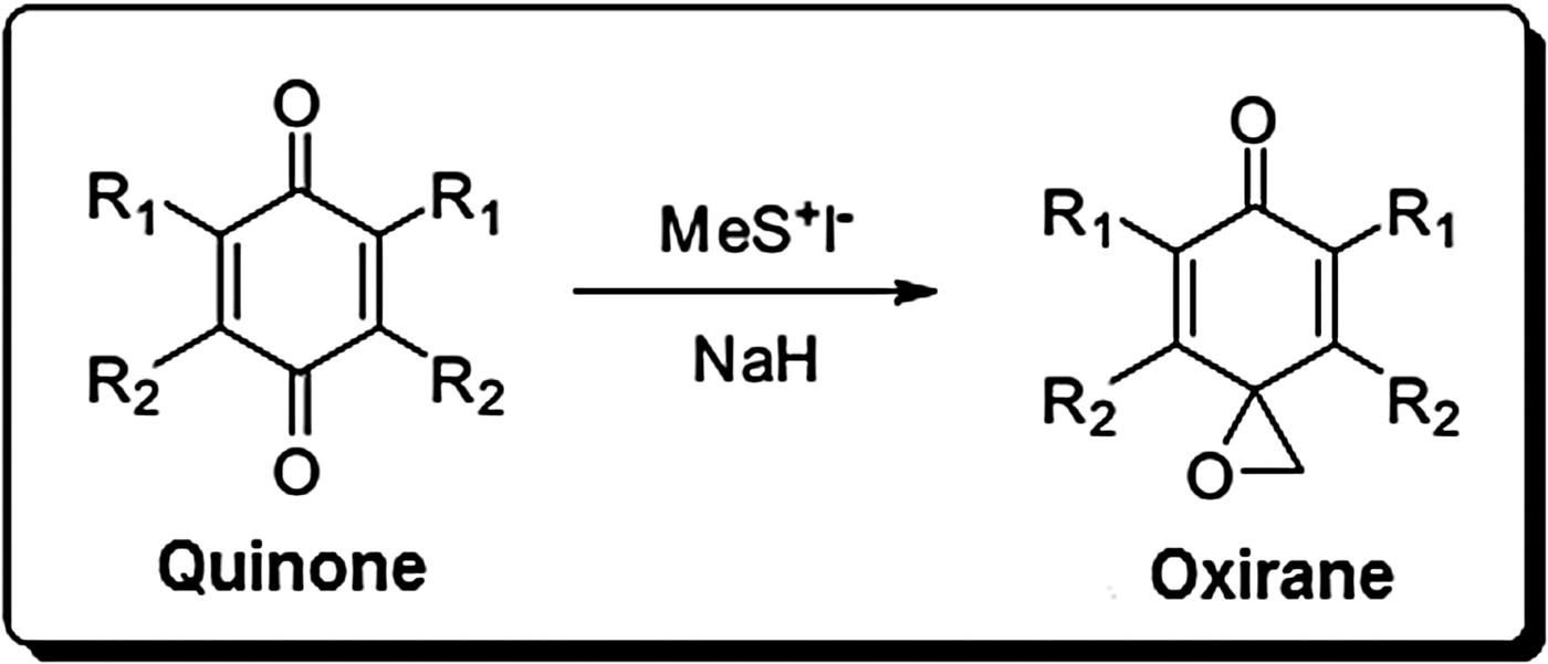
Eight naphthoxiranes were synthesized from the reaction of quinone in the presence of sodium hydride as the reducing agent in dimethyl sulphoxide (DMSO) (Sigma-Aldrich®) and further, trimethylsulphonium iodide was added under an inert atmosphere (Fig. 1) (Kubo et al. Reference Kubo, Itoh, Tsuji, Oda, Takeuchi and Itho1998).
Synthesis of oxiranes using trimethylsulphonium iodide.

The synthesis of six naphthoimidazoles and four naphthoxazoles was carried out by the condensation of β-lapachone with aromatic aldehydes (as benzaldehyde) amid aminante. The appropriate aromatic aldehyde was added (2·5 mmol) to a solution of β-lapachone (242 mg, 1·1 mmol) in glacial acetic acid (5 mL), and the mixture was gently heated to 70 °C. Ammonium acetate (1·2705 g, 16·5 mmol) was slowly added and the reaction was monitored using thin-layer chromatography. The material was then cooled to room temperature and added to water at 5 °C to precipitate. Subsequently, it was washed with a 5% solution of sodium metabisulphite to remove all the aldehyde residues, followed by rinsing in water at 5 °C until neutralization. The precipitate obtained was filtered, dried and purified using column chromatography on silica gel by using a mixture of hexane/ethyl acetate as eluent and increasing the polarity gradient. Naphthoxazoles were obtained at the polarity corresponding to 4–7% of ethyl acetate, while naphthoimidazoles were obtained at a polarity of 10–20% of ethyl acetate. The products of this reaction were recrystallized in a solvent mixture of 9:1 hexane/ethyl acetate, using acetone or pure ethanol based on the product solubility (Hooker, Reference Hooker1892; Pinto et al. Reference Pinto, Lopes, Cruz, Vasconcellos, Sampaio, Pinto and Gilbert1978; De Moura et al. Reference De Moura, Emery, Pinto, Pinto, Dantas, Salomão, Castro and Pinto2001; De Moura et al. Reference De Moura, Salomão, Menna-Barreto, Emery, Pinto, Pinto and Castro2004).
Preparation of T. canis larvae
Toxocara canis eggs were initially collected directly from the uterine tubes of female adult parasites after the treatment of young dogs with pyrantel pamoate (15 mg Kg−1). Afterwards, the eggs were incubated in a 2% formalin solution at 28 °C for 30 days with humidity >90%. By using a 5% sodium hypochlorite solution (Vetec®), the protein coating of the eggs was dissolved and the hatched T. canis larvae were collected in sterile tubes for cultivation in a Roswell Park Memorial Institute (RPMI) 1640 medium (Gibco®) supplemented with 25 mm N-(2-hydroxyethyl)piperazine-N′-(2-ethanesulfonic acid) (HEPES) (Sigma-Aldrich®), 1% glucose, penicillin/streptomycin/amphotericin B (PSF) antibiotic-antimycotic solution (Gibco®), and 0·4 µg mL−1 ofloxacin. Larvae were maintained at 37 °C and 5% CO2 with weekly change of RPMI-1640 medium, without contamination, for up to 3 months (De Savigny, Reference De Savigny1975).
Test compounds
All synthesized compounds were solubilized in DMSO at 2·5% and in sterile distilled water to achieve a concentration of 1 mg mL−1 (Mata-Santos et al. Reference Mata-Santos, Pinto, Mata-Santos, De Moura, Carneiro, Carvalho, Del Rio, Pinto, Martins, Fenalti, Da Silva and Scaini2015). Afterwards, the substances that showed larvicidal activity in 100% of larvae with the in vitro test at concentrations of 1 mg mL−1 were re-evaluated at 0·5, 0·25, 0·125 and 0·05 mg mL−1 to identify a minimal larvicidal concentration.
Larvicidal/larvistatic activity test
A 96-well flat-bottom plate was used to measure the activity of substances at a concentration of 1 mg mL−1. The tests were conducted in duplicate, using 100 T. canis larvae per well, and maintained at 37 °C for 48 h with 5% CO2. After 48 h, 0·4% trypan blue indicator (Vetec®) was added to all the wells. The larvicidal/larvistatic activity was evaluated by microscopy (100–400×), considering the impregnation of the larvae by trypan blue and larval mobility (motile, immobile but not dead, or dead) (Satou et al. Reference Satou, Horiuchi, Akao, Koike, Futija and Nikaido2005).
The immobile larvae that were not stained by trypan blue were inoculated in 7-week-old BALB/c (inbred albino strain developed originally by H.J. BAGG (Bagg albino)) healthy female mice by intraperitoneal injection. As positive control, 100 live larvae without test substances were used. A single mouse was used for each compound and each control (Mata-Santos et al. Reference Mata-Santos, Pinto, Mata-Santos, De Moura, Carneiro, Carvalho, Del Rio, Pinto, Martins, Fenalti, Da Silva and Scaini2015). This study was approved by the Ethics Committee for Animals Use of the Universidade Federal do Rio Grande, and all the experiments were performed according to the Brazilian legislation for animal experimentation. All the mice were euthanized as per animal ethics guidelines after 30 days of inoculation and examined for the recovery of the larvae by digesting the carcass, brain, liver, lungs, kidneys, heart, eyes and spleen in a solution of 1% hydrochloric acid and 1% pepsin, at 37 °C with constant agitation (Wang and Luo, Reference Wang and Luo1998). The larvicidal activity was evaluated by microscopy (100–400×). The animals were given food without antibiotics and had free access to water, and were on a 12-h light/12-h dark cycle at a room temperature of 22 ± 2 °C.
Cytotoxicity
The compounds that showed larvicidal activity (100%) of 0·5 mg mL−1 in the in vitro test were selected for the cytotoxic assay. For this assay, murine peritoneal macrophages isolated from C57BL/6 mice were used briefly, C57BL/6 mice were injected intraperitoneally with 2 mL of sterile 4% thioglycollate solution, and the peritoneal cells were collected by chilled Dulbecco's modified Eagles medium (DMEM) washes 4 days after injection to obtain the elicited macrophages. These cells were then washed and resuspended in the culture DMEM supplemented with 10% foetal bovine serum, in a 96-well flat-bottom plate. The cells (1 × 10⁶ cell well−1) were incubated in 5% CO2 for 24 h at 37 °C. After incubation, the compounds were added to the test wells (triplicate). As a control, cultures without test substances were used. The cells were incubated for 48 h in the presence of each agent, and the cell proliferation was evaluated using 3-(4,5-dimethylthiazol-2-yl)-2,5-diphenyltetrazolium bromide (MTT, Sigma-Aldrich®). MTT in phosphate-buffered saline was added to each well, and the plate was incubated in 5% CO2 for 4 h at 37 °C. The formed purple crystals of insoluble formazan were dissolved in DMSO. The cell viability was determined spectrophotometrically at 590 nm. The cell membrane integrity was evaluated by quantifying lactate dehydrogenase (LDH) via a cytotoxicity detection kit, according to the manufacturer's instructions (Doles, Brazil). Cytotoxicity was determined by averaging the results from three assays (Mata-Santos et al. Reference Mata-Santos, Dutra, Rocha, Lino, Xavier, Chinalia, Hossy, Castelo-Branco, Teodoro, Paiva and Pyrrho2014).
Evaluation in murine toxocarosis
For the experiment, we used 7-week-old BALB/c female mice that weighed 20–22 g. Compounds 1 and 3 were used for the evaluation of larvicidal activity in vivo. Based on the study carried out by Satou et al. (Reference Satou, Horiuchi, Akao, Koike, Futija and Nikaido2005), we used mice infected with 500 embryonated T. canis eggs via intragastric probe. The tested compounds were administered every 24 h at 10 mg Kg−1 of body weight intraperitoneally for 10 days between 28th and 37th day of infection. The mice were randomly divided into four groups of four animals each: groups were treated with 2·5% DMSO (Veh – negative control), albendazole (40 mg Kg−1 positive control), compound 1 and compound 3. Ten days after the last day of treatment (47th day of infection) all the mice were euthanized as per animal ethics guidelines.
The recovery of the T. canis larvae was accomplished using the technique of tissue digestion of the organs, as describe above (Wang and Luo, Reference Wang and Luo1998).
Statistical analysis
Statistical analysis was performed by analysis of variance (ANOVA) with Tukey's post-hoc test. P values of 0·05 were considered significant.
RESULTS
Larvicidal activity in vitro
Fourteen quinones, eight naphthoxiranes, seven naphthoimidazoles, four naphthoxazoles and albendazole (control) were tested against T. canis larvae.
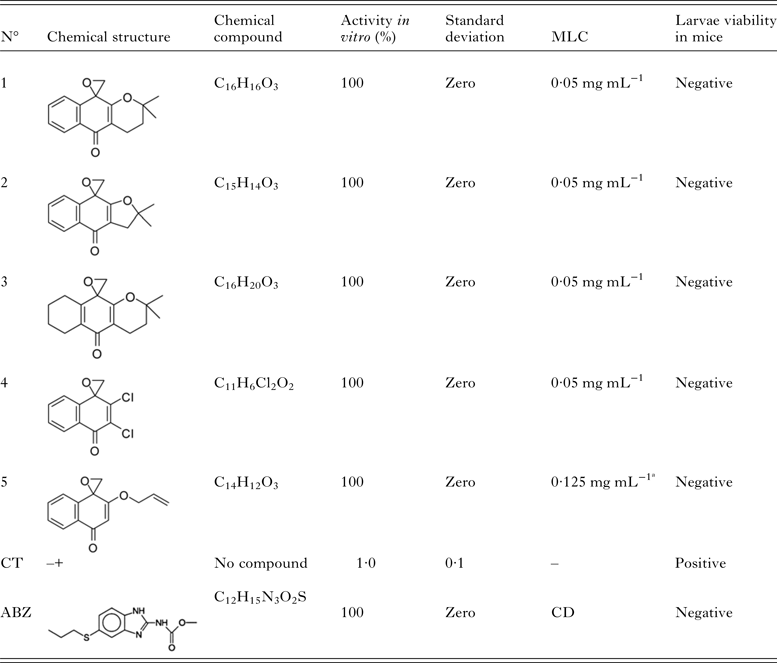
The quinones, naphthoimidazoles, naphthoxazoles and three naphthoxiranes showed activity only at 1 mg mL−1, a concentration considered inactive (Table 1). The others naphthoxiranes (1–4) showed the best results, at 0·05 mg mL−1; the compound 5 was effective at 0·125 mg mL−1 (Table 2). The compounds which, demonstrated activity below 0·5 mg mL−1, were evaluated by cytotoxic assay.
Activity in vitro of the quinone derivatives against Toxocara canis larvae (n = 27).

a Concentration considered inactive. CT, control; negative to detection of T. canis larvae in mice tissues; positive to detection of T. canis larvae in mice tissues; CD, commercial dose (40 mg Kg−1); MLC, minimal larvicidal concentration.
Larvicidal activity in vitro of the naphthoxiranes against Toxocara canis larvae (n = 5).
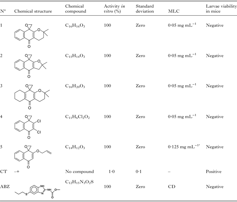
a Concentration considered inactive. CT, control; negative to detection of T. canis larvae in mice tissues; positive to detection of T. canis larvae in mice tissues; CD, commercial dose (40 mg Kg−1); MLC, minimal larvicidal concentration.
Cytotoxicity
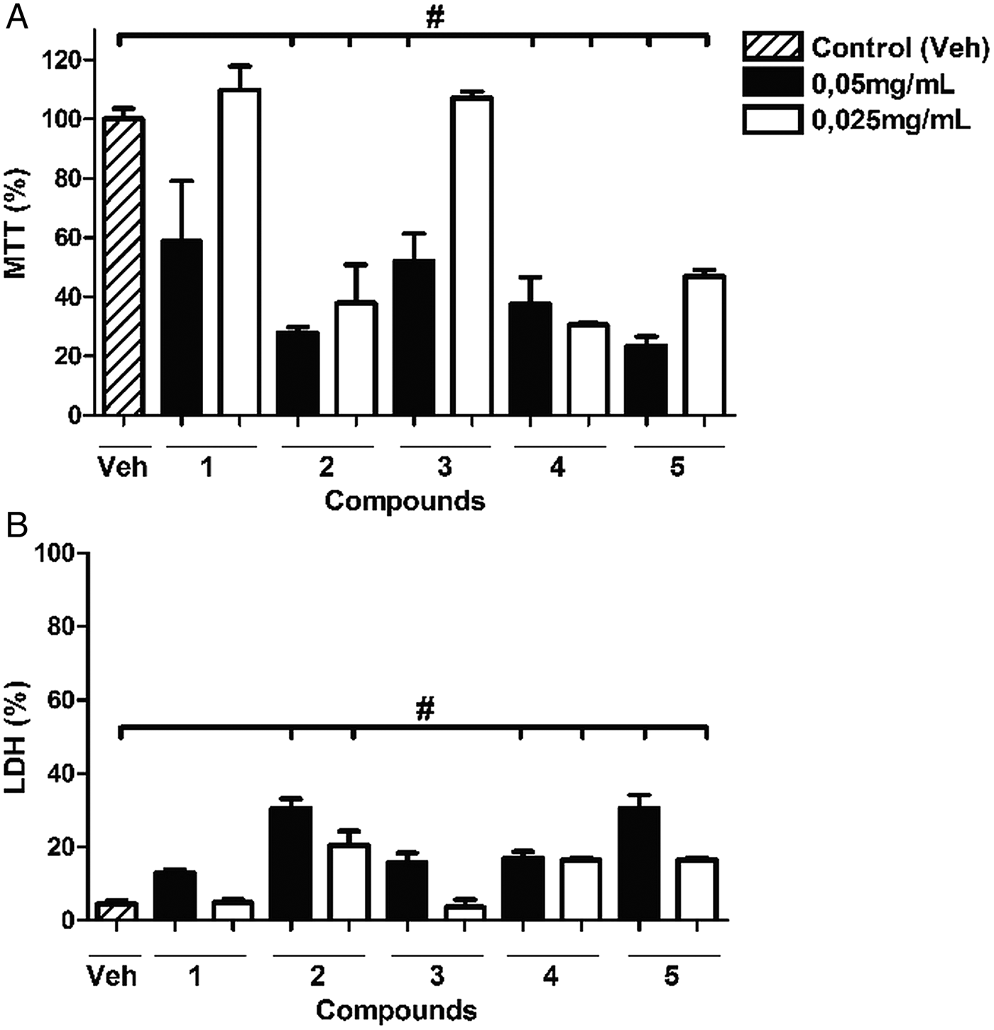
The results of MTT assay and LDH are represented in Fig. 2. The naphthoxiranes (1 and 3), were less cytotoxic than others (below 50% in MTT assay) at 0·05 mg mL−1 (in vitro effective concentrations) and considered to be acceptable for in vivo testing (Satou et al. Reference Satou, Horiuchi, Akao, Koike, Futija and Nikaido2005).
Cytotoxicity was measured by the MTT assay (A) and LDH-release (B) from murine peritoneal macrophages C57BL/6 mice treated with in vitro larvicidal compounds (Naphthoxiranes). Data represent means ± SD for triplicate values. #P < 0·05 compared with control. Abbreviations: LDH, lactate dehydrogenase; MTT, 3-(4,5-dimethylthiazol-2-yl)-2,5-diphenyltetrazolium bromide.

Evaluation in murine toxocarosis
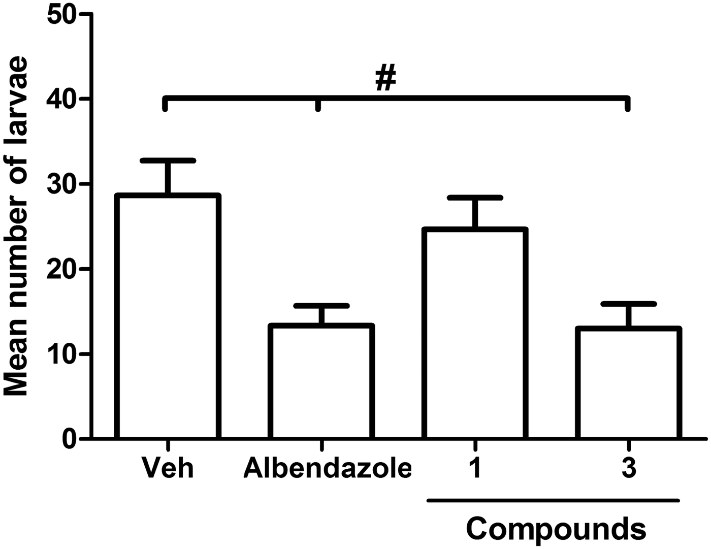
Naphthoxiranes (1 and 3 of Fig. 2) were administered intraperitoneally and evaluated at 10 mg Kg−1 of body weight for their anthelmintic activity in mice infected with T. canis eggs. As shown in Fig. 3, compound 3 was effective in reducing the intensity of infection in experimental toxocarosis and was equivalent to albendazole in efficacy.
Average number of Toxocara canis larvae recovered after treatment intraperitoneally, in the organs of mice with toxocarosis. Control group treated with 2·5% DMSO (Veh) as described in Materials and Methods. Data represent means ± SD for triplicate values. #P < 0·05 compared with control. Abbreviation: DMSO dimethyl sulphoxide.

DISCUSSION
The naphthoquinone derivatives are commonly found in nature and their biological and pharmacological activities are of great interest to the pharmaceutical industry (Araújo et al. Reference Araújo, Alencar and Neto2002). Quinones and their derivatives, known for their antiparasitic activity, are achieving prominent positions as precursors in the development of new drugs for the treatment of parasitic diseases (De Moura et al. Reference De Moura, Emery, Pinto, Pinto, Dantas, Salomão, Castro and Pinto2001; De Andrade-Neto et al. Reference De Andrade-Neto, Goulart, Da Silva, Da Silva, Pinto, Pinto, Zaliz, Carvalho and Krettli2004; Carneiro et al. Reference Carneiro, Nascimento, Pinto, Pinto, Lechuga, Santos, Júnior, Resende, Bourguignon and Ferreira2012).
In this study, the evaluated quinones and their derivatives, naphthoimidazoles and naphthoxazoles, did not prove to be effective against T. canis larvae at low concentrations. These results were unexpected since the original quinones, lapachol and β-lapachone, showed activity at concentrations 500 and 250 µg mL−1 respectively, against T. canis larvae (Mata-Santos et al. Reference Mata-Santos, Pinto, Mata-Santos, De Moura, Carneiro, Carvalho, Del Rio, Pinto, Martins, Fenalti, Da Silva and Scaini2015). In addition, naphthoimidazoles and naphthoxazoles demonstrated activity at low concentrations against Trypanosoma cruzi in other studies (Neves-Pinto et al. Reference Neves-Pinto, Dantas, De Moura, Emery, Polequevitch, Pinto, De Castro and Pinto2000; De Moura et al. Reference De Moura, Emery, Pinto, Pinto, Dantas, Salomão, Castro and Pinto2001).
Of all the compounds evaluated in this study, the naphthoxiranes demonstrated the best activity in murine toxocarosis. Previous studies have shown that oxirane derivatives are more active against T. cruzi than their origin quinones, except for β-lapachone (Jorqueira et al. Reference Jorqueira, Gouvêa, Ferreira, Da Silva, De Souza, Zuma, Cavalcanti, Araújo, Santos and Bourguignon2006). However, β-lapachone had significant toxicity, unlike naphthoxiranes that proved to be less toxic than their origin quinones (Carneiro et al. Reference Carneiro, Nascimento, Pinto, Pinto, Lechuga, Santos, Júnior, Resende, Bourguignon and Ferreira2012). The difference between activity and cytotoxicity of quinones is partly attributable to their different redox potentials, since these vary according to the anchored groups (Kumagai et al. Reference Kumagai, Shinkai, Miura and Cho2012).
The presence of the oxirane group decreased the lipophilicity, dipole moment and cytotoxicity of these compounds (Ferreira et al. Reference Ferreira, Jorqueira, Souza, Silva, Souza, Gouvêa, Rodrigues, Pinto, Castro, Santos, Araújo and Bourguignon2006).
Further studies on naphthoxiranes are warranted, either in relation to the changes in their chemical structure or as a liposome or even in combination with probiotics, which have shown immunomodulatory responses, thus reducing the intensity of infection caused by T. canis larvae (Avila et al. Reference Avila, Conceição, Telmo, Dutra, Los Santos, Martins, Berne, Da Silva and Scaini2012).
As seen with the administration of albendazole and other compounds in murine toxocarosis, an ideal drug should not only decrease the intensity of infection but also eradicate all the larvae housed in the tissues, because they can migrate to the brain and eyes (Abo-Shehada et al. Reference Abo-Shehada and Herbert1984 Delgado et al. Reference Delgado, Botto, Mattei and Escalante1989; Lescano et al. Reference Lescano, Chieffi, Neto, Ikai and Ribeiro2005). Studies are ongoing for the evaluation of compound 3 administered orally and via the intravenous route as well as for the assessment of its cytotoxicity at high concentrations.
ACKNOWLEDGEMENTS
The authors are grateful to Prof. Antônio Ventura Pinto and to Instituto de Pesquisas de Produtos Naturais (IPPN-UFRJ) for the compounds.
FINANCIAL SUPPORT
This work was supported by Coordenação de Aperfeiçoamento de Pessoal de Nível Superior (T. M-S. and J.F.) and partially supported by CNPq (Conselho Nacional de Desenvolvimento Científico e Tecnológico; P.F.C and K.C.G.d M.). Authors declare no conflict of interests.