Introduction
The olfactory epithelium consists of olfactory receptor neurons, supporting cells and basal cells. Throughout the vertebrate lifespan, there is continuous replacement of the olfactory receptor neurons, which regenerate from basal cells.Reference Graziadei and Graziadei 1 Olfactory receptor neurons also undergo replacement after neuronal injury. Many studies have examined the degeneration and regeneration of the olfactory epithelium following neuronal injury.Reference Graziadei and Graziadei 1 , Reference Suzuki 2 Our group has reported on the morphological changes of supporting cells following bulbectomy; these changes contribute to the regeneration of the olfactory epithelium.Reference Makino, Ookawara, Katoh, Ohta, Ichikawa and Ichimura 3 However, many aspects of olfactory epithelium regeneration remain unclear, including the mechanisms involved, the molecular pathways and the morphology.
Several recent studies have revealed that tissue-type plasminogen activator is closely related to neuronal injury in the central and peripheral nervous system.Reference Nagai and Matsuo 4 One report showed that tissue plasminogen activator was expressed in neuronal cells such as microglia and astrocytes, and that it played an important role in the central nervous system in response to excitotoxic injury.Reference Siao, Fernandez and Tsirka 5 Studies using ‘knock-out’ mice deficient in tissue plasminogen activator showed a decrease in neurotoxic effects in these mice, and neurons cultured from these mice were protected against hypoxia and oxidative stress.Reference Nagai, Yamamoto, Tsuboi, Ihara, Urano and Takada 6 Mice deficient in tissue plasminogen activator also showed reduced neural damage following focal cerebral ischaemiaReference Lee, Lok, Rosell, Kim, Murata and Atochin 7 and injury to the spinal cord.Reference Abe, Nakamura, Yoshino, Oya and Kimura 8 Tissue plasminogen activator has been reported to be expressed in the olfactory bulb.Reference Thewke and Seeds 9 In that particular study, it was suggested that tissue plasminogen activator played a regulatory role in the development and maintenance of the olfactory system; however, no morphological changes were identified within the olfactory epithelium.
In order to provide evidence that tissue plasminogen activator is indeed involved in the development and maintenance of the olfactory system, it is necessary to show the associated morphological changes within the olfactory epithelium. The current study therefore examined the morphological changes of the olfactory epithelium before and after injury, using mice deficient in tissue plasminogen activator.
The results of this study suggest that tissue plasminogen activator is a regulator of supporting cell function and its absence may enhance regeneration of the olfactory epithelium after bulbectomy. To our knowledge, this is the first study to examine the olfactory epithelium of mice deficient in tissue plasminogen activator using transmission electron microscopy.
Materials and methods
Animals and surgery
This study used mice (12–16 weeks old) with tissue plasminogen activator deficiency.Reference Carmeliet, Schoonjans, Kieckens, Ream, Degen and Bronson 10 These mice were originally derived from common, inbred laboratory mice type C57BL/6. All surgical procedures were carried out in accordance with the guidelines approved by the Jichi Medical University Animal Care and Concern Committee.
The mice were anaesthetised with 2.4 per cent isofluorane inhalation (180 ml/minute). After cutting the right olfactory nerve under a stereomicroscope, the right olfactory bulb was removed (bulbectomy) using vacuum aspiration with a fine stainless steel tube. The animals underwent morphological examination on days 1, 2, 3, 5 or 7 after the unilateral bulbectomy, with four mice per group (i.e. 20 mice in total). A control group was also established (n = 4); the mice in this group underwent a sham operation.
Tissue preparation for microscopy
The mice were perfused through the heart with a cold physiological saline solution, followed by 2 per cent paraformaldehyde and 2.5 per cent glutaraldehyde buffered with 0.1 M cacodylate solution (pH 7.4). Epithelial tissue from the nasal septum was carefully dissected and immersed in the same fixative for 12 hours. After washing with cold cacodylate buffer, the specimens were post-fixed with 1 per cent osmium tetroxide solution buffered with cacodylate solution for 2 hours on ice. Specimens were then treated with 0.5 per cent uranyl acetate for 2 hours at room temperature. This was followed by dehydration with an ascending ethanol series. The specimens were then embedded in an Epon mixture (TAAB Laboratories, Berks, UK). For light microscopy, Epon-embedded sections (0.5–0.7 µm thick) were stained with toluidine blue. For electron microscopy, ultrathin sections were mounted on a copper grid and stained with uranyl acetate and lead citrate. Sections were observed using a Jem-2000EX electron microscope (Tokyo, Japan).
Results
Light microscopy
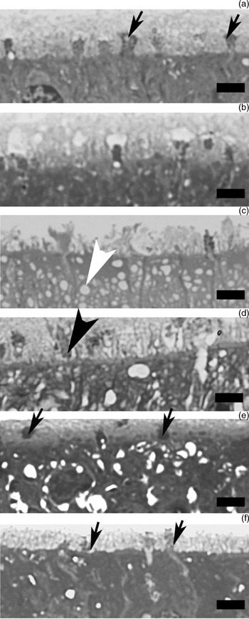
Prior to bulbectomy, the free surface of the epithelium was covered with a faintly stained brush border of supporting cells, olfactory vesicles and mucus (Figure 1a). One day after bulbectomy, the olfactory vesicles appeared to be intact (Figure 1b). However, 2 days after bulbectomy, there were a reduced number of olfactory vesicles on the free surface of the epithelium (Figure 1c). Some cytoplasmic projections were also identified. In addition, numerous small, empty vesicles were present in the apical area of the supporting cells. Three days after bulbectomy, the olfactory vesicles were barely recognisable on the free surface of the epithelium, and a large number of cytoplasmic projections were observed within the brush borders (Figure 1d). Five days after bulbectomy, the cytoplasmic projections had disappeared from the free surface of the epithelium and olfactory vesicles were found there (Figure 1e). Seven days after bulbectomy, olfactory vesicles were found on the free surface of the epithelium (Figure 1f).
Light micrographs of the olfactory epithelium. (a) Intact epithelium before bulbectomy. Olfactory vesicles (black arrows) can be seen on the free surface of the epithelium. (b) One day after bulbectomy. The staining of olfactory vesicles is unchanged. (c) Two days after bulbectomy. The number of olfactory vesicles on the free surface of the epithelium has declined. Numerous small, empty vesicles have appeared in the apical area (white arrowhead). (d) Three days after bulbectomy. Olfactory vesicles are barely recognisable. A large number of cytoplasmic projections (black arrowhead) are present in the brush borders. (e) Five days after bulbectomy. The cytoplasmic projections have disappeared and olfactory vesicles can again be seen (black arrows). (f) Seven days after bulbectomy. The epithelium is almost intact and olfactory vesicles can be seen (black arrows). (Bar = 3 µm)

Electron microscopy
At baseline
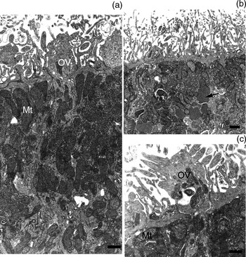
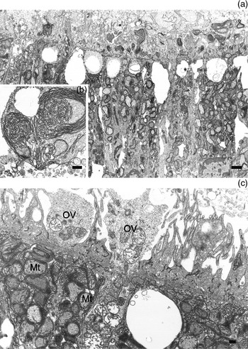
Figure 2 shows electron micrographs of the intact olfactory epithelium of mice with tissue plasminogen activator deficiency, prior to bulbectomy. These micrographs reveal the free surface of the epithelium and the underlying olfactory receptor neurons and supporting cells. Ciliated olfactory vesicles extended above the free surface of the epithelium, and distinct, tight junctions were observed between the olfactory vesicles and the supporting cells. Microvilli were detected on the free surface of the supporting cells. The apical portion of the cells contained numerous rod-shaped mitochondria and membranous structures. A uniformly fine material was observed in some regions (Figure 2b) under transmission electron microscopy, but not under light microscopy.
Electron micrographs of the intact olfactory epithelium. (a) Ciliated olfactory vesicles can be seen. There are many rod-shaped mitochondria in the supporting cells. (b) A uniformly fine material is visible in the supporting cells (black arrow). (c) Ciliated olfactory vesicles and many rod-shaped mitochondria can be seen in the supporting cells. OV = olfactory vesicles; Mt = mitochondria (bar = 1 µm)

One day after bulbectomy
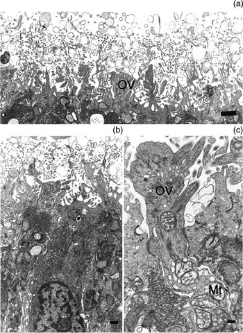
The free surface of the olfactory epithelium from mice with tissue plasminogen activator deficiency exhibited a complex structure with many vacuoles located among the microvilli (Figure 3a and 3b). These vacuoles probably resulted from the degenerating cilia. Olfactory vesicles were overlaid with the degenerating microvilli and cilia. Figure 3c shows slight degeneration of the olfactory vesicles and degenerating cilia in the olfactory epithelium.
Electron micrographs of the olfactory epithelium 1 day after bulbectomy. (a) & (b) The thickness of the microvilli from supporting cells has increased. There are many vacuoles derived from the degenerating cilia (bar = 1 µm). (c) Slight degeneration of the olfactory vesicles can be observed (bar = 200 nm). OV = olfactory vesicles; Mt = mitochondria

Two days after bulbectomy
The number of microvilli protruding from the supporting cell surfaces on the free surface of the olfactory epithelium had decreased (Figure 4a). Instead of microvilli, we observed broad cytoplasmic projections containing undefined cytoplasmic organelles protruding into the luminal side (Figure 4a). In the cytoplasm of the supporting cells, the mitochondria were swollen, there was a decrease in the electron opacity and the cristae were obscure. There were numerous degenerating olfactory vesicles, with slight mitochondrial degeneration in the olfactory epithelium (Figure 4b). A uniformly fine material protruded onto the luminal surface of the olfactory epithelium in some regions (Figure 4a).
Electron micrographs of the olfactory epithelium 2 days after bulbectomy. (a) Only a small number of microvilli and broad cytoplasmic projections can be seen. Some of the cytoplasmic projections contain unidentified cell organelles (black arrow). Materials with a uniform appearance have protruded into the luminal side (white arrow) (bar = 1 µm). (b) The mitochondria are swollen. The electron opacity of mitochondria has decreased and cristae are obscure. Olfactory vesicles with partially degenerated mitochondria (black arrow) can be observed (bar = 200 nm). OV = olfactory vesicles; Mt = mitochondria

Three days after bulbectomy
The free surfaces of the olfactory epithelium were more prominent than those at 2 days after bulbectomy (Figure 5a). The cytoplasm of the supporting cells contained swollen and degenerated mitochondria, degenerated olfactory vesicles, and receptor neuron dendrites (Figure 5c). A large lamellar and smooth endoplasmic reticulum was evident within the cytoplasmic projections of the supporting cells in the microvilli (Figure 5b).
Electron micrographs of the olfactory epithelium 3 days after bulbectomy. (a) Only a small number of microvilli and broad cytoplasmic projections are present (bar = 1 µm). (b) The projections of the supporting cells contain smooth endoplasmic reticulum (bar = 200 nm). (c) The olfactory vesicles have almost disappeared. The smooth endoplasmic reticulum and the degenerating mitochondria are closely linked (bar = 200 nm). OV = olfactory vesicles; Mt = mitochondria

Five days after bulbectomy
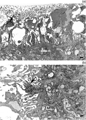
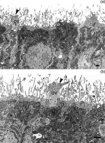
Compared with observations at 3 days, we detected marked changes in the morphology of the olfactory epithelium 5 days after bulbectomy. Morphologically, the olfactory epithelium seemed almost completely recovered. The olfactory vesicles and receptor neuron dendrites were nearly intact (Figure 6a and 6b).
Electron micrographs of the olfactory epithelium 5 days after bulbectomy. (a) Near-normal microvilli and mitochondria are present. Olfactory vesicles and receptor neuron dendrites are visible (black arrowhead). (b) Olfactory vesicles and receptor neuron dendrites are visible (black arrowhead). OV = olfactory vesicles (bar = 1 µm)

Seven days after bulbectomy
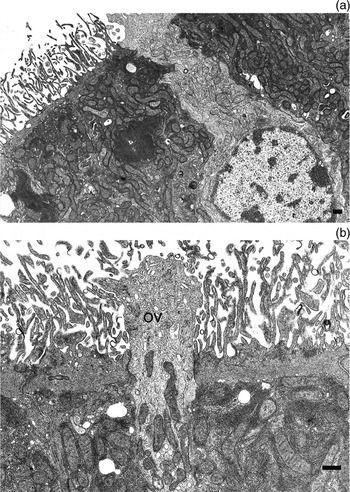
In order to determine the morphological changes in the olfactory epithelium of mice deficient in tissue plasminogen activator, we compared the olfactory epithelium of these mice with that of wild-type mice at 7 days after bulbectomy (using findings from our previous study). In mice with tissue plasminogen activator deficiency (Figure 7a and 7b), the morphological features of the olfactory epithelium were similar to those of ‘intact mice’ (i.e. the wild-type mice that underwent the sham operation). Olfactory vesicles and receptor neuron dendrites were visible and showed almost complete morphological recovery. The smooth endoplasmic reticulum and mitochondria in the cytoplasm of the apical portion of the supporting cells were nearly intact.
Electron micrographs of the olfactory epithelium 7 days after bulbectomy. (a) The olfactory epithelium has a near-normal appearance. (b) Normal olfactory vesicles and receptor neuron dendrites are visible. OV = olfactory vesicles

Discussion
Degeneration of the olfactory epithelium after neuronal injury is a significant clinical problem because it leads to irreversible olfactory disorders. Several experimental studies have reported on this problem, but many aspects remain unclear. Our recent investigations have focused on tissue plasminogen activator as a possible molecule involved in the recovery of olfactory epithelium.
The tissue plasminogen activator plasmin system is well understood. Plasmin is converted from the zymogen plasminogen by tissue plasminogen activator or urokinase-type plasminogen activator, and can degrade the extracellular matrix. These proteolytic activities of plasmin are involved in neuronal death.Reference Nagai and Matsuo 4 – Reference Nagai, Yamamoto, Tsuboi, Ihara, Urano and Takada 6 Fibrinolytic system components play important roles in the nervous system.Reference Nagai and Matsuo 4 In addition to acting as a fibrinolytic agent, it has been suggested that tissue plasminogen activator plays an important role in the brain. For instance, tissue plasminogen activator may interact with the N-methyl-D-aspartate receptor complex,Reference Nicole, Docagne, Ali, Margaill, Carmeliet and MacKenzie 11 thereby mediating microglial activation.Reference Gravanis and Tsirka 12 Furthermore, mice deficient in tissue plasminogen activator showed reduced blood–brain barrier injury following focal cerebral ischaemia.Reference Tsuji, Aoki, Tejima, Arai, Lee and Atochin 13
The potential role of the tissue plasminogen activator plasmin system in the olfactory epithelium has been demonstrated indirectly. Non-integrin laminin receptor precursor protein is expressed in the olfactory stem and progenitor cells.Reference Jang, Kim and Schwob 14 Matrix metalloproteinases 9 and 2 are expressed in the olfactory epithelium,Reference Costanzo, Perrino and Kobayashi 15 , Reference Tsukatani, Fillmore, Hamilton, Holbrook and Costanzo 16 and their expression increases in the olfactory epitheliumReference Costanzo, Perrino and Kobayashi 15 , Reference Costanzo and Perrino 17 and olfactory bulb.Reference Bakos, Schwob and Costanzo 18 Plasmin may play a role in degrading laminin and activating matrix metalloproteinases. Therefore, we hypothesised that tissue plasminogen activator and other fibrinolytic factors may be involved in the continuous neurogenesis cell proliferation, migration and/or differentiation seen within the olfactory epithelium.
In this novel study, we examined the olfactory epithelium of mice deficient in tissue plasminogen activator following bulbectomy, using light microscopy and transmission electron microscopy. We compared our findings with those of our previous study using wild-type mice treated in the same way.Reference Makino, Ookawara, Katoh, Ohta, Ichikawa and Ichimura 3 As the mice deficient in tissue plasminogen activator were derived from C57BL/6 mice, any differences between these types of mice should reflect the function of tissue plasminogen activator in the olfactory epithelium.
For our light microscopic observations, we stained sections of plastic-embedded material (0.5–0.7 µm thick) with toluidine blue. At 0–2 days after bulbectomy, we found that the morphological changes at the free surface of the olfactory epithelium were similar for both wild-type mice and mice deficient in tissue plasminogen activator. At 5–7 days after bulbectomy, the olfactory vesicles at the free surface of the epithelium were barely recognisable in wild-type mice, and numerous cytoplasmic projections were observed within the brush borders in these mice.Reference Makino, Ookawara, Katoh, Ohta, Ichikawa and Ichimura 3 In fact, it is often reported that the olfactory neurons of wild-type mice almost vanish at 5–7 days after bulbectomy.Reference Graziadei and Graziadei 1 – Reference Makino, Ookawara, Katoh, Ohta, Ichikawa and Ichimura 3 , Reference Costanzo, Perrino and Kobayashi 15 , Reference Costanzo and Perrino 17 However, in the olfactory epithelium of mice deficient in tissue plasminogen activator, olfactory vesicles were recognisable, and the cytoplasmic projections had disappeared.
Transmission electron microscopy observations of the pre-bulbectomy olfactory epithelium showed marked morphological differences in the supporting cells of mice deficient in tissue plasminogen activator, compared with those of wild-type mice. We also found that the prevalence of an unknown, uniformly fine material within the supporting cells, indicative of phagocytic ability,Reference Suzuki 2 had decreased in mice deficient in tissue plasminogen activator. This apparent decline in terms of phagocytic ability was not seen in the supporting cells of wild type mice. This suggests that the tissue plasminogen activator plasmin system regulates olfactory epithelium regeneration.
At 5 days after bulbectomy, the morphological features of the olfactory epithelium in mice deficient in tissue plasminogen activator were similar to those prior to bulbectomy. We found numerous intact olfactory vesicles and almost fully recovered supporting cells in the olfactory epithelium.
-
• The olfactory epithelium of mice deficient in tissue plasminogen activator was examined using light and transmission electron microscopy
-
• An unknown, uniformly fine material was seen in epithelial supporting cells pre-bulbectomy
-
• The olfactory neurons of wild-type mice are reported to almost vanish 5–7 days after bulbectomy
-
• The olfactory neurons of mice deficient in tissue plasminogen activator were regenerating at 5–7 days after bulbectomy
-
• The tissue plasminogen activator plasmin system may inhibit olfactory epithelium regeneration following neural injury
To further clarify the morphological changes, we compared the olfactory epithelium of wild-type (intact) mice with mice deficient in tissue plasminogen activator 7 days after bulbectomy. In the mice deficient in tissue plasminogen activator, the morphological features of the supporting cells were similar to those of the wild-type mice. However, the olfactory vesicles and receptor neuron dendrites were visible in mice deficient in tissue plasminogen activator, but not in wild-type mice. Furthermore, the olfactory epithelium was still in a severe degenerative state in the wild-type mice.
The findings of this study suggest that the olfactory receptor neurons regenerate earlier in mice with tissue plasminogen activator deficiency than in wild-type mice. It is hypothesised that the tissue plasminogen activator plasmin system might play an inhibitory role in the regeneration of the olfactory epithelium following bulbectomy.
In clinical settings, tissue plasminogen activator has been used in fibrinolytic therapy for acute myocardial infarctionReference Llevadot, Giugliano and Antman 19 and cerebral infarction.Reference Fugate, Giraldo and Rabinstein 20 Our results provide evidence that the suppression of tissue plasminogen activator activity in the olfactory epithelium may improve functional recovery following olfactory tract injury, by affecting the degeneration and regeneration of the olfactory epithelium. These findings could lead to new therapeutic strategies for the treatment of olfactory disorders.
Acknowledgments
We would like to thank T Ohmori, A Ishiwata, Y Kashiwakura, Y Noda, K Okada and S Ueshima for their advice and helpful discussion. This study was supported by an official annual grant from Jichi Medical University School of Medicine.