Introduction
Diamond has become a material of increasing interest for electronic devices over the last decade, particularly as a biologic sensing platform, due to its many attractive material properties.[Reference Nebel, Rezek, Shin, Uetsuka and Yang1] Nano-crystalline diamond (NCD) thin films can be grown by chemical vapor deposition (CVD) on various substrates to fabricate functional devices using the standard microfabrication techniques. Diamond is a wide band gap semiconductor that can be doped to give metal-like conductivity, allowing development of biocompatible electrodes, which are electrochemically robust, giving an extremely wide chemical potential window.[Reference Kraft, Gmbh and Str2] Furthermore, the wide band gap of un-doped diamond allows it to be used as an insulating material in the voltage range of biologic signals.
Microelectrode arrays (MEAs) were developed in the 1970s, and have become an indispensable tool in the field of electrophysiology for non-invasively investigating electrical activity of excitable cells, such as neurons and cardiomyocites, both in vivo and in vitro. Moreover, MEAs enable the recording of spontaneous extracellular electric signals, allowing the interactions within populations of synaptically connected neurons to be studied, including their collective response to drugs, crucial for therapeutic drug-screening. MEAs can also be used to directly stimulate neurons, in order to study evoked response and the underlying networks organization, connectivity, and response dynamics.[Reference Stett, Egert, Guenther, Hofmann, Meyer, Nisch and Haemmerle3] However, most of the MEAs show both the capacitive and injection currents. The injection currents, depending on the current used, may deteriorate the neural tissue due to the local pH changes. The capacitive current is of high interest due to absence of any non-displacement currents.
Here we report on the fabrication of MEAs using boron-doped nano-crystalline diamond (BNCD) microelectrodes with a passivation layer made of intrinsic NCD. BNCD exhibits many advantages as an electrode material compared with common gold, platinum, and TiN materials, including high chemical and mechanical stability, a 3V-wide electrochemical water potential window, and high biocompatibility.[Reference Vaitkuviene, McDonald, Vahidpour, Noben, Sanen, Ameloot, Ratautaite, Kaseta, Biziuleviciene, Ramanaviciene, Nesladek and Ramanavicius4, Reference Cottance, Nazeer, Rousseau, Lissorgues, Bongrain, Kiran, Scorsone, Bergonzo, Bendali, Picaud and Joucla5] Moreover, BNCD has been shown to be an effective electrode material for biologic sensors.[Reference Cottance, Nazeer, Rousseau, Lissorgues, Bongrain, Kiran, Scorsone, Bergonzo, Bendali, Picaud and Joucla5–Reference Meijs, Alcaide, Sørensen, McDonald, Sørensen, Rechendorff, Gerhardt, Nesladek, Rijkhoff and Pennisi9] NCD is also highly biocompatible,[Reference Vaitkuviene, McDonald, Vahidpour, Noben, Sanen, Ameloot, Ratautaite, Kaseta, Biziuleviciene, Ramanaviciene, Nesladek and Ramanavicius4, Reference Edgington, Thalhammer, Welch, Bongrain, Bergonzo, Scorsone, Jackman and Schoepfer10] as well as transparent, and insulating, making it a suitable material as a passivation layer to provide a complete diamond-sensing platform surface. Recently, full diamond MEAs were used for detection of action potential of myocardial cells, which generally have substantially higher amplitudes than for neurons.[Reference Maybeck, Edgington, Bongrain, Welch, Scorsone, Bergonzo, Jackman and Offenhäusser6] One of the limits of such technology is the high resistance of the electrodes. Our work led to electrodes with purely capacitive coupling and resistance comparable with the state-of-the-art non-diamond MEAs. Our method of fabrication combined with optimal layout and specific diamond surface functionalization allowed for extracellular recording of neural signals from hippocampal neurons.
Materials and methods
Design and fabrication of MEAs
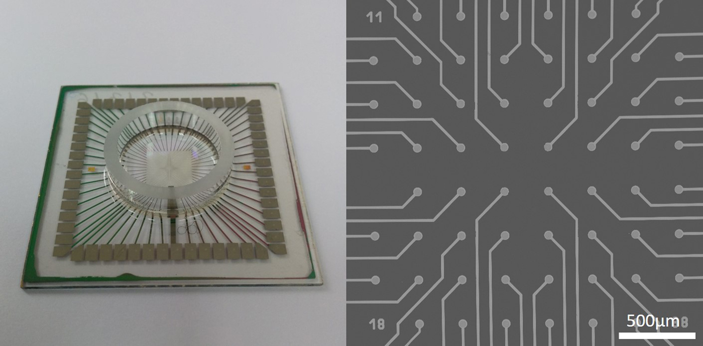
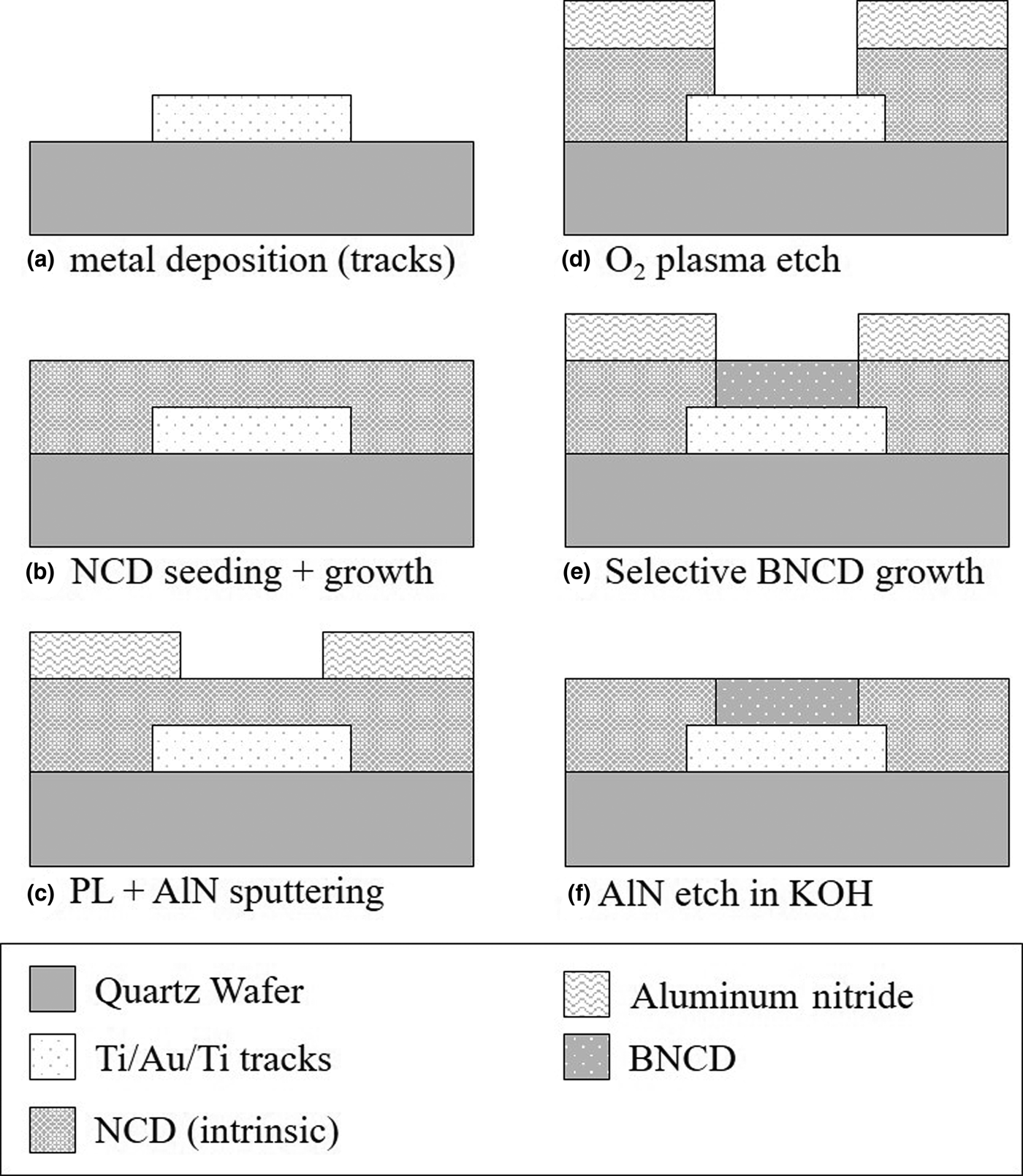
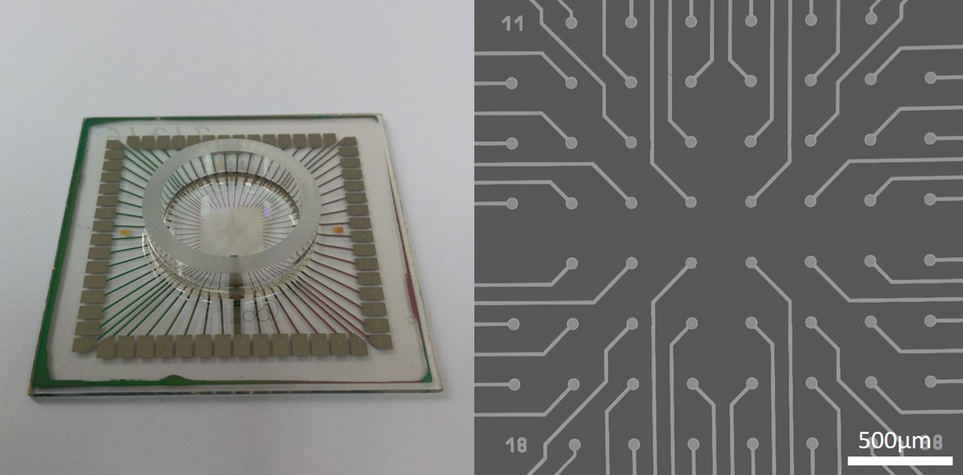
The MEA design was derived from commercial planar MEAs (MultiChannel System GmBH, Reutlingen, Germany) to allow comparison of performance with commercial MEAs. It incorporates a regular 8 × 8 arrangement of 40 µm diameter circular microelectrodes with a pitch of 200 µm, as shown in Fig. 1. Figure 2 shows the fabrication process for the MEA, which starts with a wafer of size 49 mm made from quartz glass in order to withstand the stresses induced from high-temperature diamond growth. The fabrication technique has the advantage of using a single photolithographic step to open the electrodes in the insulating NCD layer and grow the conductive BNCD layer; this allows for the insulating and conductive layers to be at the same level, with virtually no step height between them. Additionally, the MEAs are made on a transparent quartz substrate so that neurons are easily visualized using standard microscopy.
Fully packaged BNCD/NCD MEA (left) and SEM image of the active sensing area (right).
Fabrication process of MEAs with BNCD electrodes and NCD passivation layer. (a) a Ti/Au/Ti metal tri-layer is deposited using lift-off photolithography to form the metal tracks, (b) the surface is seeded with diamond nanoparticles and 180 nm NCD layer is grown by MPECVD, (c) photolithography is used to define a hard mask made of AlN, (d) the NCD layer is etched through to the underlying metal track by oxygen plasma, (e) the surface is selectively seeded with diamond nanoparticles and the BNCD electrode is grown, (f) AlN is etched in a KOH solution.

Quartz substrates were initially prepared by standard RCA 1 and RCA 2 cleaning.[Reference Kern11] Metal tracks were defined on the substrate using standard photolithography lift-off techniques. A thin (~5 nm) titanium adhesion layer was sputtered, followed by 300 nm gold and a second (~20 nm) adhesion layer of titanium. The surface was seeded for diamond growth by spin coating a 0.33 g/L solution of detonation diamond nanoparticles on the substrate (NanoAmando®B, NanoCarbo Research Institute, Japan).[Reference Pobedinskas, Degutis, Dexters, Janssen, Janssens, Conings, Ruttens, D'Haen, Boyen, Hardy, Van Bael and Haenen12] The NCD passivation layer was then grown using a gas mixture of 3% methane and 97% hydrogen in a MPECVD (Microwave Plasma Enhanced Chemical Vapor Deposition) growth reactor (Seki Technotron corp. AX6500). NCD was grown at a plasma temperature of 600–650 °C, by maintaining a pressure of 15 Torr and 2000 W with total gas flow of 300 sccm. A second lift-off process was used to sputter a hard mask of aluminum nitride (AlN), which defined the electrode area. Oxygen reactive ion etching (RIE) was used to open holes in the passivation layer and expose titanium/gold electrodes below. The substrate was then selectively seeded with detonation diamond nanoparticles, which attached to the electrode while being repelled by the surface charge of AlN.[Reference Pobedinskas, Degutis, Dexters, Janssen, Janssens, Conings, Ruttens, D'Haen, Boyen, Hardy, Van Bael and Haenen12] BNCD electrodes were then grown with a gas mixture of hydrogen, methane, and trimethylboron in a MPECVD growth reactor (Seki Technotron corp. AX6500). BNCD was grown at plasma temperature of 750 °C at 30 Torr and 3500 W with total gas flow of 500 sccm. Finally, the contacts were opened by placing a silicon wafer over the center area and etching the diamond covering the electrodes with an oxygen plasma treatment.
Additionally, a set of 1 cm × 1 cm NCD coated quartz substrates were prepared to test if the fabrication procedure left any artifacts that could have negative effects on the electrophysiological properties of the cells. Substrates went through all the same fabrication steps as MEAs, except for metal track deposition or photolithography to define microelectrode-opening holes.
Rat primary cortical neurons cultures
Prior to cell seeding, BNCD-MEAs and commercial indium tin oxide (ITO)-based glass MEAs (Multichannel Systems GmBH), employed as control, were treated with 70% ethanol for 10 min and air-dried overnight. Quartz substrates and glass coverslips used for patch clamp experiment were autoclaved upon high-pressure saturated steam at 121 °C for 30 min. MEAs, substrates, and coverslips were then coated with polyethylenimine (PEI) by incubation under 0.1% (w/v) of PEI in milli-Q water at room temperature for at least 8 h; samples were then rinsed thoroughly with milli-Q water and air dried.[Reference Vaitkuviene, McDonald, Vahidpour, Noben, Sanen, Ameloot, Ratautaite, Kaseta, Biziuleviciene, Ramanaviciene, Nesladek and Ramanavicius4, Reference Ojovan, McDonald, Rabieh, Shmuel, Erez, Nesladek and Spira13] The PEI treatment is based on our previous results, in which we demonstrated the PEI or poly-D-lysine is necessary pre-condition for vital neuron growth.[Reference Ojovan, McDonald, Rabieh, Shmuel, Erez, Nesladek and Spira13]
Rat primary cortical cultures were obtained by postnatal (P0) Wistar rats (Charles River, France) using standard methods and following the guidelines of the Ethics Committee of the University of Antwerp. Cortices were dissected, sliced, digested in a trypsin solution, mechanically dissociated, centrifuged, and suspended in Modified Essential Medium supplemented with 5% horse serum, 50 µg/mL gentamycin, 200 mM L-glutamine, and 2 M glucose.
Cells were seeded at nominal densities of 6500 cells/mm2 on MEAs, and of 1500 cells/mm2 on glass coverslips and quartz substrates. All chemicals were purchased by Sigma Aldrich (Belgium).
Electrophysiology
Cellular electrophysiology
Whole-cell patch-clamp recordings were carried out at 34 °C from neuronal somas after 9 days in vitro (DIV) using an Axon Multiclamp Amplifier (Molecular Devices, USA) controlled by LCG software.[Reference Linaro, Couto and Giugliano14] During recordings, glass coverslips and BNCD-coated quartz substrates were kept in the artificial cerebrospinal fluid, containing (in mM): 145 NaCl, 5 Hepes, 5 Glucose, 4 KCl, 2 CaCl2, 2 Na-pyruvate, and 1 MgCl2; pH adjusted to 7.4 with NaOH.
Patch pipettes were pulled from borosilicate glass and filled with intracellular solution, containing (in mM): 135K-gluconate, 10 Hepes, 10 PC-Na2, 4 NaCl, 4 ATP-Mg2+, 0.3 GTP-Na2 and 0.2 EGTA; pH adjusted to 7.3 with KOH. Electrical impedance of patch pipettes was 6–8 MΩ. Recordings were analyzed using custom MATLAB scripts (The Math-works, USA). Statistical analysis was performed using the Liliefors test, to assess normality of data sets, and the Student's t test to evaluate statistical significance; data are presented as mean ± standard error of the mean (SEM).
Network electrophysiology
Neuronal network activity was recorded at 17, 19, 21, 24, and 28 DIV from two control ITO-MEAs and 7 BNCD-MEAs, mounted into a recording amplifier (Multichannel System GmBH). Spontaneous electrical activity was recorded for 30 min; traces were acquired and stored on disk for offline analysis by MCRack (Multichannel Systems, Germany) and subsequently analyzed by QSpike Tools.[Reference Mahmud, Pulizzi, Vasilaki and Giugliano15]
Individual microelectrodes of a MEA were defined as active, when the frequency of occurrence of detected biologic events (spikes) at that single electrode exceeded 0.05 Hz.
Results and discussion
Morphological characterization
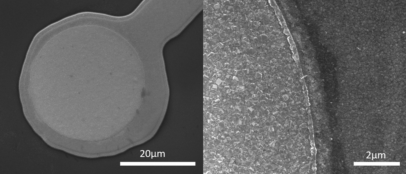
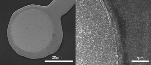
Important characteristics of functional MEA electrodes are the impedance and capacitance. These parameters depend on the material and geometrical properties used for the construction of the functional electrodes as well as for the isolating layer. Diamond has the advantage of being able to be used both as a conductive and an isolating material. While undoped diamond is highly isolating, the conducting diamond is obtained by p-type doping with boron. The thickness of the doped and undoped diamond layers determines the capacitive and isolating properties, respectively. The thickness of the diamond layers is also important from the fabrication point of view, for definition of the lithographic and etching steps. The thickness of diamond passivation layer was therefore carefully monitored by in situ interferometry and was optimized to approximately 180 nm, which was sufficient to insulate the metal tracks. BNCD electrodes were between 180 and 200 nm, which was found by using profilometry to measure the step height above the diamond passivation layer. The average electrode diameter was approximately 30 µm, giving an active area of ~707 µm2. Scanning electron microscopy was used to measure the morphology and grain-size of the diamond layers. NCD and BNCD crystals are highly faceted, with an average grain size of ~100 and ~200 nm, respectively (Fig. 3).
SEM image of BNCD electrode (left) and SEM image showing fine grain structure of BNCD electrode versus NCD passivation layer.

The grain size is an important parameter for obtaining an optimal diamond film surface in terms of crystal quality (and consequently good electrical properties) and surface roughness of the electrode surface. Consequently, the grain size was controlled by the growth parameters such as the methane concentration in the gas mixtures during growth. A concentration of 3% methane was used for the NCD growth to provide a smoother continuous film with low stress, to ensure no pinholes or cracking in the layer.[Reference Vandevelde, Nesladek, Quaeyhaegens and Stals16] A methane concentration of 1% was used in the gas mixture for BNCD to obtain a larger diamond grain size in order to maximize the conductivity.[Reference Janssens, Pobedinskas, Vacik, Petráková, Ruttens, D'Haen, Nesládek, Haenen and Wagner17] A boron-to-carbon ratio of 9000 ppm in the gas mixture was used to ensure sufficient boron-doping levels for high electrode conductivity. The resultant layers had conductivities of about 400 Ω/square.
Electrochemical characterization
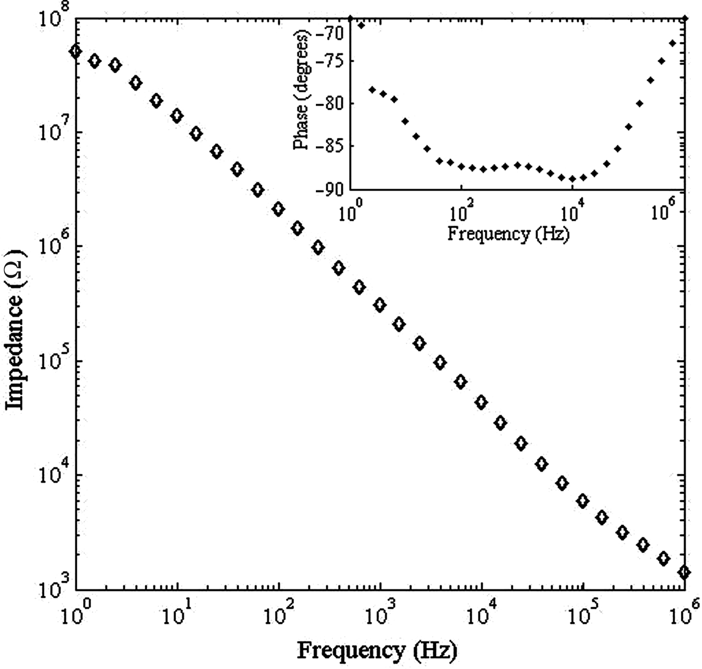
One of the critical requirements for microelectrodes to detect multi-unit neuronal activity is to have a low-enough impedance in the frequency range of 0.1–1 kHz, enabling adequate spatial selectivity and sensitivity.[Reference Merrill and Tresco18] Impedance of BNCD electrodes is expressed in terms of the equivalent Randal circuit and depends on the BNCD impedance (doping and the electrode geometry) as well as on forming the electric charge double-layer on the electrode immersed in bio-analyte. To simulate the working conditions, impedance spectroscopy (IS) was used to perform the electrical characterization of the devices prior to cell seeding. IS was performed in 1 M phosphate buffer in frequency sweep mode with 0.1 V peak-to-peak sine wave from 1 MHz to 1 Hz (Solartron SI1260 Impedance/Gain Analyzer). Electrodes show typical shape and characteristics of diamond electrodes[Reference Honda, Rao, Tryk, Fujishima, Watanabe, Yasui and Masuda19]; with impedance values around 300 kΩ at 1 kHz (Fig. 4). The lowest obtained impedance was ~100 kΩ for electrodes with 30 µm diameter active area (identical to commercial MEAs) Interestingly, IS showed −80° to −90° phase angle, in the range of 100–10,000 Hz (Fig. 5), which shows an almost completely capacitive coupling of the electrical current, ideal for the stimulation of cells.[Reference Fromherz and Stett20] Most electrode materials, such as gold,[Reference Cogan, Ludwig, Welle and Takmakov21] ITO,[Reference Morin, Takamura and Tamiya22] or carbon nanotubes,[Reference Gerwig, Fuchsberger, Schroeppel, Link, Heusel, Kraushaar, Schuhmann, Stett and Stelzle23] have a charge injection behavior that is a combination of Faradic and capacitive currents. When MEAs are used for stimulation, charge injection through Faradic currents can result in damage to the surrounding cell tissues,[Reference Cogan, Ludwig, Welle and Takmakov21] Diamond is a highly inert material, which has a large electrochemical window (~3 eV) and low double-layer capacitance (about 7 µF/cm2), resulting in an almost perfect capacitive charge injection.[Reference Frank, Petrák and Tarábková24] The capacitive coupling is ideal in terms of the neural interfaces stability as no active ionic, i.e., Faradic currents flow between the cell membrane and the electrode, which would otherwise impact on electrochemical degradation of the cell tissue.
Bode plot of a BNCD electrode with active area or 707 µm2. Insert shows the frequency-phase response of the electrode.

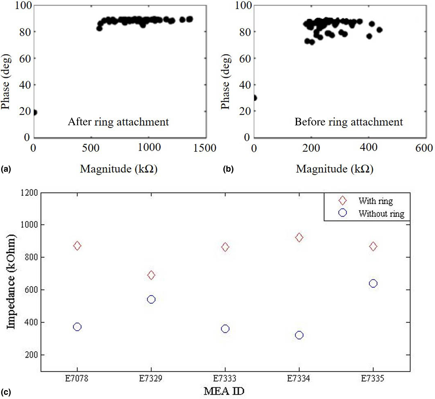
(a) Plot of impedance magnitude and phase of all microelectrodes before and after (b) attaching a glass ring as a culture chamber. (c) Impedance magnitudes for 5 MEAs, averaged over all their microelectrodes.
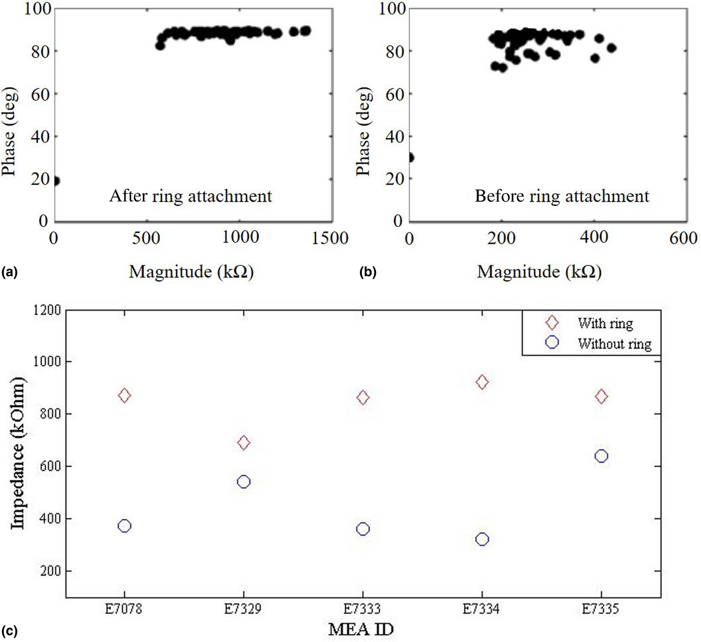
Additionally, impedances of all MEAs were tested at 1 kHz using an impedance testing device MEA-IT (Multichannel Systems GmBH). Impedance values were measured before and after attachment of a glass ring with a polydimethylsiloxane (PDMS)-based glue (Sylgard 184, Dow Corning) for creating a culture chamber on the MEA. Figures 5(a) and 5(b) show the impedance magnitude and phase for all of the 59 electrodes on a representative MEA, showing that the average impedance increases drastically after exposure to PDMS, while the average phase stays relatively constant. The average impedance value for each MEA in the set, before and after ring attachment, is reported in Fig. 5(c). Though the standard ring mounting procedure was followed, using a PDMS-based elastomer, impedance dramatically increased, and reaching values up to 1250 kΩ. Reasons for this increase might lie in the fact that PDMS has been known to outgas monomers that can diffuse across the substrate surface and significantly impact electrode performance.[Reference Regehr, Domenech, Koepsel, Carver, Ellison-zelski, Murphy, Schuler, Alarid and David25, Reference Nagarah26] Diamond surface is extremely sensitive on forming a strong bond to charged molecules or polymers[Reference Krysova, Vlckova-Zivcova, Barton, Petrak, Nesladek, Cigler and Kavan27] that might lead to formation of an isolating layer on the surface, leading to impedance enhancement. Though the thickness of the polymer layer can be reduced significantly by liquid chemistry, the last monolayer of polymer is strongly bound chemically to diamond surface and practically irremovable by for example, stripping in liquid sodium dodecyl sulfate. Partial electrode performance was recovered by performing a cleaning in CF4 plasma to try to remove PDMS residues.[Reference Garra, Long, Currie, Schneider, White and Paranjape28] This procedure led to a lower electrode impedance, though still higher when compared with those of MEAs before ring attachment.
Cellular electrophysiology and functional tests
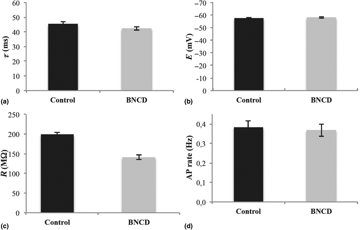
To assess whether NCD affects neuronal viability, single-cell, or network's properties, whole-cell patch clamps recordings were performed under the voltage-clamp configuration on neurons grown on NCD substrates. Results (Fig. 6) show that neurons grown on NCD substrates have membrane passive properties (i.e., time constant, input resistance, and membrane potential) and active response firing properties comparable with control conditions (n = 6 cells).
Effect on neurons cell properties by culturing on BNCD films compared with control condition (n = 6). (a) Time constant τ, (b) membrane resting potential E, (c) input resistance R, and (d) action potential rate. Error bars correspond to standard deviation of the data.

Functional tests
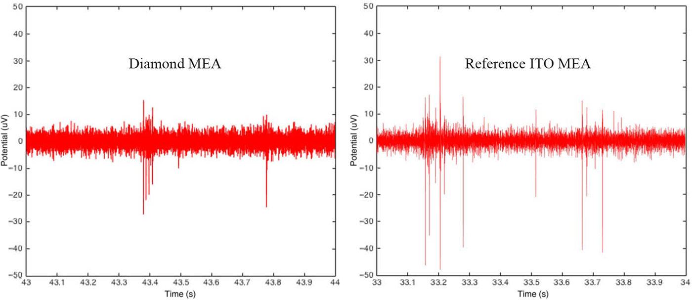
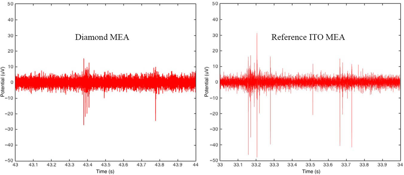
To devise functionality of MEAs, a set of experiments is currently undergoing with the diamond MEAs, involving the extracellular recording and stimulation of rat primary cortical neuronal cultures. Preliminary results indicate that BNCD electrodes, with NCD passivation layer, can detect single spikes and bursts with amplitudes in the 20–30 µV range with noise levels of about ±6 µV (see Fig. 7). Comparison among the functional BNCD channels to ITO reference electrodes shows similar noise levels, as well as similar spike amplitudes, though ITO references performed slightly better; however, ITO electrodes are a mix of capacitive and resistive Faradic behavior.[Reference Morin, Takamura and Tamiya22] ITO references electrodes showed amplitudes in the range of 40–50 µV with a noise level of about ±6 µV. Furthermore, it appears that spike detection may be better on the reference electrodes, though further analysis is required to determine whether this is due to lower spike detection, or a physiological change due to the diamond surface. We attribute this result to the high impedance caused by the PDMS during ring attachment, leading to a lower spike amplitude and lower spike detection, rather than any change to the network function. The electrode resistance rises up from ~400 to ~1250 Ω after the ring attachment. After CF4 plasma cleaning, the resistance decreased to ~800 Ω. Currently, we are developing plasma technologies to remove the polymer monolayers from the diamond surface without any re-deposition of etched organic molecules to the surface.
Spiking activity recorded on a diamond MEA compared with a control MEA with ITO electrodes.
While diamond has continuously been shown to be a good substrate for neuron growth,[Reference Vaitkuviene, McDonald, Vahidpour, Noben, Sanen, Ameloot, Ratautaite, Kaseta, Biziuleviciene, Ramanaviciene, Nesladek and Ramanavicius4] we have demonstrated here that it can also be used as an effective isolation layer at thicknesses of <180 nm. This allows the cell culture to be in direct contact with only one material minimizing any contact potential as well as provides homogenous neuron coverage. This property is important for studying neural networks as it is known that neural growth is strongly dependent on the material on which the neural cultures are cultivated.[Reference Fattahi, Yang, Kim and Abidian29] PEI is a polymer commonly used in the pretreatment of glass for cell culturing as it adds surface charges that promote neural adhesion. Poly-L-lysine (PEI) was also used as a pretreatment to cell culturing on diamond MEAs as it was found to also promote strong neural adhesion and growth on NCD substrates.[Reference Vaitkuviene, McDonald, Vahidpour, Noben, Sanen, Ameloot, Ratautaite, Kaseta, Biziuleviciene, Ramanaviciene, Nesladek and Ramanavicius4]
Furthermore, we show that it is possible to fabricate MEAs on optically transparent substrates and produce highly homogenous layers with negligible leakage current from cracks or pinholes. Our new fabrication process has been designed to allow a variable height of the electrode compared to the isolation layer, from ~100 nm below to >1 µm above. An electrode height close to the thickness level of the passivation layer minimized the cleft size between the cell and device. This improves coupling when the cell is tightly bound to the passivation layer, by bringing the electrode as close to the cell as possible. A greater signal-to-noise ratio (SNR) should be possible by resolving the issue of increased impedance from gluing glass rings to the MEA and a strategy for this is being developed. The impedance could further be improved by using nanostructuring techniques to increase the surface area; this could easily be added into the current fabrication process with no additional lithography step needed.[Reference Piret, Hébert, Mazellier, Rousseau, Scorsone, Cottance, Lissorgues, Heuschkel, Picaud, Bergonzo and Yvert30] Recently, we have already achieved ~10× increase of the BNCD capacitance, making it attractive for ultrasensitive MEAs based on purely capacitive coupling.[Reference Alcaide, Taylor, Fjorback, Zachar and Pennisi7] In summary, this fabrication process can be used to make functional MEAs from a fully diamond interface on transparent substrates for neural recordings that perform similarly to ITO reference MEAs; with room in the process to enhance the electrode interface.
In conclusion, we have developed a novel fabrication for MEAs made of thin diamond films on transparent substrates, which allow the detection of neural signals with advantage of diamond biocompatibility, surface neutrality, capacitive coupling, and excellent electrochemical characteristics.
Acknowledgments
Financial support from the FP7 of the European Commission (NMP project “MERIDIAN” no. 280778-02, IEF “INCA-NANEP” no. 328214, ITN “NAMASEN” no. 264872), the Flanders Research Foundation (FWO, contract no. G088812N) is acknowledged. We also thank Mr. D. Van Dyck and Mr. M. Wijnants for technical assistance.