I. INTRODUCTION
Tuning the properties of materials in nanoregime by manipulating their size and shape serves immense application possibilities. Noble metal nanomaterials, in particular, are enjoying the well-deserved attention steaming from their size- and shape-dependent properties that make them desirable for applications such as surface-enhanced Raman scattering spectroscopy,Reference Rycenga, Kim, Camargo, Cobley, Li and Xia1–Reference Eustis and El-Sayed3 catalysis,Reference Rupprechter and Rioux4 and cancer therapy.Reference Huang, Jain, El-Sayed and El-Sayed5 One-dimensional noble metal materials, nanowires, are particularly alluring due to their special optical, thermal, and electrical properties. Having a high surface area and only one unconfined direction, which is available for electrical conduction, makes nanowires attractive for applications where direct electrical conduction rather than tunneling transport is preferred such as sensitive electronic biosensors.Reference Liu and Searson6, Reference Yogeswaran and Chen7 Also, nanowires with very high aspect ratios will provide production of transparent and conductive networks for the fabrication of electrodes for organic solar cells and light-emitting diodes.Reference Lee, Connor, Cui and Peumans8–Reference Rosamond, Gallant, Atherton, Petty, Kolosov and Zeze10
Various methods have been developed to synthesize silver (Ag) or gold (Au) nanowires.Reference Coskun, Aksoy and Unalan11–Reference Kim, Sohn, Wu and Huang20 However, preparation of high aspect ratio Au nanowires, in particular, with high structural purity and yield is rather challenging, and preparation routes, in general, are hampered by difficult purification steps, impractical production methods, and polydispersity of the products. For instance, template-assisted preparation is one of the most widely applied techniques for the synthesis of Au nanowires.Reference Kim, Cha, Shin, Jho and Lee21 The method can be achieved using various templates such as porous alumina films, etched ion track membranes, mesoporous silica, and biotemplates (i.e., DNA).Reference Forrer, Schlottig, Siegenthaler and Textor17, Reference Liu, Duan, Toimil-Molares, Karim, Cornelius, Dobrev, Yao, Sun, Hou, Mo, Wang and Neumann22–Reference Patolsky, Weizmann, Lioubashevski and Willner24 However, the major drawback of this method is the difficulty in the removal of the template.Reference Tsuji, Hashimoto, Nishizawa and Tsuji19 Other methods such as electrochemicalReference Pérez‐Juste, Liz‐Marzán, Carnie, Chan and Mulvaney25 or photochemical synthesisReference Esumi, Matsuhisa and Torigoe26 require complicated experimental setups and their products generally lack uniformity.Reference Kim and Song27 A solution-based route called “seed-mediated growth” is another route that was first developed by Jana et al.Reference Jana, Gearheart and Murphy28 and later modified by Kim et al.Reference Kim, Sohn, Wu and Huang20 to synthesize Au nanowires with high aspect ratio around 200. However, the method does not yield products with high structural purity, thus requires time-consuming postpurification steps. The production of Au nanowires with “modified hydrothermal (MHT) process”, which is developed by Sinha et al.,Reference Sinha, Basu, Sarkar, Pradhan and Pal29 provides a promising way to overcome these difficulties. The process involves two steps: (i) the deposition of Au (III) ions on the anion exchange resin and (ii) the reduction of Au (III) in the presence of a surfactant at high temperature and pressure. It is a rather new Au nanowire synthesis method that provides simple process setup and solution-based alternative that enables production of micrometer-long very pure Au nanowires in gram quantities. The hydrothermal process, in general, embodies strongly interacting physical and chemical parameters, which play crucial roles on the quality of materials produced. The implication of these parameters can be depicted by systematic monitoring the reaction.
Herein, we report on a parametric study of Au nanowire synthesis via modified MHT process. A simple modification, altering the heating method, provided a firm control over the reaction temperature, which is one of the most important parameters in the hydrothermal process. This control resulted in an enhancement of structural purity of the products and synthesis of nanowires with controllable diameters. The parameters, such as surfactant type and concentration, temperature, and reaction time, were investigated to elucidate the optimum conditions for the synthesis of Au nanowires with very high aspect ratios, structural purity, and yield.
II. EXPERIMENTAL SECTION
A. Materials
All chemicals, such as HAuCl4·3H2O, ethylenediaminetetraacetic acid (Na2-EDTA), hexamethylenetetramine (HMTA), and Amberlite IRA-400 (Cl) ion exchange resin were purchased from Sigma-Aldrich (St. Louis, MO) and used without further purification. Deionized (DI) water (18 MΩ) was used throughout all experiments.
B. Methods
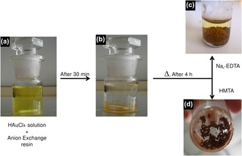
A solution of 1 mM HAuCl4 was prepared by dissolving 0.039 g of Au (III) chloride trihydrate (HAuCl4·3H2O) in 10 mL DI water. Au (III) ions were immobilized on anion exchange resin by adding 0.125 g Amberlite IRA-400 (anion exchange resin) to the Au solution and mixing for 30 min. The color of the solution turned from yellow to colorless and colorless beads became yellowish indicating that the anion exchange process was completed successfully, as shown in Fig. 1. Next, resin beads were washed seven times with 5 mL DI water to remove the excess AuCl4− that were not immobilized on surface of resin. Synthesis of desired Au nanowires was achieved at optimum conditions derived after the parametric study. Briefly, 0.03 M Na2-EDTA (0.3 M for HMTA) was added to the test tube and heated for 4 h in an oil bath at 125 °C (110 °C for HMTA). Formation of thick brown colored layer indicated the synthesis of Au nanowires (Fig. 1). The parameters used in the experiments were (i) concentration of surfactant molecules (0.03–0.5 M of HMTA and 0.01–0.3 M of Na2-EDTA), (ii) reaction temperature (70–125 °C), and (iii) reaction time (10 min to 5 h for both surfactant molecules; HMTA and Na2-EDTA). Throughout the parametric study, only one parameter was changed at a time, while the others were kept constant.
Photographs of (a) mixture of HAuCl4 solution (yellow colored) with anion exchange resin (colorless beads); (b) gold ions are deposited onto anion exchange resin (color became orange) after 30 min of stirring; as-synthesized gold nanowires in the presence of (c) Na2-EDTA and (d) HMTA after 4 h of heating.

C. Characterization
Scanning electron microscopy (SEM) studies were acquired using FEI Nova Nano SEM 430 microscope (FEI Company, Hillsboro, OR) operated at 10 kV. High-resolution transmission electron microscopy (HRTEM) was performed on a JEOL transmission electron microscopy (TEM) 2100F microscope (JEOL Ltd., Tokyo, Japan) operating at 200 kV. X-ray diffraction (XRD) measurements were done on a Rigaku D/Max-2000 PC diffractometer (Rigaku Americas Corporation, The Woodlands, TX) with Cu Kα radiation (λ = 1.54 Å) operating at 40 kV. PHI-5000 VersaProbe [Physical Electronics (PHI) Chanhassen, Minneapolis, MN], equipped with Al Kα at 1486.92 eV source, was used to perform x-ray photoelectron spectroscopy (XPS) analyses.
III. RESULTS AND DISCUSSION
Au nanowires were synthesized on anion exchange resin matrix with MHT procedure. First, AuCl4− anions were immobilized by exchanging the anion with the Cl− anions on resin matrix, which consists of a polymer with quaternary ammonium cationic head group, as shown in Fig. 1. Next, synthesis of Au nanowires was achieved by the reduction of Au (III) ions by surfactant molecules, which are also acting as structure-directing agent (Fig. 1). This method is adapted from the work of Sinha et al.Reference Sinha, Basu, Sarkar, Pradhan and Pal29 through some modifications. In this study, we have modified their method by changing the way of heating the reaction vessel and using a different surfactant molecule (HMTA) besides the one (Na2-EDTA) they have studied. The use of HMTA as a structure-directing agent was previously reported extensively for the synthesis of ZnO nanowires through hydrothermal route.Reference Akgun, Kalay and Unalan30–Reference Savu, Parra, Joanni, Jancar, Eliziario, de Camargo, Bueno, Varela, Longo and Zaghete34 However, there are a few reports on the use of the molecule in Au nanoparticle synthesis.Reference Hu, Li, Wang, Han, Zhang, Yuan and Niu35, Reference Ali Umar and Oyama36 To the best of our knowledge, this is the first time where HMTA is used as a surfactant for the synthesis of Au nanowires. To obtain uniform heat distribution, we have performed our experiments within a temperature-controlled oil bath instead of using a light bulb for heating. Precise control over the reaction temperature was achieved and significant increase in the reaction yield following this simple modification was observed. In addition, the effects of surfactant concentration, temperature, and reaction time for both surfactant molecules, HMTA and Na2-EDTA, were investigated. It was observed that these parameters play crucial roles on the morphology, yield, and structural purity of the materials produced.
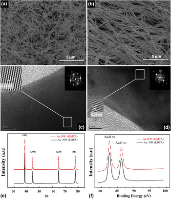
SEM, XRD, XPS, and TEM analysis results for Au nanowires prepared with HMTA are shown in Fig. 2. These results are for the nanowires synthesized under optimum reaction conditions that are determined by the parametric study. The conditions are 0.3 M for HMTA, 0.03 M for Na2-EDTA, 110 °C for HMTA, 125 °C for Na2-EDTA, and 4 h of reaction time for both molecule. SEM images of nanowires prepared in the presence of either surfactant molecules reveal the formation of very high aspect ratio nanowires in high structural purity [Figs. 2(a) and 2(b)]. The diameters of nanowires are in 50–70 nm range for HMTA and 90–110 nm range for Na2-EDTA. The length of the nanowires could not be measured since they are bundled and efforts to isolate individual nanowire resulted in their breakage. Therefore, attempts to derive exact aspect ratios were not successful, but they are estimated to be much higher than 200 based on the SEM images.
Characterization of Au nanowires: SEM images of Au nanowires synthesized with (a) HMTA and (b) Na2-EDTA. HRTEM of (c) HMTA- and (d) Na2-EDTA-directed Au nanowires on a holey carbon-coated Cu grid. Left inset shows inverse FFT image indicating the distance between (111) planes; right inset shows the FFT pattern. (e) XRD pattern for the nanowires synthesized with HMTA (dashed line) and Na2-EDTA (solid line). (f) XPS spectra for the nanowires synthesized with HMTA (dashed line) and Na2-EDTA (solid line).

HRTEM images of nanowires synthesized with both surfactant molecules and their fast Fourier transform (FFT) patterns are shown in Figs. 2(c) and 2(d). FFT analysis of the images revealed that interplanar spacing for lattice fringe is 0.24 nm for both HMTA and Na2-EDTA, correspond to the (111) lattice plane of face-centered cubic Au.Reference Whetten, Khoury, Alvarez, Murthy, Vezmar, Wang, Stephens, Cleveland, Luedtke and Landman37–Reference Zhang, Zhang, Lei, Zhao and Mao39 Strong signals of Au were observed in energy dispersive spectroscopy analyses (see Fig. S1)
XRD analysis confirmed the crystal structure of Au nanowires. Figure 2(f) illustrates XRD patterns of Au nanowires prepared with HMTA and Na2-EDTA. Both exhibited diffraction peaks (at 2θ: 38.40°, 44.60°, 64.78°, and 77.74°), which were indexed to (111), (200), (220), and (311) planes of Au cubic phase, respectively (JCPDS Card No. 65-2870).Reference Li, Su, Lv, Xia and Wang40 The lattice parameter of Au nanowires was calculated by UNIT CELL software and found as 4.073 Å, which is very close to that of bulk Au (JCPDS, No. 65-2870, α0= 4.078 Å).Reference Zhang, Ding, Chow, Ran and Yi41
XPS analysis demonstrates the characteristic spin–orbit doublets of Au (4f 7/2) and Au (4f 5/2) at 82.5 and 86.2 eV for HMTA-supported nanowires and 82.9 and 86.5 eV for Na2-EDTA-supported nanowires, as shown in Fig. 2(e). C (1s) at 284.9 eV was used as an internal reference. The results agree well with the previously reported XPS results for pure metallic Au.Reference Lü, Ji, Qian, Qi, Wang and Suo42
The synthesis of Au nanowires with MHT method requires systematic optimization of reaction parameters such as type and concentration of the reactants, temperature, and reaction time systematically to synthesize high quality nanowires. The results of the study on the reaction parameters are presented in the following sections.
A. Effect of surfactant
Surfactants are essential in nanomaterials synthesis. They provide a dynamic and protective shell, which stabilizes the nanomaterial against aggregation and mediates nucleation and growth processes.Reference Xiao and Qi43, Reference Burda, Chen, Narayanan and El-Sayed44 Therefore, using the right surfactant in appropriate amount is crucial for controlling the size and the shape of nanomaterials as well as for preparing high quality materials. In this study, two different surfactant molecules, HMTA and Na2-EDTA, were used. Both molecules have amine groups acting as an amine base reducing Au (III) ions and forming a complex ion with Au. These complex formations initiate nucleation processes since the molecules are also serving as structure-directing agents during the nanowire growth. The growth and final size of the nanowires depend strongly on the concentrations of reactants, i.e., Au ions and surfactant. In the procedure used, the concentration of Au ions is limited by the ion exchange capacity of the resin (3.5 mmol/g for AuCl4− ions).Reference Sinha, Basu, Sarkar, Pradhan and Pal29 Therefore, the surfactant concentration has a dominant effect among the reactants in the synthesis and strongly influences the fate of the nanowire growth. Since the growth process will continue until the depletion of the Au supply, high enough concentration of surfactant is needed for the synthesis of nanowires in desired size, shape, and purity.
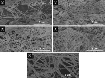
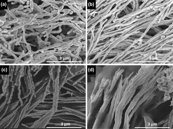
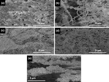
SEM images of Au nanowires synthesized with HMTA at concentrations of 0.03, 0.05, 0.1, 0.3, and 0.5 M are given in Figs. 3(a)–3(e), respectively. SEM images of Au nanowires synthesized with Na2-EDTA in 0.01, 0.03, 0.1, and 0.3 M concentrations are in Figs. 4(a)–4(d), respectively. The nanowire morphology was observed for all concentrations investigated with both surfactant molecules; however, morphology, yield, and structural purity vary significantly with surfactant concentration. This change was more notable when HMTA was used [Figs. 3(a)–3(e)]. Low amount of nanowire production, nonuniform diameter, and low structural purity were observed at low concentrations of HMTA (0.01 and 0.05 M) [Figs. 3(a) and 3(b)]. The observation suggested that inadequate amount of surfactant was available in the reaction media to direct the nanowire formation. This deficiency resulted in the formation of undesired Au structures. The nanowire synthesis was improved considerably with an increase in surfactant concentration [Fig. 3(c)]. The highest yield and structural purity were obtained at a HMTA concentration of 0.3 M, which was determined as the optimum concentration for synthesis of Au nanowires [Fig. 3(d)]. Increase of the concentration to 0.5 M led to significant increase in diameter of nanowires (average ∼60 nm) and change in their surface morphology [Fig. 3(e)]. Smooth surface of nanowires become spiky at this concentration. This was most likely due to the continuation of the growth with the additional supply of surfactant and dissolution of some of the Au nanoparticles contributing to the further growth of nanowires. This allowed branching on the nanowire surface with a mechanism known as the Ostwald ripening.Reference Pei, Mori and Adachi45
SEM images of Au nanostructures synthesized at HMTA concentrations of (a) 0.03 M, (b) 0.05 M, (c) 0.1 M, (d) 0.3 M, and (e) 0.5 M.

Small, nonuniform, and rod-like structures were formed when 0.01 M concentration of Na2-EDTA was used, as shown in Fig. 4(a). As the concentration was increased to 0.03 M, the welding or directional assembly of these short structures was observed leading to the formation of longer nanowires [Fig. 4(b)]. The formation of nanowires on the negatively charged resin with dipole–dipole attraction was suggested by Sinha et al.Reference Sinha, Basu, Sarkar, Pradhan and Pal29 The suggested mechanism is that the Au nanoparticles are produced on resin surface and polarized by the resin as a result of electrostatic force field. The polarization initiates the directional assembly of nanoparticles and growth of nanowires via dipole–dipole attraction.Reference Sinha, Basu, Sarkar, Pradhan and Pal29 This growth process is assisted by the surfactant molecules, and assembly or growth process could not be completed and desired nanowires cannot be synthesized unless there is sufficient amount of surfactant molecules. At Na2-EDTA concentrations of 0.1 and 0.3 M, adhesion of nanowires and formation of micrometer-sized structures were observed, as shown in Figs. 4(c) and 4(d), respectively. The electrostatic interaction created by the surfactant molecules in excess is the most likely reason for the edge to edge assembly of nanowires. The change in the morphology of nanostructures with the use of different surfactant molecules at concentrations higher than the optimum values is presumably because of differences in the chelating properties as well as electronic natures of the surfactant molecules; Na2-EDTA has anionic counterparts, while HMTA is neutral.
SEM images of Au nanostructures synthesized at Na2-EDTA concentration of (a) 0.01 M, (b) 0.03 M, (c) 0.1 M, and (d) 0.3 M.

B. Effect of temperature
Temperature plays an essential role in the synthesis of high quality nanowires with the MHT method. It affects the kinetics of the product formation and thermodynamic stability of the final product.Reference Burda, Chen, Narayanan and El-Sayed44, Reference Knauth and Schoonman46 The vapor pressure is higher and the structure of water is different at elevated temperatures. This affects the properties (i.e., solubility and reactivity) of reactants and leads to the formation of nanocrystals, which cannot be obtained at room temperature.Reference Burda, Chen, Narayanan and El-Sayed44, Reference Knauth and Schoonman46 Crystallization, in general, is governed by two main factors: (i) the degree of supersaturation, which has significant impact on the rate of nucleation and crystal growth, and (ii) dynamic equilibrium of dissolution and crystallization processes.Reference Burda, Chen, Narayanan and El-Sayed44, Reference Knauth and Schoonman46 Manipulation of these kinetic and thermodynamic factors by temperature can lead to the formation of desired crystals in hydrothermal process. In this study, we have observed that reaction temperature has a profound effect on the morphology, structural purity, and uniformity of the synthesized nanocrystals. Also, increase in the quality (structural uniformity and purity) and yield of nanowires was observed with uniform heating, which is obtained simply by keeping the reaction vessel in a temperature-controlled oil bath. This modification in the experimental setup allowed us to control the temperature and study the effect of temperature. Figures 5(a)–5(e) demonstrate the SEM images of nanostructures formed by using HMTA at temperatures of 70, 90, 110, 125, and 150 °C, respectively. While there was no nanowire formation at 70 °C [Fig. 5(a)], some started to form at 90 °C [Fig. 5(b)]. However, nonuniform crystal formation was observed at both temperatures. Low rate of crystal growth along with a high supersaturation at this temperature were most likely the reasons for the unwanted crystal growth. A significant increase in the number of wires was observed as the temperature was increased to 110 °C [Fig. 5(c)]. The nanowires have diameters in the range of 45–70 nm. Despite the formation of some nanocrystals with different morphologies, the highest yield and structural purity were obtained at this temperature. This suggests that the optimum kinetic and thermodynamic level was reached at 110 °C for growing desired nanocrystals. As the temperature increased to 125 °C, crystallization of nanostructures started sooner, and an increase in the diameter of nanowires (between 75 and 105 nm) was observed [Fig. 5(d)]. Considerable structural change and formation of anisotropically grown micrometer-sized structures were observed upon further increase in the temperature to 150 °C [Fig. 5(e)]. This is most likely due to the disruption of dissolution–crystallization dynamic equilibrium through high dissolution rate at this temperature.
SEM images of Au structures synthesized in the presence of HMTA at temperatures of (a) 70 °C, (b) 90 °C, (c) 110 °C, (d) 125 °C, and (e) 150 °C.

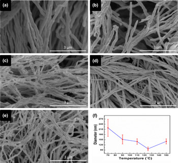
On the other hand, unlike HMTA, nanowire synthesis was observed at all studied temperatures when Na2-EDTA was used as a surfactant. SEM images of Au nanowires synthesized in the presence of Na2-EDTA at temperatures of 70, 90, 110, 125, and 150 °C are shown in Figs. 6(a)–6(e), respectively. Significant variation in the diameter of Au nanowires was noted. The change in the average diameter of nanowires with temperature was illustrated at a plot provided in Fig. 6(f). Average diameter distribution was very broad at low temperatures, ranging from 165 to 270 nm and from 120 to 180 nm for the synthesis conducted at 70 and 90 °C, respectively [Figs. 6(a) and 6(b)]. Diameters of the nanowires decreased and diameter distribution became narrower as the reaction temperature was increased [Figs. 6(c) and 6(f)]. Nanowires with the smallest diameter (average 100 nm) with narrow distribution (between 90 and 110 nm) were obtained when the reaction was carried out at 125 °C [Figs. 6(d) and 6(f)]. Further increase in the temperature to 150 °C resulted in an increase in both, as shown in Fig. 6(e). This suggests that the optimum degree of supersaturation and dynamic equilibrium were reached at 125 °C for the synthesis of nanowires with desired properties. As in use of HMTA, here too, further increase of temperature increases the dissolution rate and thus disturbs the equilibrium between dissolution and crystallization processes. The shift of the equilibrium in favor of crystallization resulted in unwanted increase of nanowire diameter.
SEM images of Au nanowires synthesized in the presence of Na2-EDTA at different temperatures (a) 70 °C, (b) 90 °C, (c) 110 °C, (d) 125 °C, and (e) 150 °C. (f) Changes in nanowire diameter with reaction temperature.

C. Effect of reaction time
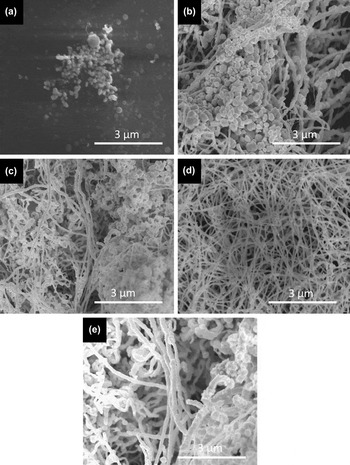
Time is another important parameter since the morphology and structural uniformity of the nanomaterials are strongly depending on the length of nucleation and growth processes. This effect was investigated at optimum temperatures of 110 and 125 °C for HMTA and Na2-EDTA, respectively. Reaction times of 10 min, 30 min, 2 h, 4 h, and 5 h were investigated and the corresponding SEM images of the synthesized Au nanowires with HMTA are provided in Figs. 7(a)–7(e), respectively. Only spherical nanoparticles were formed at the end of 10 min of reaction time as shown in Fig. 7(a). Formation of small nanoparticles with low thermodynamic stability occurred at the early stages of reaction, and they had a tendency to grow into larger particles as a result of large free energy driving force.Reference Burda, Chen, Narayanan and El-Sayed44 Nanowire formation was observed only after 30 min, and the amount of nanowires was increased considerably as the reaction was extended to 2 h [Figs. 7(b) and 7(c)]. The highest yield and structural purity were achieved when the reaction was carried out for 4 h, as shown in Fig. 7(d). At longer reaction times such as 5 h, diameter of nanowires increases as well as their distribution [Fig. 7(e)]. This was most likely due to a decrease in the saturation degree as the reactants depleted. This initiates the Ostwald ripening and particles smaller than the critical nuclei size dissolved to contribute to the growth of larger particles.
SEM images of Au nanostructures synthesized in the presence of HMTA at 110 °C for (a) 10 min, (b) 30 min, (c) 2 h, (d) 4 h, and (e) 5 h.

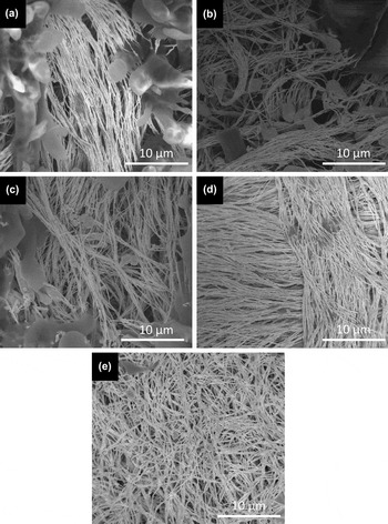
Figures 8(a)–8(e) show SEM images of nanostructures obtained at different times of 10 min, 30 min, 2 h, 4 h and 5 h, respectively, in the presence of Na2-EDTA. Nanowire formation was observed in early stages of the reaction, only after 10 min, compared to the reaction where HMTA was used as a surfactant [Fig. 8(a)]. Micrometer-sized structures as well as nanowires were formed within 10 min. The amount of nanowires got enormously increased as the reaction time was prolonged to 4 h. Further extension of reaction time resulted in a slight disruption of nanowire morphology and reformation of micrometer-sized particles. As a result, the optimum time for the reactions carried out with both surfactant molecules was determined as 4 h. A decrease in the reaction time for the production of high yield Au nanowires was observed compared to the one reported by Sinha et al.Reference Sinha, Basu, Sarkar, Pradhan and Pal29 This is probably because of our modification in the experimental setup that provides a precise control of reaction temperature and even heat distribution in the reaction vessel.
SEM images of Au nanostructures synthesized in the presence of Na2-EDTA at 125 °C for (a) 10 min, (b) 30 min, (c) 2 h, (d) 4 h, and (e) 5 h.

IV. CONCLUSIONS
The Au nanowires were synthesized with hydrothermal process on ion exchange resin matrix in aqueous media. Significant increase in the yield and structural purity of nanowires was observed with a simple modification in the MHT procedure. The significance of parameters such as surfactant concentration, reaction temperature, and reaction time on properties of products was demonstrated for HMTA and Na2-EDTA surfactant molecules, which are functioning as both structure-directing and reducing agents. The optimum reaction conditions were determined to achieve the synthesis of Au nanowires with high aspect ratio, structural purity, and yield for both surfactant molecules. Higher yield of nanowires were obtained when Na2-EDTA was used. On the other hand, HMTA, which is used for the first time in production of Au nanowires to our knowledge, resulted in the formation of thinner nanowires with narrower distribution of diameters compared to the nanowires obtained in the presence of Na2-EDTA. As a result, both molecules allow the synthesis of Au nanowires with high aspect ratio, yield, purity, and controllable diameter at demonstrated optimum conditions. These Au nanowires that can be synthesized easily and quickly would be great candidates to be used in various applications where enhanced optical properties or direct electrical conduction in nanoscale are needed.
Supplementary Material
Supplementary materials can be viewed in this issue of the Journal of Materials Research by visiting http://journals.cambridge.org/jmr.
ACKNOWLEDGMENTS
We thank Assistant Professor Dr. H. Emrah Unalan and Assistant Professor Dr. Eren Kalay for helpful discussions. We also acknowledge the support from METU-BAP Project No. 07-02-2012-101.