Introduction
The majority of malignant neoplasms in the head and neck region are squamous cell carcinomas (SCCs), and the treatment of these malignancies is well defined. However, there are tumour subtypes which are less common and which have a different clinical course, and for which there is no general consensus on appropriate treatment.
Adenosquamous carcinoma is defined by the World Health Organization as ‘a malignant tumour with histological features of both adenocarcinoma and squamous cell carcinoma’.Reference Pindborg, Smith and Van der Wall1 Adenosquamous carcinoma is known as an uncommon tumour in the head and neck region; it is extremely rare in the paranasal sinuses, with only a few cases reported in the English literature.Reference Keelawat, Liu, Roehm and Barnes2 In the head and neck region, this malignancy has an aggressive behaviour associated with a poor outcome.Reference Napier, Gormely, Newlands and Ramsay-Baggs3, Reference Sheahan, Fitzgibbon, Lee and O'Leary4 Prognosis has remained poor despite aggressive treatment, and an effective treatment course remains to be established. However, this will be a difficult task given the paucity of cases.
We report a case of adenosquamous carcinoma occurring in the left maxillary sinus, which had an aggressive clinical course but a complete response to S-1. The drug 1 M Tegafur (FT)-0.4 M 5-chloro-2, 4-dihydroxypridine (CHDP)-1 M potassium oxonate (Oxo) is a modulated oral fluoropyrimidine pro-drug which in phase II studies prompted the highest response rate in cases of unresectable, advanced carcinoma, compared with many other oral anticancer agents.Reference Schoffski5 A discussion of the clinical course and treatment of adenosquamous carcinoma is presented, along with a review of the pertinent literature.
Case report
A 77-year-old man was referred to our institute due to increasing pain in his left cheek over one month. The patient was a non-smoker and a social drinker. His past medical history was unremarkable.
Physical examination of the patient's face was unremarkable.
Upon nasal endoscopy, an exophytic tumour was identified in the left nasal cavity. The tumour originated from the lateral nasal wall in the area of the middle meatus. There was no nasopharyngeal or palatal extension and no clinical evidence of intraorbital or cranial nerve involvement. There were no palpable lymph nodes on neck examination.
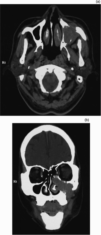
Computed tomography (CT) scanning revealed a tumour occupying and expanding the left maxillary sinus, with bony destruction of the anterior and posterior sinus walls and suspicious extension into the pterygoid musculature (Figure 1a). The superior border of the tumour extended to the orbital floor, with no evidence of intraorbital extension. The tumour also extended into the nasal cavity and left ethmoid sinus (Figure 1b). Computed tomography scans of the chest and abdomen were negative for metastasis.
Computed tomography scan showing tumour in the left maxilla. (a) Axial view showing the expanding tumour, with bony destruction and suspicious extension into the pterygoid musculature. (b) Coronal view showing the tumour occupying and expanding the left maxillary sinus.

Transnasal biopsy was performed and was initially interpreted as showing invasive SCC. The final clinical and radiological staging for this tumour was T3 N0 M0 (stage III).
Neoadjuvant chemoradiation to the left maxilla and upper cervical region with carboplatin and fluorouracil was performed, with a total dose of 40 Gy of radiation.
A left total maxillectomy was carried out via a Weber–Ferguson approach. A split-thickness skin graft was harvested from the lateral thigh and used to line the maxillary defect.
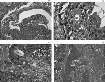
Further examination of the resection specimen showed the presence of two distinct malignant components: a glandular component consisting of adenocarcinoma (Figure 2a, b and c) and a keratinising SCC (Figure 2d). These two components were generally found to occur in separate, distinct areas. Thus, the histopathological diagnosis was adenosquamous carcinoma. There was evidence of vascular and lymphatic invasion.
Photomicrographs showing: (a) adenocarcinoma component (H&E; ×200); (b) adenocarcinoma component (H&E; ×400); (c) adenocarcinoma component (H&E; ×1000); and (d) squamous cell carcinoma component (H&E; ×100).

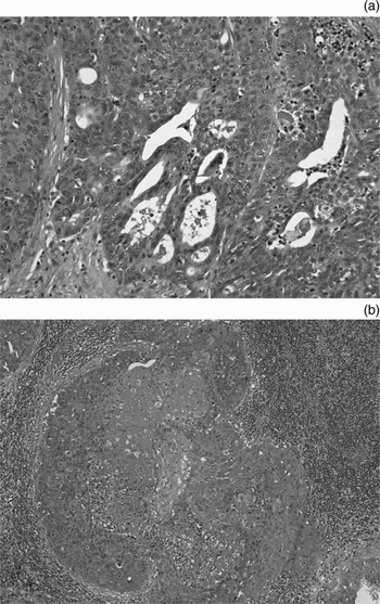
The patient's initial healing was satisfactory. However, six weeks after surgery, an enlarged left submandibular lymph node was detected. A left modified radical neck dissection was performed with preservation of the accessory nerve and internal jugular vein. Examination of resected lymph nodes showed evidence of cervical metastases in levels II and V, with evidence of glandular and squamous differentiation (Figure 3).
Photomicrographs showing: (a) adenocarcinoma component (H&E; ×200); and (b) squamous cell carcinoma component in a metastatic lymph node (H&E; ×20).

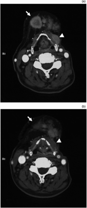
Eleven months after the neck dissection, rapidly enlarging, ipsilateral, deep neck lymph nodes and contralateral submandibular lymph nodes were detected (Figure 4a). The patient was reluctant to undergo any further surgery or hospitalisation. Therefore, a 50 mg, twice daily dose of S-1 was administrated orally on a four-week cycle followed by a two-week rest.
Axial computed tomography scans showing metastatic neck lesions before and after S-1 treatment. (a) Before treatment; scan shows expanding lymph node with central necrosis in the right submandibular area (arrow) and a left deep cervical metastatic lesion (arrow head). (b) After treatment; scan shows a remarkable reduction in the neck lesions (arrow and arrow head).

Four weeks after initiation of S-1 administration, both lymph nodes significantly decreased in size and could not be detected by CT (Figure 4b). During the ensuing 18 weeks of S-1 treatment, locoregional control was achieved, with good quality of life. However, neck metastases then recurred and S-1 administration was ceased.
The patient died 34 months after initial presentation, having survived for 15 months after re-presentation with bilateral neck metastases.
Discussion
Adenosquamous carcinoma is one of the most uncommon neoplasms in the head and neck region. Since Gerughty reported 10 cases of adenosquamous carcinoma of the head and neck in 1968, approximately 60 cases have been added in the English literature.Reference Gerughty, Hennigar and Brown6, Reference Som, Silvers, Catalano, Brandwein and Khorsandi7 Keelawat et al. analysed the clinical features of adenosquamous carcinoma in the head and neck region by studying 58 cases; they reported that these tumours mainly occur in men and that the mean age at presentation was within the sixth decade. The commonest sites of occurrence within the head and neck would appear to be the larynx and oral cavity. Cases have also been reported in the oropharynx, hypopharynx, nasal cavity and lip. Occurrence of adenosquamous carcinoma in the paranasal sinuses is extremely rare; Keelawat et al. reported only one case (1.7 per cent) amongst their 58.Reference Keelawat, Liu, Roehm and Barnes2 Adenosquamous carcinoma in the head and neck is considered extremely aggressive. Pain due to perineural invasion is often the only symptom at presentation, and local bony invasion is not uncommon.Reference Sheahan, Fitzgibbon, Lee and O'Leary4, Reference Som, Silvers, Catalano, Brandwein and Khorsandi8 The prognosis is very poor, with reported local recurrence and distant metastasis rates of 64.7 and 23.1 per cent, respectively, and a five-year survival rate of 13 per cent.Reference Keelawat, Liu, Roehm and Barnes2
Due to the paucity of cases, the treatment of adenosquamous carcinoma of the head and neck remains controversial. To date, surgical treatment has been the mainstay for this malignancy. Radiation has also been employed in some cases but there is no clear consensus on its use.Reference Keelawat, Liu, Roehm and Barnes2, Reference Yoshimura, Mishima, Obara, Yoshimura and Maruyama9 Only a few cases have been treated with chemotherapy.Reference Keelawat, Liu, Roehm and Barnes2, Reference Sheahan, Toner and Timon10–Reference Evans12 These chemotherapies were administered as part of multimodal treatment regimes, the majority of which failed to control the malignancy. The distinct efficacy of chemotherapy for adenosquamous carcinoma of the head and neck has yet to be determined.
There is unanimous agreement that adenosquamous carcinoma is an extremely aggressive and treatment-resistant tumour.Reference Keelawat, Liu, Roehm and Barnes2, Reference Sheahan, Fitzgibbon, Lee and O'Leary4, Reference Alos, Castillo, Nadal, Caballero, Mallofre and Palacin7 Therefore, it is reasonable to believe that chemotherapy may have a role as adjuvant treatment in such cases. The role of chemotherapy in the treatment of adenosquamous carcinoma in the head and neck has been widely ignored, with little justification. The historical association of adenosquamous carcinoma with mucoepidermoid carcinoma, a chemoresistant tumour, could in part explain the lack of popularity of chemotherapy as a treatment modality. In 1984, Evans clearly categorised adenosquamous carcinoma as a distinct neoplasm.Reference Evans12
Another controversy involves the cells of origin of adenosquamous carcinoma. Nowadays, many researchers consider that adenosquamous carcinoma arises from epithelial cells and variants of SCC.Reference Napier, Gormely, Newlands and Ramsay-Baggs3, Reference Abdelsayed, Sangueza, Newhouse and Singh13–Reference Neville, Damm, Allen and Bouquot15 However, others have stated that adenosquamous carcinoma originates from minor salivary glands.Reference Yoshimura, Mishima, Obara, Yoshimura and Maruyama9 This may be one reason why chemotherapy has not been employed in adenosquamous carcinoma treatment (as chemotherapy is not effective in the treatment of other salivary gland tumours).
• Adenosquamous carcinoma is a very rare tumour, characterised pathologically by the simultaneous presence of distinct areas of adenocarcinoma and squamous cell carcinoma
• In the head and neck, these tumours have a very poor prognosis
• A case is described of adenosquamous carcinoma arising in the maxillary sinus of a 77-year-old man. Despite surgical treatment and chemoradiotherapy to the primary site, he developed bilateral neck metastases
• The patient was treated with S-1, a novel oral fluoropyrimidine anticancer agent, with a complete (albeit finite) response
Yoshimura et al. analysed 19 cases of head and neck adenosquamous carcinoma and stressed the importance of correct diagnosis prior to treatment initiation.Reference Yoshimura, Mishima, Obara, Yoshimura and Maruyama9 Diagnosis of adenosquamous carcinoma is not easy, especially in small biopsy samples due to the admixture of the two different types of malignancies (SCC and adenocarcinoma). Some of Yoshimura and colleagues' adenosquamous carcinoma cases were only diagnosed after radical resection and whole block examination. Analysis revealed that 11 of the 17 cases had been diagnosed as other carcinoma types (SCC in seven cases, adenocarcinoma in two cases and mucoepidermoid carcinoma in two cases) prior to initial treatment. The survival of cases diagnosed as adenosquamous carcinoma prior to initial treatment was better than that of the misdiagnosed cases. Yoshimura et al. attributed this difference to greater treatment aggressiveness when the diagnosis of adenosquamous carcinoma was confirmed prior to treatment initiation.
In the present case, adenosquamous carcinoma arose from the maxillary sinus. Our patient's age at presentation, initial symptom of facial pain and aggressive metastases were all consistent with previous reports. However, the sensitivity to S-1 in this case should be noted. Treatment with S-1 chemotherapy for our patient's neck metastases achieved a notable response. Even though the tumour ultimately recurred, locoregional control was achieved for 18 weeks of S-1 therapy, while maintaining excellent functional status. The efficacy of S-1 in the palliative setting raises the possibility of improved prognosis if used as adjuvant treatment for first line therapy.
Adenosquamous carcinoma is a rare and aggressive tumour in the head and neck, and diagnosis can be challenging. Even if the initial biopsy does not reveal adenosquamous carcinoma, one should be aware of the possibility of this diagnosis, especially in clinically aggressive tumours. Adenosquamous carcinoma cases should be treated with multimodal treatment. Surgical treatment with wide local resection and radiotherapy are recommended for advanced cases. In addition, chemotherapy, especially using S-1, should be considered as adjuvant treatment.
Acknowledgement
The authors thank Dr Apostolos Christpoulos for his invaluable help in proofreading the manuscript, and Drs Masataka Edo and Kuniaki Mihara for providing clinical information.