Introduction
The siluriform fish commonly known as catfish comprise more than 2800 species with worldwide distribution (Mendoza-Palmero et al., Reference Mendoza-Palmero, Blasco-Costa and Scholz2015). Catfish, especially silurids and clarids, are very much appreciated by fishermen and are used for local consumption. The dactylogyridean monogenean fauna of catfish is very diverse, including 379 species belonging to 31 genera (Lim et al., Reference Lim, Timofeeva and Gibson2001). Catfish monogeneans are included in the subfamilies Ancylodiscoidinae Gusev (Reference Gusev1961) and Ancyrocephalinae Bychowsky (Reference Bychowsky1937) within the Dactylogyridae. The phylogenetic relationships within Dactylogyridae have been a matter of discussion for several decades. The status of all genera infecting siluriform fish was reviewed by Lim et al. (Reference Lim, Timofeeva and Gibson2001) and raised Ancylodiscoidinae to the familial level. The present study is a small effort to figure out the phylogenetic relationships of Bychowskyella spp. with other dactylogyrids.
Members of Bychowskyella Akhmerov (Reference Akhmerov1952) have well-developed dorsal anchors without roots, with long recurved patches; dorsal connecting bar wide and fenestrated; onchium absent; ventral anchors small, without roots, without patches and similar types of sclerotized reproductive structures. To date, 32 species have been attributed to Bychowskyella and its synonyms, but only 24 species are considered valid (Lim et al., Reference Lim, Timofeeva and Gibson2001). Morphometric comparison of monogenean specimens in our collection with the three species of Bychowskyella Akhmerov, 1952, as B. fossilisi Majumdar & Agarwal, Reference Majumdar and Agarwal1989, B. tchangi Gusev, Reference Gusev1976 and B. wallagonia (Jain, Reference Jain1959a) Gusev, Reference Gusev1961, led to the conclusion that these three species are parasitic in fish belonging to the families Heteropneustidae (B. fossilisi from Heteropneustes fossilis), Clariidae (B. tchangi from Clarias batrachus) and Siluridae (B. wallagonia from Wallago attu) in India. The huge morphological similarities among these species of the genus Bychowskyella stimulated our study to revise them.
The aim of the present study was to clarify the taxonomical status and validation of these species on the basis of morphological and molecular analysis.
Materials and methods
Morphology
Siluriform fish (W. attu, H. fossilis and C. batrachus) were collected from October 2011 to December 2012 from three different localities, viz. Meerut (29°01′N, 77°45′E) and Hastinapur (29°09'N, 77°59'E) in the state of Uttar Pradesh, and Guwahati (26°11′N, 91°44′E) in the state of Assam, India. For the collection of parasites from Guwahati, Assam, India, gills were removed from hosts and stored in 5% formalin until processing. Fish were identified using Srivastava (Reference Srivastava1980) and Froese & Pauly (Reference Froese and Pauly2010). Gills were removed carefully from freshly killed fish and placed in a Petri dish with saline water to get rid of excess gill mucus. The monogenean parasites were collected and examined using a binocular dissecting microscope.
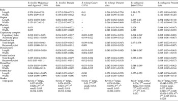
For light microscopy examination, specimens were cleared gradually in water and mounted in glycerin, then identified to the species level following the morphotype based on the data from previous studies. A light microscope (Motic SMZ-168, Xiamen, People's Republic of China) equipped with a digital image analysis system (Motic Image Plus 2.0 for Windows) and drawing attachment was used for morphometric analysis. For comparison, we studied five specimens of B. fossilisi, four specimens of B. tchangi and four specimens of B. wallagonia deposited in the Museum of the Department of Zoology, Chaudhary Charan Singh University, UP, India, as indicated in the respective taxonomic accounts. Comparative measurements are given in table 1. All measurements are in millimetres. Specimens of monogeneans were fixed in 95% ethanol in individual vials for molecular analysis.
Comparative measurements (in mm) of Bychowskyella fossilisi Majumdar & Agarwal (Reference Majumdar and Agarwal1989), B. tchangi Gusev (Reference Gusev1976) and B. wallagonia (Jain, Reference Jain1959a) Gusev, Reference Gusev1961; ranges given in brackets.

Measurements and morphotypes used to characterize anchors, bars and hooks were according to the guidelines suggested by Gusev (Reference Gusev and Bauer1985) for Dactylogyridae. The morphotypes used in the description of species are designated as: wunderoid type – anchors with sharply recurved point and comparatively small roots diverging, outer root is smaller than inner one; wunderoid-juvenile type – anchors with recurved point but without roots; vastator type – transverse bar having rough massive piece; definitive type – hooks with thick end and oblong spindle-shaped handle; and initial type – male copulatory organ (MCO) consists of a straight or curved cirrus and accessory piece is like a tape connected with the initial part of the cirrus and finished by a claw or extension.
Molecular analysis
Genomic DNA extraction was conducted using the QIAGEN DNeasy™ tissue kit (Qiagen, Hilden, Germany) according to the manufacturer's instructions. The partial 18S rDNA fragments were amplified using the primers Worm A: GCGAATGGCTCATTAAATCAG; 1270R: CCGTCAATTCCTTTAAGT; 930 F: GCATGGAATAATGGAATAGG; and Worm B: CTTGTTACGACTTTTACTTCC (Littlewood & Olson, Reference Littlewood, Olson, Littlewood and Bray2001). Polymerase chain reaction (PCR) amplifications were performed in 25 μl using an AG22331 Thermocycler (Eppendorf, Hamburg, Germany). Each amplification reaction contained 3 μl DNA, 1 μl Taq polymerase (1 U; Biotools, Madrid, Spain), 2.5 μl 10× Taq buffer (Biotools), 3.4 μl deoxynucleoside triphosphates (dNTPs), 0.9 μl of each primer and 13.3 μl water. PCR conditions were: an initial denaturation at 94°C for 3 min; 40 cycles at 94°C for 40 s, 57°C for 45 s and 72°C for 1 min; and a final extension at 72°C for 10 min. Amplified PCR products were electrophoresed in 1% agarose gel in Tris–acetate–EDTA buffer, the gel was stained with 1% ethidium bromide and purified with the Purelink™ Quick Gel Extraction and PCR Purification Combo Kit (Invitrogen, Löhne, Germany). For sequencing, the samples were sequenced with the above primers using the Big Dye Terminator v. 3.1 cycle sequencing kit in an ABI 3130 Genetic Analyzer, Applied Biosystems (Foster City, California, USA).
Amplified forward and reverse sequences for a particular species were aligned, edited using Bioedit (Hall, Reference Hall1999) and then submitted to GenBank. Based on the BLAST search, the sequence showing maximum similarity with the newly generated sequences was downloaded from GenBank. DNA pairwise distances were calculated using the p-distance model. For phylogenetic analysis, generated 18S rDNA sequences of monogeneans belonging to the Dactylogyridae were retrieved from GenBank. Nucleotide sequences were aligned using default parameters in CLUSTAL W (Thompson et al., Reference Thompson, Higgins and Gibson1994) implemented in MEGA v. 6 (Tamura et al., Reference Tamura, Stecher, Peterson, Filipski and Kumar2013). Before phylogenetic analysis, a nucleotide substitution model was estimated with Modeltest v. 3.07 (Posada & Crandall, Reference Posada and Crandall1998).
Phylogenetic analyses were conducted by using three approaches, neighbour joining (NJ) and maximum likelihood (ML) using MEGA v. 6 (Tamura et al., Reference Tamura, Stecher, Peterson, Filipski and Kumar2013), and Bayesian inference (BI) using Topali 2.5 (Milne et al., Reference Milne, Lindner, Bayer, Husmeier, McGuire, Marshall and Wright2008). The NJ tree was inferred under a best-fit model. ML topologies were inferred by model testing of the data, model of best fit by the Akaike Information Criterion and the GTR + I + G was chosen using MEGA 6.06. Bootstrap values based on 1000 resampled datasets were generated for ML. For Bayesian inference analyses, substitution models were tested by the Bayesian information criterion and GTR + I + G was chosen. BI analysis was run for 1,000,000 generations, sampling every 100th tree and discarding as ‘burn in’ the first 25% of the sampled tree. The phylogenetic analysis involved the 15 published 18S rDNA sequences of Dactylogyrus falciformis, Dactylogyridae spp. 1 and 2, Protogyrodactylus amacleithrium, P. johnstonettiegsi, Euryhaliotrematoides triangulovagina, E. grandis, E. microphallus, Euryhaliotrema sp. 3, E. johnii, E. perezponcei, E. triangulovagina, Haliotrema kurodai and H. eukurudai (see accession numbers in fig. 3) that showed 89–91% maximum identity with three newly generated 18S rDNA sequences of Bychowskyella species. Haliotrema kurudoi was chosen as an out-group in the final alignment.
Results
The examination of gills showed three species of the genus Bychowskyella based on morphological studies and identified as B. fossilisi, B. tchangi and B. wallagonia.
Bychowskyella fossilisi Majumdar & Agarwal (Reference Majumdar and Agarwal 1989 )
Host
Heteropneustes fossilisi (Bloch, 1794).
Locality
River Ganga, Hastinapur, Uttar Pradesh (UP), India.
Site of infection
Gill lamellae.
Specimens studied
Five.
Material deposited
Specimen slides have been deposited in the Museum of the Department of Zoology, Chaudhary Charan Singh University, UP, India, under the voucher number HS/monogenea/2015/07.
Representative sequence
The 18S rDNA sequence (1800 bp) for B. fossilisi is deposited in GenBank under accession number KT852454.
Re-description
Body very small with bluntly lobed haptoral end. Cephalic region having four pairs of glandular head organs. Eyespots found to be dispersed. Pharynx spherical and muscular. Oesophagus short. Intestinal caeca simple, bifurcate and confluent posteriorly above haptor. Haptor discoidal (figs 1a and 2a). Dorsal anchors wunderoid-juvenile type with small recurved point but without roots, shaft supported with sleeve sclerites. Two long recurved patches on dorsal anchors. Dorsal transverse bar straight, fenestrated in middle. Ventral anchors small, having strong recurved point. Ventral bar paired, each supported with sleeve sclerites. Marginal hooks seven pairs. One pair large, definitive-type; handle distinguished into two portions, a thin shaft and a swollen, large, bag-shaped handle at the distal end; blade, 0.003 (0.003–0.004); shaft, 0.007 (0.006–0.007); handle, 0.015 (0.014–0.015). Six pairs small, ‘larval type’; blade, 0.003 (0.003–0.004); handle, 0.008 (0.007–0.009). Sickle filament loop attached at the distal part of blade on ventral side. MCO simple and initial type (fig. 1b). Comprised of cirrus proper and an accessory piece. Cirrus proper curved, double-walled chitinoid tube. Accessory piece having clawed proximal end. Vitelline follicles follicular and extend between the confluent end of intestinal caeca. Vaginal armature not found.
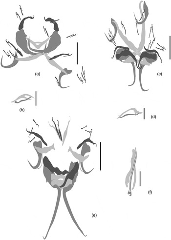
Morphology of (a, b) Bychowskyella fossilisi to show (a) the haptoral armature and (b) the copulatory complex; (c, d) Bychowskyella tchangi to show (c) the haptoral armature and (d) the copulatory complex; and (e, f) Bychowskyella wallagonia to show (e) the haptoral armature and (f) the copulatory complex. Scale bars: (a, c, e, f) 0.05 mm, (b, d) 0.01 mm.

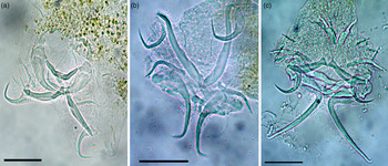
Microphotographs of haptoral armature of (a) Bychowskyella fossilisi; (b) Bychowskyella tchangi; (c) Bychowskyella wallagonia. Scale bars: (a–c) 0.02 mm.

Remarks
Majumdar & Agarwal (Reference Majumdar and Agarwal1989) erected B. fossilisi for the first time from H. fossilisi in Raipur state, India. The species is distinguished from other species by its long dorsal patch, which is slightly curved; a fenestrated dorsal bar; and an accessory piece clawed at its proximal end. It differs from B. tchangi in the shape of the male copulatory organs, dorsal bar and size of dorsal patches.
Bychowskyella tchangi Gusev (Reference Gusev 1976 )
Host
Clarias batrachus (Linnaeus, 1758).
Locality
River Brahmaputra, Guwahati, Assam, India.
Site of infection
Gill lamellae.
Specimens studied
Four.
Material deposited
Specimen slides have been deposited in the Museum of the Department of Zoology, Chaudhary Charan Singh University, UP, India, under the voucher number HS/monogenea/2015/08.
Synonyms
Clariotrema tchangi (Gusev, Reference Gusev1976) Long, Reference Long1981.
Representative sequence
The 18S rDNA (1700 bp) sequence for B. tchangi is deposited in GenBank under accession number KT852455.
Re-description
Body small to moderate size, maximum width attained at the level of gonads. Cephalic region equipped with four pairs of well-developed head organs. Eyespots may or may not be compact, numerous pigmented granules present. Pharynx spherical in outline, muscular structure. Intestinal caeca simple, bifurcated and confluent posteriorly. Haptor markedly offset from the body proper (figs 1c and 2b). Dorsal anchors wunderoid-juvenile type with strong recurved point but without roots, armed with articulated finger-like knob on inner side of the basal part, recurved point 0.011, supported by sleeve sclerites. Dorsal patch very long and curved. Dorsal transverse bar vastator type, widened and somewhat oval fenestrated at middle, articulate knob present in the middle. Ventral anchors wunderoid type with strong shaft and smoothly recurved point, base without root, smooth recurved point. Ventral connective bar is in two pieces; bar stick-like, wide at the middle portion with narrow outer ends. Marginal hooks seven pairs. One pair large, definitive type; blade, 0.006 (0.005–0.006); shaft, 0.009 (0.009–0.010); handle, 0.025 (0.024–0.026). Six pairs small, larval type; blade, 0.005 (0.004–0.006); handle, 0.010 (0.009–0.011). MCO consists of cirrus proper and an accessory piece (fig. 1d). Cirrus proper smoothly curved, double-walled tube with widened proximal part, almost cylindrical for its remaining part, having obliquely cut-off ends. Accessory piece chitinoid and like a tape going from the initial part of cirrus proper, and forming a two-horned widening part at its ends.
Remarks
Bychowskyella tchangi was first described by Gusev (Reference Gusev1976) from C. batrachus in Lucknow, India and subsequently from C. batrachus in Peninsular Malaysia (Lim & Furtado, Reference Lim and Furtado1983). Bychowskyella tchangi was further reported from the new host, Clarias macrocephalus in Peninsular Malaysia (Lim, Reference Lim1991). This species is easily characterized by the presence of seven pairs of hooks, in which six pairs are larval type and one pair has a long, widened handle and thickened pivot of handle (only one pair is twice as large as the others); articulated knob present at the middle part of the dorsal bar; distinctive dorsal patch, curved at one end and broad at the other end; accessory piece is clawed at both ends, distal as well as proximal, and in the shape of the ventral bar.
Bychowskyella wallagonia (Jain, Reference Jain 1959a ) Gusev, Reference Gusev 1961
Host
Freshwater shark, Wallago attu (Bloch and Schn., 1801).
Locality
River Ganga, Meerut, Uttar Pradesh (UP), India.
Site of infection
Gill lamellae.
Specimens studied
Four.
Material deposited
Specimen slides have been deposited in the Museum of the Department of Zoology, Chaudhary Charan Singh University, UP, India, under the voucher number HS/monogenea/2015/09.
Synonyms
Sprostonia wallagonia Jain, Reference Jain1959a; Neosprostonia wallagonia (Jain, Reference Jain1959a) Jain, Reference Jain1959b.
Representative sequence
The 18S rDNA sequence (1120 bp) for B. wallagonia is deposited in GenBank under accession number KT852456.
Re-description
Body extremely large, divided into cephalic, trunk, peduncle and haptor region. Cephalic region bears four pairs of head organs and two pairs of eyespots, one anterior and the other posterior, the latter being larger due to presence of a larger number of melanistic granules. Pharynx muscular. Intestine bifurcated and crura confluent posteriorly, anterior to haptor. Gonads tandem and inter-caecal. Testis median, post-equatorial, post-ovarian, oblong, 0.137 (0.135–0.140) × 0.074 (0.070–0.079). Seminal vesicle very short, hardly visible, pyriform and lies medially. Haptor discoidal to hexagonal (figs 1e and 2c). Haptoral armament consists of a pair of dorsal and a pair of smaller ventral anchors, a dorsal and a ventral bar, a pair of stout dorsal supporting bars and seven pairs of marginal hooks. Dorsal anchors very well developed, roots undifferentiated, strong recurved point. Dorsal bar stout, transverse. Dorsal patch conical in shape with rounded proximal part and a pointed distal end. Ventral anchor consists of a slightly bifurcate base, a shaft and a fine, recurved point; shaft supported by sleeve sclerite. Ventral transverse bar is in two pieces; bar yoke-shaped, shallow groove anteriorly and wide groove posteriorly. Seven pairs of marginal hooks, all similar in shape but dissimilar in size. Each hook consists of a swollen handle, a short shaft and a crochet-shaped blade or sickle. Dimensions of hooks are as follows. First large pair: blade, 0.006 (0.005–0.009); shaft, 0.022 (0.022–0.025); handle 0.016 (0.016–0.018). Second ventral large pair: blade, 0.005 (0.005–0.006); shaft, 0.012 (0.011–0.014); handle, 0.018 (0.017–0.021). Third medium-sized pair: blade, 0.007 (0.007–0.009); shaft, 0.007 (0.007–0.008); handle, 0.013 (0.012–0.018). Fourth to seventh pairs: blade, 0.005 (0.005–0.006); shaft, 0.005 (0.005–0.006); handle, 0.004 (0.011–0.007). MCO consists of an accessory piece and cirrus proper, both articulated firmly at the base (fig. 1f). Accessory piece long, having the shape of an inverted Y. Cirrus thick walled and stout, terminating in a fine point. At the base of cirrus, two prostate glands are present, one filled with hyaline fluid and the other with highly granular fluid. Ovary oval, post-equatorial, pre-testicular and median.
Remarks
Jain (Reference Jain1959a) described B. wallagonia from W. attu at Lucknow. It can be differentiated from all other species of Bychowskyella by the male copulatory organ and the morphology of anchors and bars, although the male copulatory organ of B. wallagonia somewhat resembles that of Bychowskyella chauhani Venkatanarasaiah (Reference Venkatanarsaiah1989), described from the same host in Andhra Pradesh, India, in the shape and approximate length of the cirrus tube. However, an onchium is present in B. chauhani that does not exist in B. wallagonia. This species differs significantly from B. tchangi in the shape of the MCO, the cirrus is tube like and smoothly curved in B. tchangi whereas in B. wallagonia it is long, pyriform, stout and thick walled, terminating proximally with the extension and inverted Y-shaped accessory piece in B. wallagonia, which is thin, tape like and forked at its end in B. tchangi. Bychowskyella wallagonia is characterized by the shape of the cirrus and the accessory piece, the absence of projections on the ventral anchors, a long dorsal patch, conical in shape and forming a ‘V’ with arms wide apart, and the conspicuously smaller size of the ventral bar.
Molecular analyses
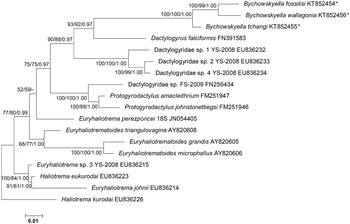
Three 18S rDNA sequences of lengths 1800, 1700 and 1120 bp were recovered during molecular screening of these monogeneans. The newly generated sequences were aligned together with 15 published sequences belonging to the family Dactylogyridae retrieved from GenBank (see accession numbers in fig. 3). The partial 18S rDNA sequences used for phylogenetic reconstruction were Haliotrema kurodai, H. eukurodai, Euryhaliotrema sp. 3, E. perezponcei, E. johnii, Euryhaliotrematoides microphallus, E. triangulovagina, E. grandis, Protogyrodactylus johnstonettiegsi, P. amacleithrium, Dactylogyrus falciformis, Dactylogyridae sp. FS-2009, Dactylogyridae sp. 1, Dactylogyridae sp. 2 and Dactylogyridae sp. 4. Sequences were aligned using default parameters in CLUSTAL W. Phylogenetic analysis was generated by neighbour joining (NJ), maximum likelihood (ML) and Bayesian inference (BI) analysis. The concatenated 18S rDNA sequences of three Bychowskyella species revealed >96% sequence identity with each other, followed by 89–91% similarity with other dactylogyrid monogeneans represented by the BLAST search. The topology of the phylogenetic analysis using NJ, ML and BI was identical; therefore we present only the ML tree here. In the phylogenetic tree, all three newly generated sequences belonging to Bychowskyella clustered together and formed a well-supported monophyletic clade (fig. 3). The phylogenetic analyses of the concerned species of Bychowskyella proved that their sequences are very much similar with each other and formed a single clade; they were placed on the same branch, supported by a high bootstrap value of 100% (fig. 3), providing congruent strong support for their status. Pairwise comparison between Bychowskyella species shows 0.02% intraspecific sequence divergence with each other. Regarding the 18S rDNA sequences of Bychowskyella, apart from the three described species, no other sequences of this gene are available on the database for comparison. Only one 28S rRNA sequence of Bychowskyella, i.e. B. pseudobagri (EF100541) from China, is available on GenBank, showing the scarcity of molecular sequences from this group.
Phylogenetic tree showing the relative position of Bychowskyella spp. from India obtained in this study (shown by asterisks). The topology of tree was generated by maximum likelihood (ML) analysis of the 18S rDNA sequences of these monogeneans. Numbers separated by slashes near the nodes represent the bootstrap support values, as NJ/ML/BI respectively. Bootstrap values (1000 replications) and posterior probabilities are shown at the nodes. Nodes unsupported by BI are marked with a hyphen, and accession numbers are given next to species names.

Discussion
Akhmerov (Reference Akhmerov1952) created Bychowskyella to accommodate B. pseudobagri from Pelteobagrus (= Pseudobagarus) in the Amur-Chinese region. Tripathi (Reference Tripathi1959) erected Silonditrema, with S. cauveryi from Silonia silondia, India. Jain (Reference Jain1959a) proposed Sprostonia but later replaced it with Neosprostonia. However, Jain (Reference Jain1959b), in the same publication, synonymized Neosprostonia and Silonditrema with Bychowskyella. Long (Reference Long1981) created Clariotrema to accommodate species of Bychowskyella bearing only one pair of marginal hooks with enlarged handles, rather than three pairs, and with dispersed pigment granules as eyespots. Later he re-assigned all three Bychowskyella species to this genus, i.e. B. tchangi, B. austrofujianensis and B. meridionalis, and assigned C. tchangi (Gusev, Reference Gusev1976) as the type species. Ma et al. (Reference Ma, Wang and Li1983) erected Neobychowskyella to accommodate N. glyptothoraci, having two onchia. Clariotrema and Neobychowskyella are considered junior synonyms of Bychowskyella. Until now, of the 32 species attributed to Bychowskyella and its synonyms, 24 species were considered valid. Five nominal species of Bychowskyella, i.e. B. chauhani Venkatanarsaiah (Reference Venkatanarsaiah1989), B. singhi Rajeswari & Kulkarni (Reference Rajeswari and Kulkarni1983), B. tripathi Kumar & Agarwal (Reference Kumar and Agarwal1981), B. wallagonia (Jain, Reference Jain1959a) Gusev, Reference Gusev1961 and Silonditrema tripathai Kumar & Agarwal (Reference Kumar and Agarwal1977) from W. attu, one species, i.e. B. fossilisi from H. fossilisi and one species, i.e. B. tchangi from C. batrachus, have been erected in India. Lim et al. (Reference Lim, Timofeeva and Gibson2001) assigned B. singhi in species inquirendae and S. tripathai and B. tripathii in nomen nudum due to the unavailability of formal descriptions. The taxonomic status of Indian species belonging to genus Bychowskyella was not clear up until now. Furthermore, it was difficult to establish the validity of most of the species, as no morphological and molecular descriptions were available. Morphological studies combined with molecular biology may aid in making definitive taxonomic determination when dealing with suspected species, as so much confusion exists regarding the validation and substantiation of monogenean fauna. Such an approach is essential to evaluate phylogenetic relationships (Boeger & Kritsky, Reference Boeger, Kritsky, Littlewood and Bray2001; Šimková et al., Reference Šimková, Plaisance, Matějusová, Morand and Verneau2003; Pouyaud et al., Reference Pouyaud, Desmarais, Deveney and Pariselle2006; Mendoza-Palmero et al., Reference Mendoza-Palmero, Blasco-Costa and Scholz2015).
Based on the phylogenetic analysis, the species of genus Bychowskyella are grouped together with species of the family Dactylogyridae, in agreement with the classification by Lim et al. (Reference Lim, Timofeeva and Gibson2001) based on morphological characteristics. The 18S rDNA sequences of Bychowskyella species from India formed a monophyletic clade in the phylogram. These nucleotide variations (0.02%) prompt us to interpret that all three species show individual existence, which was further supported by morphological methods of identification at the species level. Monogeneans parasitizing the related catfish from India were recovered as phylogenetically distant in molecular analyses. We further concluded from the results of both morphological and molecular methods that the three species concerned seem to share some similarities in their morphology that placed them in the genus Bychowskyella, and the similarities in their nucleotide sequences placed them in a monophyletic clade in the phylogram. The molecular analysis supported the validation of three Bychowskyella species, but the phylogeny of this group is still unclear because no data from other representatives of the genus are available.
For developing a detailed interpretation, it is necessary to obtain more data from other species belonging to the genus Bychowskyella, and different genes are needed for further analysis, to compare and establish an exact relationship among dactylogyrids. In conclusion, this study contributes to a baseline understanding of some Indian Bychowskyella species, concerning refinement of species description and phylogenetic analysis, to establish the relationship among dactylogyrids.
Acknowledgements
The authors thank the Head of the Department of Zoology, Chaudhary Charan Singh University, Meerut, for providing laboratory facilities. We thank Dr Haren Ram Chiary, from the Department of Zoology, Kirorimal College, University of Delhi, North Campus, New Delhi, Delhi, India, for collecting the host C. batrachus from Guwahati, Assam, India.
Financial support
This study was partially supported by grants from the UGC (University Grants Commission), India, under the senior research fellowship (RGNF; F.14-2(SC)/2010 (SA-III)) to C.V. and post-doctoral fellowship ((F.15-191/2012 (SA-II)) to A.C.
Conflict of interest
None.