INTRODUCTION
Cryptosporidia are tiny, unicellular parasites that infect a wide variety of organisms. Cryptosporidium parvum is one of the most important causes of heavy, watery diarrhoea in calves leading to high economical losses. Calves can shed high numbers of oocysts (up to 1010 per animal) and thus they are one of the main sources of dispersal stages. Humans are mainly affected by C. parvum and Cryptosporidium hominis where they cause heavy self-curing diarrhoea. Nevertheless, Cryptosporidium is still an issue mainly due to the small oocysts which are difficult to detect in and to remove from drinking water resulting in several outbreaks in the last decades. They can also become a life-threatening disease in immunosuppressed patients. With the introduction of HAART against HIV Cryptosporidium has become less important in industrialized countries but is still a problem in third world countries, although a systematic evaluation of the current situation is missing. The phylogenetic classification of Cryptosporidium is still being debated as they share characteristics with apicomplexan coccidia as well as with apicomplexan gregarines (Barta and Thompson, Reference Barta and Thompson2006). Cryptosporidia possess an apical complex but have lost their apicoplast while retaining some of their genes. Like coccidia they attach to the cell surface and undergo gliding motility before they start to enter the cell. However, it seems that unlike coccidia they do not invade the cell actively but rather trigger the cell to embrace them with host cell-derived membrane. As a result cryptosporidia do not fully invade the cell but stay in an epicellular location. At the cell parasite interface Cryptosporidium forms an actin-rich disk, a feeder organelle that is thought to be responsible for nutrition intake, and a small channel funnelling into the host cell cytoplasm. Phylogenetic analyses of the SSU RNA place cryptosporidia as a separate branch next to the gregarines (Carreno et al. Reference Carreno, Martin and Barta1999). This evolutionary distance might explain why cryptosporidia are resistant to the common anti-coccidial drugs.
Cryptosporidium, in contrast to Toxoplasma, cannot continuously be propagated in vitro and all attempts to establish a continuous cell culture have failed so far. This might be the reason why there are no tools available for the genetic manipulation of these parasites although they are urgently needed to explore the host-parasite interaction. This review aims to summarize the progress in molecular research on host-parasite interaction in recent years. Most of the studies used C. parvum as a model and therefore the vast majority of the data refer to this parasite.
BIOLOGY OF C. PARVUM
The life cycle
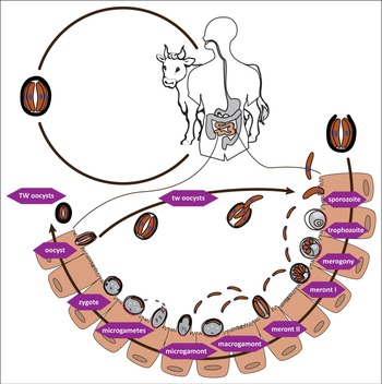
Cryptosporidium parvum infects humans and a variety of animals but their main hosts are calves at the age of 7–12 days, where it causes watery, yellowish and foul-smelling diarrhoea. It is a common disease in calves, affecting up to 100% of the young animals. The calves become infected through oral uptake of the oocysts. Due to the temperature rise and the changed environment, four sporozoites excyst from each oocyst in the small intestine where they start to invade enterocytes. The host cell is triggered to embrace the parasite with membrane protrusions thereby forming a parasitophorous vacuole (PV). At the same time an actin disc is forming underneath the attached parasite. The sporozoite then develops into a trophozoite that starts to replicate, leading to meronts with six to eight merozoites. Merozoites are released from the PV and start to infect circumjacent enterocytes to undergo another replication cycle resulting in meronts with four merozoites. Merozoites II are thought to be programmed to develop into gametocytes upon infection of further enterocytes. Fertilized macrogamonts develop to either thick- or thin-walled oocysts containing four sporozoites. Thin-walled oocysts are believed to cause auto-infection whereas thick-walled oocysts are released with the faeces to infect new hosts (see Fig. 1).
Life cycle of C. parvum. Sporulated oocysts are released with the faeces containing four infectious sporozoites. The sporozoites excyst in the small intestine and start to invade enterocytes. During invasion the sporozoite is embraced by protrusions of the host cell forming the parasitophorous vacuole. Underneath the forming trophozoite emerges a host cell-derived actin disc (shown in red). After asexual replication a meront type 1 develops that contains 6–8 merozoites. Type 1 merozoites infect adjacent epithelial cells either developing to another trophozoite or into a type 2 meront. Merozoites originating from a type 2 meront are thought to start the sexual replication cycle. Upon infection of another cell they either develop to macrogamonts or microgamonts. Microgamonts then release microgametes that fertilize a macrogamont. The resulting diploid zygote differentiates into four haploid sporozoites and subsequently the oocyst wall is formed around them. Two forms of oocysts are described. Thin-walled oocysts are described to lead to autoinfection whereas the thick-walled oocysts are released with the faeces to infect new hosts. TW: thick-walled oocyst, tw: thin-walled oocyst.

The oocyst and the excystation process
In vitro studies revealed that Cryptosporidium, in contrast to other coccidia such as Eimeria or Toxoplasma, need only a single stimulus to start excystation (Fayer and Leek, Reference Fayer and Leek1984). This would explain why Cryptosporidium is able to settle in extra-intestinal locations such as the lungs of immunosuppressed hosts. However, there are several stimuli supporting the excystation of sporozoites. Firstly, temperature is a major factor. Low temperatures (4 °C) prevent hatching of sporozoites but just raising the temperature to 20 °C resulted in excystation rates of up to 13% and up to 91% when the temperature was 37 °C. Secondly, bile salts and digestive enzymes trigger the release of sporozoites (Reduker and Speer, Reference Reduker and Speer1985; Forney et al. Reference Forney, Yang, Du and Healey1996a ). Finally, a corrosive treatment (NaOCl, HCl) supports excystation probably by facilitating the access of bile salts into the oocysts (Reduker and Speer, Reference Reduker and Speer1985; Robertson et al. Reference Robertson, Campbell and Smith1993). However, there are publications reporting different outcomes of pre-treatment of oocysts varying from a rather lethal impact on oocyst viability to amplifying effects on the excystation process. There are also reports on differences between the strains and isolates used (Robertson et al. Reference Robertson, Campbell and Smith1993).
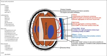
Upon activation of the sporozoites within the oocyst they start to produce a panel of proteases crucial for the excystation process. The overall protease activity in oocysts homogenates was shown to increase within the first hour of excystation (Forney et al. Reference Forney, Yang and Healey1996b ). Using different protease inhibitors it turned out that mainly serine and cystein proteases are expressed in activated oocysts. Blocking the activity of serine proteases by different inhibitors led to a strong although not complete inhibition of excystation emphasizing the importance of this class of proteases whereas the inhibition of cystein proteases did not lead to a significant inhibition of excystation (Forney et al. Reference Forney, Yang and Healey1996b ). Furthermore, an arginine aminopeptidase (RAP), expressed on the sporozoite surface, was shown to be involved in excystation (Okhuysen et al. Reference Okhuysen, Chappell, Kettner and Sterling1996; Padda et al. Reference Padda, Tsai, Chappell and Okhuysen2002). Altogether, the data of these in vitro studies suggest that upon uptake of the oocysts the rise in temperature activates the sporozoites leading to the production of proteases and an increased motility. At the same time, the outer layer of the oocyst, consisting mostly of glycoproteins, is degraded by the corrosive environment of the stomach leading to higher permeability of the oocyst wall. The infiltrating bile salts promote the activation of the sporozoites. Digestive enzymes such as trypsin in the intestinal environment as well as proteases produced by the parasite start to degrade oocyst wall proteins, mostly in the area of the suture leading to the rupture of the suture (Reduker et al. Reference Reduker, Speer and Blixt1985). The oocyst wall structure is not fully understood but supposed to be less complex than plant or fungal cell walls (Chatterjee et al. Reference Chatterjee, Banerjee, Steffen, O'Connor, Ward, Robbins and Samuelson2010) (see Fig. 2). Nevertheless, oocysts are highly resistant against chemical and environmental influences probably due to the layered structure of the oocyst wall consisting of a fragile glycocalyx, a lipid and glycoprotein layer. This protects against liquid intrusion as well as against mechanical stress. The inner proteinaceous layer mainly consists of Cryptosporidium oocyst wall protein (COWP) 1 and to a minor extent of COWP8. COWPs are a family (COWP1-9) of cysteine-rich proteins which, except for COWP1 and COWP8, are little characterized so far. The cysteines are thought to form disulphide bonds leading to the rigidity of the inner layer (Spano et al. Reference Spano, Puri, Ranucci, Putignani and Crisanti1997; Templeton et al. Reference Templeton, Lancto, Vigdorovich, Liu, London, Hadsall and Abrahamsen2004a ; Bushkin et al. Reference Bushkin, Motari, Carpentieri, Dubey, Costello, Robbins and Samuelson2013). The oocyst architecture is summarized in Fig. 2.
Architecture and composition of the oocyst. Schematic drawing of the current structural model of the oocyst. The oocyst is covered by glycocalyx that alters the electrophoretic mobility and is shown to be immunogenic. The high resistance is provided by a lipid bilayer protecting against liquid intrusion and a protein rich layer that confers mechanical resilience. The sporozoites are connected to the OW by filaments that contain glycoproteins which play a role in host cell attachment and invasion. COWP: Cryptosporidium oocyst wall protein, GalNac: N-acetylgalactosamine, gp: glycoprotein, OW: oocyst wall.

Attachment to the host cell
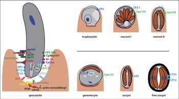
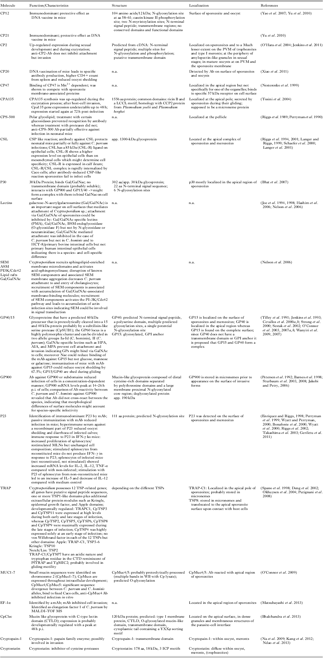
Attachment to the host cell is mediated by a variety of molecules that are expressed on the sporozoite surface and that can bind to host cell receptor or surface structures. The known molecules involved in attachment and/or invasion are summarized in Table 1 and Fig. 3. A genomic search for 32 widely conserved surface domains resulted in the finding of 51 proteins probably expressed on the zoite surface. Intriguingly many of them have a lineage-specific architecture different from Plasmodium and Toxoplasma indicating they have evolved separately. Many of the surface domains (such as Notch/Lin1, Neurexin-Callagen-domain) are only found in animals other than apicomplexans which hints towards a lateral transfer of genes of animal origin. This notion is supported by the presence of a previously detected protein O-linked glycosylation pathway in Cryptosporidium but not in Plasmodium that was found only in animals (Templeton et al. Reference Templeton, Iyer, Anantharaman, Enomoto, Abrahante, Subramanian, Hoffman, Abrahamsen and Aravind2004b ).
Molecules involved in Cryptosporidium attachment and invasion of the host cell. Most data are based on studies with C. parvum. Some molecules were also detected in other developmental stages than the sporozoite. They might have additional functions and/or be involved in merozoite invasion.

Cryptosporidium molecules possibly involved in host cell attachment and invasion

The first contact of the sporozoite is with the top mucus layer of epithelial cells. The mucus layer constitutes an efficient barrier against microbes. It reaches considerable thickness from 120 μm (small intestine) up to 800 μm (colon) (Atuma et al. Reference Atuma, Strugala, Allen and Holm2001) which has to be penetrated by the sporozoite (3 μm). Whether Cryptosporidium utilizes proteases to digest the mucus as shown for Entamoeba (Moncada et al. Reference Moncada, Keller and Chadee2003; Lidell et al. Reference Lidell, Moncada, Chadee and Hansson2006), simply forces its way through the mucus by movement or uses other mechanisms is so far unclear.
Obviously Cryptosporidium expresses at least one lectin on the sporozoite surface that specifically binds to galactose-N-acetylgalactosamine (Gal/GalNAc) but not to other sugars (Chen and LaRusso, Reference Chen and LaRusso2000). Preincubation with Gal/GalNAc or bovine submaxillary mucin (BSM) can prevent the attachment and/or invasion of cells in vitro (Joe et al. Reference Joe, Verdon, Tzipori, Keusch and Ward1998; Chen and LaRusso, Reference Chen and LaRusso2000; Hashim et al. Reference Hashim, Mulcahy, Bourke and Clyne2006). Recently, a 30 kDa Gal/GalNAc lectin (p30) has been identified that is predominantly located in the apical region of sporozoites. Attachment inhibition assays with native and recombinant p30 revealed that p30 plays at least in vitro a substantial role in attachment. However, p30 is a soluble protein that lacks a GPI anchor or a transmembrane domain and is exocytosed during excystation leading to the question how it mediates binding. It was found out that p30 is associated with gp40 and gp900, two glycoproteins also involved in attachment, and therefore hypothesized that these molecules form an adhesion complex (Bhat et al. Reference Bhat, Joe, PereiraPerrin and Ward2007).
The parasite lectins of course need Gal/GalNAc-protein counterparts on the host cell surface. Nelson and colleagues could show that Cryptosporidium leads to the formation of sphingolipid-enriched membrane microdomains (SEMs) on the host cell membrane that attract Gal/GalNAc epitope containing glycoproteins. Chemical inhibition of SEM formation as well as knock-down of acid-sphingomyelinase (ASM), which is important for the aggregation of the microdomains, reduced accumulation of Gal/GalNAc-glycoproteins and attachment and invasion of C. parvum in vitro. Moreover, recruitment and phosphorylation of PI-3K (phosphatidylinositol 3-kinase) and subsequent accumulation of cdc42 and actin was inhibited, indicating that SEMs might act as signal transducers for actin rearrangement (Nelson et al. Reference Nelson, O'Hara, Small, Tietz, Choudhury, Pagano, Chen and LaRusso2006). Another well characterized attachment glycoprotein is the gp40/15 complex. Gp40 and gp15 originate from a 60 kDa (gp60) precursor that is proteolytically cleaved, probably by a subtilisin-like serine protease (CpSUB1) (Cevallos et al. Reference Cevallos, Zhang, Waldor, Jaison, Zhou, Tzipori, Neutra and Ward2000a ; Strong et al. Reference Strong, Gut and Nelson2000; Wanyiri et al. Reference Wanyiri, O'Connor, Allison, Kim, Kane, Qiu, Plaut and Ward2007, Reference Wanyiri, Techasintana, O'Connor, Blackman, Kim and Ward2009). Whereas gp15 is anchored in the membrane by a GPI anchor, gp40 is supposed to be a soluble protein (Cevallos et al. Reference Cevallos, Zhang, Waldor, Jaison, Zhou, Tzipori, Neutra and Ward2000a ; O'Connor et al. Reference O'Connor, Kim, Khan and Ward2003). Nevertheless, gp40 is shown to bind to cells in a dose-dependent and saturable manner and antibodies against gp40 reduce infection by up to 82% in vitro (Cevallos et al. Reference Cevallos, Zhang, Waldor, Jaison, Zhou, Tzipori, Neutra and Ward2000a ). Furthermore, gp40 is shed in trails and co-localizes with gp15 indicating they might form a complex (O'Connor et al. Reference O'Connor, Wanyiri, Cevallos, Priest and Ward2007b ). Gp40 is a highly glycosylated mucin-like protein (Cevallos et al. Reference Cevallos, Bhat, Verdon, Hamer, Stein, Tzipori, Pereira, Keusch and Ward2000b ), a feature also exhibited by gp900 (Barnes et al. Reference Barnes, Bonnin, Huang, Gousset, Wu, Gut, Doyle, Dubremetz, Ward and Petersen1998). Gp900 is an immunodominant protein identified by antibodies from hyperimmune colostrums of cows (Petersen et al. Reference Petersen, Gut, Doyle, Crabb, Nelson and Leech1992). It has a putative transmembrane domain indicating it might be anchored to the cell membrane upon release from the micronemes (Barnes et al. Reference Barnes, Bonnin, Huang, Gousset, Wu, Gut, Doyle, Dubremetz, Ward and Petersen1998). Prevention of gp900 binding to cells resulted in reduced infections. Interestingly, the structure and probably the attachment sites of gp900 as well seem to differ between C. parvum and C. hominis. Some antibodies raised against peptide and carbohydrate epitopes of either C. parvum or C. hominis gp900 turned out to be not cross-reactive between the two species. Hence, gp900 might be one molecule through which Cryptosporidium defines host specificity. The circumsporozoit-like glycoprotein (CSL) is shown to preferentially bind to epithelial cell lines but less to mesenchymal cells. CSL binding is mediated by an 85 kDa protein (designated as CSL-R) on the host cell. CSL binding was also confirmed in a bovine intestine indicating its relevance for the in vivo situation.
There are a number of other molecules identified mostly due to their ability to inhibit invasion in vitro or by their immunodominant properties. They are summarized in Table 1.
Gliding motility
Gliding motility is a unique feature of Apicomplexa that was mainly studied in Toxoplasma and Plasmodium. The current model assumes that upon attachment micronemes release a panel of molecules that mediate attachment. One of these molecules is TRAP in Plasmodium or MIC2 in Toxoplasma. TRAP attaches via the TSP (Thrombospondin) and vWF-like A domains to the host cell and links the receptor through the cytoplasmic tail domain (CTD) with the motor complex. The CTD is coupled to an actin filament via an aldolase tetramer. Locomotion is created by myosin (MyoA) anchored to the inner membrane complex (IMC). It pushes the actin filament backwards thereby creating a force that drives the parasite forward. The TRAPs are thereby translocated to the posterior end of the zoites where they are cleaved within the transmembrane part by a rhomboid protease leaving trails of shed material behind them (reviewed in Baum et al. Reference Baum, Papenfuss, Baum, Speed and Cowman2006; Morahan et al. Reference Morahan, Wang and Coppel2009). However, there is not much research on gliding motility of Cryptosporidium, an apicomplexan parasite that does not have extra-intestinal stages and does not invade the host cell completely. Although it is considered that the machinery of gliding motility is highly conserved between Apicomplexa, there might be some variability. In Cryptosporidium, 12 TSP1 containing proteins have been identified in silico. In contrast to Plasmodium, CpTSPs bear Apple or Kringle domains, which are thought to replace the vWF domain. This might be due to the fact that Cryptosporidium attaches to epithelial cells instead of hepatocytes or erythrocytes and thereby facilitates different attachment molecules (Naitza et al. Reference Naitza, Spano, Robson and Crisanti1998; Spano et al. Reference Spano, Putignani, Naitza, Puri, Wright and Crisanti1998; Deng et al. Reference Deng, Templeton, London, Bauer, Schroeder and Abrahamsen2002). In Cryptosporidium, two TSPs, TRAP-C1 and TSP7 have the canonical CTD with the conserved tryptophan. TRAP-C1 is a single-copy gene with the most structural similarities to TgMIC2. Its expression (mRNA) varies during the life cycle peaking at 12 h and 48 h in the cell culture indicating that it is a highly regulated protein (Naitza et al. Reference Naitza, Spano, Robson and Crisanti1998; Deng et al. Reference Deng, Templeton, London, Bauer, Schroeder and Abrahamsen2002). Infection of human volunteers with C. parvum revealed that TRAP-C1 is an immunogenic protein (Okhuysen et al. Reference Okhuysen, Rogers, Crisanti, Spano, Huang, Chappell and Tzipori2004). For TSP2-12 an expression profile has been generated showing different expression patterns for the different TSPs making it likely they fulfil different tasks during development (Deng et al. Reference Deng, Templeton, London, Bauer, Schroeder and Abrahamsen2002). Nevertheless, Cryptosporidium relies on gliding motility shown by time-lapse microscopy and the inhibition of the conserved actin-myosin motor complex. Depolymerization of actin or inhibition of myosin by specific chemical drugs prevented movement and the active penetration of but not attachment to the host cell (Chen et al. Reference Chen, O'Hara, Huang, Nelson, Lin, Zhu, Ward and LaRusso2004c ; Wetzel et al. Reference Wetzel, Schmidt, Kuhlenschmidt, Dubey and Sibley2005).
Invasion of the host cell
Upon attachment to the cell and gliding around, the sporozoites somehow force their way into the cell. Toxoplasma sporozoites do this by forming a tight junction (moving junction) with the cell membrane through which they squeeze themselves into the nascent PV inside the host cell. This mechanism, which includes the release of the contents of the rhoptries, is well understood for Toxoplasma and Plasmodium (reviewed in Baum et al. Reference Baum, Gilberger, Frischknecht and Meissner2008; Sibley, Reference Sibley2010). However, Cryptosporidium is different in so far as it does not fully invade the cell but rather stops half way through, which is called intracellular but extra-cytoplasmic. Moreover, histological and some molecular studies suggest an engulfment of the parasite rather than an active invasion.
Calcium is an important messenger
Most studies on the molecular processes during Cryptosporidium invasion address changes in the host cell rather than the processes in the sporozoites. From Toxoplasma and Plasmodium it is known that calcium is an important messenger that regulates many processes during invasion. The cytosolic calcium concentration orchestrates several transporters in the acidocalcisomes, the endoplasmatic reticulum, the mitochondria and the cell membrane (Moreno and Docampo, Reference Moreno and Docampo2003). Cryptosporidium possesses a Ca2+-ATPase located at the sporozoite apical and perinuclear regions (Zhu and Keithly, Reference Zhu and Keithly1997). The function of this ATPase has not been identified yet and shows no homology with ATPases of the plasma membrane or the sarcoplasmic-endoplasmic reticulum of other parasitic protozoa (Moreno and Docampo, Reference Moreno and Docampo2003). The essentiality of Ca2+ for the invasion of Cryptosporidium was proven by using the intracellular calcium chelator BAPTA-AM that prevented the apical organelle discharge, an effect that could be partially reverted by raising the intracellular calcium content (Chen et al. Reference Chen, O'Hara, Huang, Nelson, Lin, Zhu, Ward and LaRusso2004c ). The rise in intracellular calcium can be partially controlled by bile salts. While sporozoites show an immediate increase in intracellular calcium and degranulation upon excystation in the absence of bile salts, they show a delayed increase in calcium and less degranulation in the presence of bile salts or natrium taurocholate, indicating a more controlled secretion (King et al. Reference King, Keegan, Phillips, Fanok and Monis2012). Knock-out studies in Toxoplasma revealed an essential function of calcium-dependent protein kinase (CDPK) 1 in the calcium-regulated exocytosis of micronemes (Lourido et al. Reference Lourido, Shuman, Zhang, Shokat, Hui and Sibley2010). CDPKs are protein kinases that have their ancestral origin in plants and are rare in animal cells, making them attractive targets for therapeutic intervention. Cryptosporidium contains seven CDPK family members which might be important in regulatory processes (Billker et al. Reference Billker, Lourido and Sibley2009). Indeed, using a specific inhibitor for CpCDPK1, Murphy and colleagues demonstrated that CDPK1 is essential for cell invasion in vitro (Murphy et al. Reference Murphy, Ojo, Larson, Castellanos-Gonzalez, Perera, Keyloun, Kim, Bhandari, Muller, Verlinde, White, Merritt, Van Voorhis and Maly2010). The importance of calcium signalling is also supported by a comparative analysis of eukaryotic genomes showing that compared with yeast (having a comparable number of protein-coding genes) Cryptosporidium and Plasmodium have a remarkably higher number of calcium-binding EF hand domains (Templeton et al. Reference Templeton, Iyer, Anantharaman, Enomoto, Abrahante, Subramanian, Hoffman, Abrahamsen and Aravind2004b ).
Formation of the PV and host cell actin rearrangement
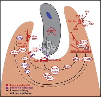
Whatever drives the essential exocytosis of the apical organelles, the events following attachment and the start of internalization differ greatly from what is known from Toxoplasma or Plasmodium. Valigurová et al. performed extensive electron microscopy studies supporting the notion that Cryptosporidium is engulfed by the host cell. The authors suggested naming this developing compartment a parasitophorous sac (PS) instead of PV as there are considerable differences. The PS consists of a host cell membrane fold that can be incompletely fused thereby leaving openings to the environment. The membrane fold contains a thin layer of host cell cytoplasm. Between the host cell membrane fold and the parasite remains a small gap. In the apical region of the parasite an electron-dense band separates the cell cytoplasm from the modified part of the membrane fold (Valigurová et al. Reference Valigurová, Jirků, Koudela, Gelnar, Modrý and Slapeta2008). Another difference is the formation of a so-called feeder organelle at the parasite-host interface. This area of extensively folded membrane is thought to function as a site of substance exchange, which is supported by data showing the localization of a predicted ABC-transporter at this site (Perkins, Reference Perkins1999; Striepen et al. Reference Striepen, Pruijssers, Huang, Li, Gubbels, Umejiego, Hedstrom and Kissinger2004). In other apicomplexan parasites the PV consists of one or more membranes that are highly decorated with parasite proteins that guarantee the exchange of nutrients and other molecules (Beyer, Reference Beyer2002; Spielmann et al. Reference Spielmann, Montagna, Hecht and Matuschewski2012). If this is the case in Cryptosporidium as well or if the feeder organelle is solely responsible for substance exchange remains to be elucidated. Early trophozoites of Cryptosporidium seem to have a direct connection to the host cell cytoplasm through a small channel that appears in transmission electron microscopy (TEM) pictures (Huang et al. Reference Huang, Chen and LaRusso2004b ; Valigurová et al. Reference Valigurová, Jirků, Koudela, Gelnar, Modrý and Slapeta2008). It is likely that Cryptosporidium secretes molecules into the host cell through this tunnel connection to manipulate the host cell. The export of parasite-derived molecules to the parasite-host interface has been proven by immunogold staining (Huang et al. Reference Huang, Chen and LaRusso2004b ). The host cell undergoes a massive reorganization during the infection. How this reorganization is triggered by the parasite is hardly understood. However, two mechanisms through which the host cell might be forced to produce the epithelial protrusions that embrace the parasite have been proposed (see also Fig. 4). One is based on the observation that Cryptosporidium attracts the water channel aquaporin 1 (AQP1) in combination with a Na+/glucose cotransporter (SGLT1) at the site of infection. The uptake of glucose goes along with a water influx that might support the increase of volume of the protrusions. Inhibition of SGLT1 and AQP1 reduced infectivity of C. parvum in cholangiocytes (Chen et al. Reference Chen, O'Hara, Huang, Splinter, Nelson and LaRusso2005). SGLT1 aggregation is also shown to be dependent on myosin IIB of cholangiocytes (O'Hara et al. Reference O'Hara, Gajdos, Trussoni, Splinter and LaRusso2010).The second mechanism is based on the massive actin rearrangement that has been found in infected cells. Actin rearrangement might be the leading force that creates the membrane envelope around the parasite but at the same time it leads to the formation of a solid actin disc underneath the attachment site. Recently integrin α2 (ITGA2) and integrin ß1 (ITGB1) have been identified as potential receptors that transduce the parasite signal into the host cell (Zhang et al. Reference Zhang, Guo and Zhu2012b ). Intriguingly, integrins are key players controlling the dynamics and structures of actin-based processes in lamellipodia and filopodia in cells (Martin et al. Reference Martin, Slack, Boerner, Martin and Parsons2002). Several authors studied the mediators involved in actin signalling on the host cell site. Notably, the signalling cascade very much resembles the signalling pathway found in the formation of lamellipodia/filopodia suggesting Cryptosporidium utilizes the same signal cascades. It was shown that Cryptosporidium uses the PI3K/Cdc42/WASP (Phosphatidylinositide 3-kinases/Cell division control protein 42/Wiskott–Aldrich Syndrome Protein) pathway to activate the Arp2/3 (Actin-Related Proteins) complex which leads to the branching of the actin filaments (Forney et al. Reference Forney, DeWald, Yang, Speer and Healey1999; Chen et al. Reference Chen, Splinter, Tietz, Huang, Billadeau and LaRusso2004b ). The involvement of further mediators of the Rho (Ras homologue gene) family (RhoA, Rac1) was ruled out indicating that only the Cdc42 pathway is utilized by Cryptosporidium (Chen et al. Reference Chen, Splinter, Tietz, Huang, Billadeau and LaRusso2004b ). This notion was supported by the finding that the downstream effectors of Cdc42, N-WASP and p34-Arc also accumulate at the host-cell parasite interface (Chen et al. Reference Chen, Huang, Splinter, Orth, Billadeau, McNiven and LaRusso2004a ). Nevertheless, inhibition of Cdc42 reduced Cryptosporidium invasion only up to 80% but never blocked it completely, making it likely that an alternative pathway exists. Indeed, Cryptosporidium induces the accumulation of the tyrosin kinase Src and a subsequent phosphorylation of cortactin, which in its activated stage recruits the Arp2/3 complex to open up another potential signalling cascade (Chen et al. Reference Chen, Huang, Splinter, Cao, Zhu, McNiven and LaRusso2003). Most recently, the involvement of calpain in actin plaque formation was demonstrated. Vital in contrast to heat-inactivated sporozoites could induce autolytic activation of calpains. Inhibition of calpain 1 and 2 led to reduced infectivity and diminished actin plaque formation (Perez-Cordon et al. Reference Perez-Cordon, Nie, Schmidt, Tzipori and Feng2011). Calpains are a family of calcium-controlled cystein proteases shown to interact with many molecules involved in actin remodelling and are suggested to regulate membrane protrusions (Franco and Huttenlocher, Reference Franco and Huttenlocher2005; Lebart and Benyamin, Reference Lebart and Benyamin2006). Another kinase that might be involved in the regulation of actin rearrangement is protein kinase Cα (PKCα), which has been shown to regulate actin condensation during E. coli invasion. Inhibition of PKCs in the Cryptosporidium in vitro model with different inhibitors reduced the number of infected cells (Hashim et al. Reference Hashim, Mulcahy, Bourke and Clyne2006). However, PKCs regulate a diverse array of cell functions and might inhibit Cryptosporidium invasion through alternative pathways. The hypothesis of actin-driven envelopment is supported by the accumulation of villin and ezrin, two molecules that link F-actin to the membrane of microvilli at the PVM although F-actin was only found in rather small amounts at this site (Bonnin et al. Reference Bonnin, Lapillonne, Petrella, Lopez, Chaponnier, Gabbiani, Robine and Dubremetz1999). Further enzymes that might be involved in host cell invasion are phospholipase (PLA) family members. For Toxoplasma it was demonstrated that cytosolic PLA2 and the secreted form are necessary for infection and it was speculated that PLA might alter the host cell membrane fluidity (Bonhomme et al. Reference Bonhomme, Bouchot, Pezzella, Gomez, Le Moal and Pinon1999). Similar results were obtained when C. parvum sporozoites were incubated with the PLA inhibitor p-BPB or an anti-PLA2 antibody and then used to infect HT-29 or Caco-2 cells. p-BPB as well as the antibody reduced infection in a dose-dependent manner when sporozoites were pre-treated but not when host cells were pre-treated with p-BPB (Pollok et al. Reference Pollok, McDonald, Kelly and Farthing2003). However, the same publication describes exogenous PLA2 isolated from cobra venom as supportive for the infection with C. parvum in vitro, whereas a recent publication showed the opposite effect using honey bee-derived PLA2 (Carryn et al. Reference Carryn, Schaefer, Imboden, Homan, Bremel and Riggs2012). This might be attributed to the diverse array of functions fulfilled by the PLA2 family.
Actin rearrangement and protrusion formation in the host cell induced by Cryptosporidium. Upon attachment Cryptosporidium triggers the host cell to undergo massive structural changes. During invasion microneme and rhoptry content are released. The transmitters and receptors are fairly unknown. Two possible receptors that might transduce a signal into the cell have been identified. Sphingolipid-enriched membrane microdomains in association with GalNac containing glycoproteins accumulate at the site of infection. In conjunction with likewise attracted PI3K and cdc42 this signal cascade might be how Cryptosporidium induces actin remodelling. Furthermore integrins (ITGA2/ITGB1) could pass a signal via Src kinase although this link has not been established. The activation of the PI3K/cdc42/N-WASP pathway leads in turn to the activation of the Arp2/3 complex and subsequently to the elongation and branching of actin filaments and formation of protrusions. The enlargement of the protrusions is supported by an increase in volume as a result of the attraction of the water channel aquaporin (AQP1). Several other molecules involved in actin rearrangement have been identified but the link between them has yet to be established. AQP: Aquaporin, Arp: actin releated proteins, ASM: acid-sphingomyelinase, ITGA: integrin α, ITGB: integrin β, cdc: cell division control protein, CDPK: calcium dependent protein kinase, FAK: focal adhesion kinase, Gal/GalNac: galactose-N-acetylgalactosamine, PI3K: phosphatidylinositol 3-kinase, PKC: protein kinase C, MIC: microneme, N-WASP: neural Wiskott–Aldrich syndrome protein, Rac: Ras-related C3 botulinum toxin substrate, RhoA: Ras homologue gene family, member A, Rhop: rhoptry, SEM: sphingolipid-enriched membrane microdomains, SGLT: sodium-glucose linked transporter, VASP: vasodilator-stimulated phosphoprotein.

Survival in the host cell
After Cryptosporidium has settled down in its niche the parasite faces new challenges. First, it has to obtain nutrients, second, make sure the host cell is not trying to eliminate it and third, it has to escape the immune response. We will focus on the interaction between parasite and host cell in the following paragraphs.
Nutrition
Cryptosporidium has a comparatively small genome that also lacks considerable parts of the metabolic machinery constraining the parasite to acquire nutrients from its host cell. Consequently, the C. parvum genome contains at least 69 transporters for different substrates (Abrahamsen et al. Reference Abrahamsen, Templeton, Enomoto, Abrahante, Zhu, Lancto, Deng, Liu, Widmer, Tzipori, Buck, Xu, Bankier, Dear, Konfortov, Spriggs, Iyer, Anantharaman, Aravind and Kapur2004). Direct acquisition of nutrients from the cytosol is hindered by the PV making it necessary to integrate appropriate transporters into the PV membrane, a strategy that is for example described for Plasmodium (Spielmann et al. Reference Spielmann, Montagna, Hecht and Matuschewski2012). Cryptosporidium differs in that it does not reside in an intracellular PV but in an epicellular location, limiting the contact area to the cytosol. This might be partially reversed by the feeder organelle that is thought to be responsible for the uptake of nutrients through increasing the contact area due to multiple membrane folds. However, ultimate proof of such a material transfer by the feeder organelle and involved transporters is still missing. The epicellular location of the parasite results in an exposure to the gut lumen that could be another source of nutrients. The zoites might directly endocytose material from the intestinal lumen if they are only partially embraced. Otherwise they need to facilitate transporters that ensure an active transport through the PV membrane.
Cholesterol is an essential lipid in membranes of eukaryotic cells that controls the membrane fluidity. Cryptosporidium is not able to de novo synthesize cholesterol and therefore relies on host cell-derived cholesterol. Ehrenman and co-workers showed that Cryptosporidium is auxotroph for plasma Low Density Lipoprotein (LDL) but not High Density Lipoprotein (HDL) and that it derives its cholesterol from these proteins. Moreover, the cholesterol incorporated by Cryptosporidium did not originate from de novo synthesis of the host cell but from micelles imported via the NPC1L1 transporter into the cell (Ehrenman et al. Reference Ehrenman, Wanyiri, Bhat, Ward and Coppens2013). The way in which it enters the parasite, however, remains elusive.
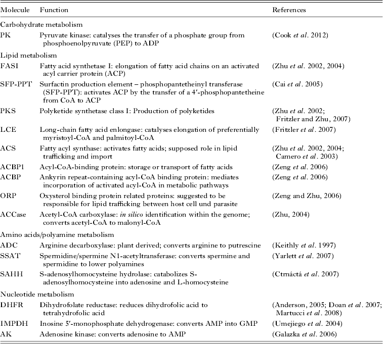
Fatty acids are produced by fatty acid synthetase (FAS) I (common in mammals) or FAS II (common in bacteria). FAS I was found in C. parvum but its preferred substrate is a C16 palmitic acid and is therefore not involved in de novo synthesis of parasite fatty acids (Zhu et al. Reference Zhu, Li, Cai, Millership, Marchewka and Keithly2004). Since Cryptosporidium lacks the FAS II (which is for example present in Toxoplasma) it was hypothesized that Cryptosporidium is generally not able to use de novo synthesis of fatty acids and instead relies on scavenging fatty acids from the host cell (Zhu, Reference Zhu2004). Just recently, it has been demonstrated that polyketide synthetase (PKS) 1 of Toxoplasma and Eimeria are highly abundant in oocysts and therefore proposed to play a crucial role in oocyst wall lipid synthesis. Similarly, FAS I of C. parvum is highly expressed during late time points in cell culture when the oocyst walls are made (Bushkin et al. Reference Bushkin, Motari, Carpentieri, Dubey, Costello, Robbins and Samuelson2013). Cryptosporidium parvum possesses a PKS class I. The function of the PKS for Cryptosporidium is so far unknown but it was speculated that the produced polyketides might function to inhibit bacteria within the niche of Cryptosporidium (Zhu et al. Reference Zhu, LaGier, Stejskal, Millership, Cai and Keithly2002). A few other enzymes involved in fatty acid metabolism have been identified and experimentally addressed (see Table 2). Since the genome of Cryptosporidium lacks any enzymes for ß-oxidation, fatty acids obviously cannot be used as an energy source. The C. parvum genome also lacks all genes of the Krebs cycle and most of the important genes for the respiratory chain of mitochondria. Cryptosporidium parvum is capable of catabolizing mono-sugars and can synthesize, store and catabolize polysaccharides indicating that it predominantly relies on glycolysis (Abrahamsen et al. Reference Abrahamsen, Templeton, Enomoto, Abrahante, Zhu, Lancto, Deng, Liu, Widmer, Tzipori, Buck, Xu, Bankier, Dear, Konfortov, Spriggs, Iyer, Anantharaman, Aravind and Kapur2004). However, the enzymes involved in the carbohydrate metabolism identified in the genome of C. parvum have not been studied biochemically.
Experimentally addressed molecules involved in the metabolism of Cryptosporidium
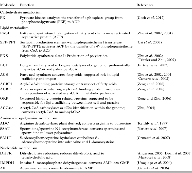
Cryptosporidium is capable of interconversion of several amino acids but not of de novo synthesis (Rider and Zhu, Reference Rider and Zhu2010) and seems to have a large set of 11 amino acid transporters (Abrahamsen et al. Reference Abrahamsen, Templeton, Enomoto, Abrahante, Zhu, Lancto, Deng, Liu, Widmer, Tzipori, Buck, Xu, Bankier, Dear, Konfortov, Spriggs, Iyer, Anantharaman, Aravind and Kapur2004). Biochemical approaches identified a plant-derived enzyme that converts arginine into polyamines. Polyamines such as spermidine and spermine can interact with negatively charged molecules such as DNA, RNA, phospholipids and acidic proteins and modulate various cellular functions. In C. parvum the conversion was accomplished by a plant-derived arginine decarboxylase (ADC) instead of an ornithine decarboxylase (ODC) that is utilized by mammalian cells (Keithly et al. Reference Keithly, Zhu, Upton, Woods, Martinez and Yarlett1997). Furthermore, Cryptosporidium is able to convert scavenged spermidine and spermine to lower polyamines by a spermidine/spermine N1-acetyltransferase (SSAT) (Yarlett et al. Reference Yarlett, Wu, Waters, Harp, Wannemuehler, Morada, Athanasopoulos, Martinez, Upton, Marton and Frydman2007). Very recently, it was demonstrated that C. parvum also induces the human SSAT that in turn provides acetylspermine that can be scavenged by the parasite (Morada et al. Reference Morada, Pendyala, Wu, Merali and Yarlett2013). Nucleotides are another important class of molecules. The thymine synthesis pathway has become an important antimalarial drug target by inhibiting dihydrofolate reductase (DHFR). Interestingly, Cryptosporidium is resistant against pyrimethamine treatment targeting DHFR. Crystal structures of C. hominis DHFR and a comparison to resistant and non-resistant Plasmodium DHFR revealed three amino acid positions that might confer resistance (Anderson, Reference Anderson2005). Cryptosporidium parvum has a greatly simplified metabolism of nucleotides that mainly relies on salvage and interconversion (Abrahamsen et al. Reference Abrahamsen, Templeton, Enomoto, Abrahante, Zhu, Lancto, Deng, Liu, Widmer, Tzipori, Buck, Xu, Bankier, Dear, Konfortov, Spriggs, Iyer, Anantharaman, Aravind and Kapur2004).
All in all, Cryptosporidium has a highly streamlined metabolism. The metabolic pathways have been mainly identified by analysing genomic data. Only a few biochemical approaches have been made, which are summarized in Table 2.
Apoptosis
Programmed cell death is well known to serve as a defence mechanism against microbial pathogens. Consequently, pathogens are under evolutionary pressure to regulate apoptosis in a way that promotes their survival either by inducing apoptosis to eliminate e.g. immune cells or by preventing apoptosis to keep the intracellular niche intact (Lamkanfi and Dixit, Reference Lamkanfi and Dixit2010). Protozoan parasites are no exception in this regard and are shown to modulate the host cell apoptosis pathways (Lüder et al. Reference Lüder, Gross and Lopes2001). The first observation that Cryptosporidium might induce apoptosis was published by Chen et al. who reported on DNA condensation and nucleus fragmentation of infected cells in vitro (Chen et al. Reference Chen, Levine, Tietz, Krueger, McNiven, Jefferson, Mahle and LaRusso1998). Induction of apoptosis by Cryptosporidium depends on caspases and the extracellular death receptor (FasR) because blockage of these pathways by inhibitors or antibodies prevents apoptosis (Chen et al. Reference Chen, Gores, Paya and LaRusso1999; Ojcius et al. Reference Ojcius, Perfettini, Bonnin and Laurent1999). As a consequence of Cryptosporidium infection, cells up-regulate FasR and Fas ligand (FalL) expression. Co-culture experiments demonstrated that FasL-sensitive Jurkat T cells undergo apoptosis when co-cultured with infected epithelial cells, indicating an autocrine and paracrine activation of apoptosis. Increased amounts of soluble FasL (mediating paracrine activation) could be detected in supernatants of infected cells by western blot, whereas other apoptosis-associated mediators such as IL-1β, TNF-α, and TGF-β remained unchanged (Chen et al. Reference Chen, Gores, Paya and LaRusso1999). Unchanged levels of TNF-α and IFN-γ were also reported by Gong et al. They demonstrated the up-regulation of the programmed cell death ligand 1 (PD-L1, B7-H1) in infected epithelial cells while the expression of the negative regulator of PD-L1 miR-513 was reduced. Among other things, PD-L1 is involved in the homoeostasis of activated T cells by inducing apoptosis. Co-cultivation of infected epithelial cells with activated T cells led to the induction of apoptosis in the latter. This might enable the parasite to actively modulate the immune response of the host (Gong et al. Reference Gong, Zhou, Hu, Liu, Sosnowska, Drescher, Dong and Chen2010). In contrast another publication reported on the absence of paracrine activation as it was found that most cells undergoing apoptosis had to be directly infected with Cryptosporidium (McCole et al. Reference McCole, Eckmann, Laurent and Kagnoff2000). Interestingly, these authors also found NF-κB-dependent inhibition of apoptosis by Cryptosporidium. Moreover, Cryptosporidium infections reduced the number of apoptotic cells if cells were treated with apoptosis-inducing agents in comparison with non-infected cells (McCole et al. Reference McCole, Eckmann, Laurent and Kagnoff2000). Using an ex vivo piglet model, Foster et al. suggested that the modulation of apoptosis in C. parvum-infected animals is rather a retention mechanism of the host than an active modulation by the parasite. Ex vivo samples of the ileum of infected piglets were positive for cleaved (activated) caspase-3 and showed increased enterocyte shedding predominantly at the tip of the villi, indicating an activation of the apoptotic pathway. The finding that the inhibitors of apoptosis XIAD and survivin were up-regulated in the epithelium of infected animals together with the finding that inhibition of XIAD or the proteasome could revert the anti-apoptotic effect demonstrated that apoptosis is inhibited in infected cells. Moreover, NF-κB, as a negative regulator of apoptosis, was up-regulated in cells of infected animals. Within the villous epithelium there was no difference in NF-κB activation between infected and non-infected enterocytes but cells in the process of shedding appeared NF-κB negative. This would be in agreement with the finding that most apoptotic cells were detected rather in the intestinal lumen than in the epithelial layer of the villi. The results led to the hypothesis that the host itself represses apoptosis of enterocytes in order to maintain the epithelial barrier function until the enterocytes are shed and undergo apoptosis (Foster et al. Reference Foster, Stauffer, Stone and Gookin2012). An induction of apoptosis has also been shown in an in vivo mouse model (Sasahara et al. Reference Sasahara, Maruyama, Aoki, Kikuno, Sekiguchi, Takahashi, Satoh, Kitasato, Takayama and Inoue2003) but whether apoptosis is actively altered by Cryptosporidium, by the epithelial cell itself or a combination of both, and how far this favours the parasite or the host remains obscure. If and to what extent Cryptosporidium intervenes with the host cell apoptotic pathway can probably only be comprehensively answered through the characterization and inhibition of defined parasite-derived molecules involved in apoptosis modulation.
Egress from the host cell
Many studies on parasite host-cell egress have been conducted for Toxoplasma and Plasmodium identifying the different strategies of and many molecules involved in parasite egress (reviewed in Roiko and Carruthers, Reference Roiko and Carruthers2009; Sibley, Reference Sibley2010). Egress from the host cell is an essential step to complete the life cycle. Unlike Toxoplasma and Plasmodium, Cryptosporidium does not reside in an intracellular PV and might therefore utilize other escape strategies. Toxoplasma and Plasmodium utilize various proteases and pore-forming proteins (PFP) to escape from the host cell. PFPs have been identified in many apicomplexan parasites except Cryptosporidium (Kafsack et al. Reference Kafsack, Pena, Coppens, Ravindran, Boothroyd and Carruthers2009). This indicates that Cryptosporidium either uses different proteins to disrupt the PVM or relies on a mechanic disruption. Cryptosporidium might utilize the apoptotic pathways of the host cell. Although it is not entirely clear if Cryptosporidium alters the apoptotic pathways, current research shows that apoptosis is induced and regulated in infected enterocytes. The studies of Foster et al. demonstrated that apoptosis is induced in enterocytes but dampened until they reach the tip of the villus. This might be a mechanism with which the host ensures the integrity of the epithelium, but it might also be used by the parasite to escape from the host cell at a certain point in its life cycle progression. A family of proteases that could be involved in egress are the calpains. Calpains are involved in cell migration and thought to destabilize the adhesion to the extracellular matrix (ECM) (mainly calpain 1 and 2) (Franco and Huttenlocher, Reference Franco and Huttenlocher2005). Just recently it was demonstrated that Plasmodium and Toxoplasma exploit host cell calpain 1 to egress from their respective host cells (Chandramohanadas et al. Reference Chandramohanadas, Davis, Beiting, Harbut, Darling, Velmourougane, Lee, Greer, Roos and Greenbaum2009). One role of calpain during invasion of Cryptosporidium has been shown (Perez-Cordon et al. Reference Perez-Cordon, Nie, Schmidt, Tzipori and Feng2011) but a possible function in egress still needs to be investigated. There are no experimental data on the egress of Cryptosporidium and as such the discussed points are hypothetical.
Merogony/gamogony
Although we know details on sporozoites, particularly attachment and invasion, much less is known about the molecular processes following cell invasion. There are hardly any molecular data on merogony, gamogony and oocyst formation. Eimeria gametocyte antigens (GAM proteins), some of which are key components of the oocyst wall, have been studied and used to generate the first subunit vaccine against Eimeria in broilers (Belli et al. Reference Belli, Witcombe, Wallach and Smith2002; Sharman et al. Reference Sharman, Smith, Wallach and Katrib2010). The example of Eimeria might emphasize that understanding of the molecular aspects of the entire life cycle is not only of academic interest but could ultimately lead to the development of treatment strategies.
OMICS
Genome
The full description of the genomes of both C. parvum and C. hominis was an important step to uncover the biology of Cryptosporidium (Abrahamsen et al. Reference Abrahamsen, Templeton, Enomoto, Abrahante, Zhu, Lancto, Deng, Liu, Widmer, Tzipori, Buck, Xu, Bankier, Dear, Konfortov, Spriggs, Iyer, Anantharaman, Aravind and Kapur2004; Xu et al. Reference Xu, Widmer, Wang, Ozaki, Alves, Serrano, Puiu, Manque, Akiyoshi, Mackey, Pearson, Dear, Bankier, Peterson, Abrahamsen, Kapur, Tzipori and Buck2004). It immediately became clear that both species rely on an extremely streamlined metabolism. The inability to synthesize many indispensible molecules de novo hinges the parasite on its host, but the lack of certain pathways, especially the apicoplast-associated pathway, also makes it resistant to common treatments against protozoan parasites (Abrahamsen et al. Reference Abrahamsen, Templeton, Enomoto, Abrahante, Zhu, Lancto, Deng, Liu, Widmer, Tzipori, Buck, Xu, Bankier, Dear, Konfortov, Spriggs, Iyer, Anantharaman, Aravind and Kapur2004). Both genomes consist of eight chromosomes of less than 10 Mb. A plastid or mitochondrial genome as found in other apicomplexans such as Plasmodium is missing although some reminiscent genes have been introduced into the parasite genome. The genomes somehow look like a mosaic since they contain many genes that are most likely of bacterial or plant origin, introduced by lateral gene transfer. In the genome study of C. parvum, 15 plant- and 14 bacteria-derived genes were identified (Abrahamsen et al. Reference Abrahamsen, Templeton, Enomoto, Abrahante, Zhu, Lancto, Deng, Liu, Widmer, Tzipori, Buck, Xu, Bankier, Dear, Konfortov, Spriggs, Iyer, Anantharaman, Aravind and Kapur2004). Huang et al. analysed the C. parvum genome in more detail and found as much as 783 high probability (<10−7) hits for plant- or bacteria-like genes. Using these genes for a more stringent analysis resulted in 31 genes of which some have been experimentally validated as originating from plants or bacteria (Huang et al. Reference Huang, Mullapudi, Lancto, Scott, Abrahamsen and Kissinger2004a ). The small genome size is a result of a reduced repertoire of metabolic genes and the absence of variant surface antigen families. Moreover, there are only a few introns (5% in C. parvum, 5–20% in C. hominis) and the intergenic regions are comparatively small, a feature Cryptosporidium shares with Theileria but that separates it from other apicomplexans (mean intergenic length: C. parvum – 566 bp, Theileria parva – 405 bp, Plasmodium falciparum – 1694 bp) (Abrahamsen et al. Reference Abrahamsen, Templeton, Enomoto, Abrahante, Zhu, Lancto, Deng, Liu, Widmer, Tzipori, Buck, Xu, Bankier, Dear, Konfortov, Spriggs, Iyer, Anantharaman, Aravind and Kapur2004; Xu et al. Reference Xu, Widmer, Wang, Ozaki, Alves, Serrano, Puiu, Manque, Akiyoshi, Mackey, Pearson, Dear, Bankier, Peterson, Abrahamsen, Kapur, Tzipori and Buck2004; Gardner et al. Reference Gardner, Bishop, Shah, de Villiers, Carlton, Hall, Ren, Paulsen, Pain, Berriman, Wilson, Sato, Ralph, Mann, Xiong, Shallom, Weidman, Jiang, Lynn, Weaver, Shoaibi, Domingo, Wasawo, Crabtree, Wortman, Haas, Angiuoli, Creasy, Lu, Suh, Silva, Utterback, Feldblyum, Pertea, Allen, Nierman, Taracha, Salzberg, White, Fitzhugh, Morzaria, Venter, Fraser and Nene2005). The genomes of C. parvum and C. hominis are nearly identical and Xu et al. therefore concluded that phenotypic differences are due to polymorphism and gene regulatory variations. This notion is supported by a study that analysed the presence of ten genetic loci which were thought to be species specific for either C. parvum or C. hominis. Nine out of ten genes were present in both genomes indicating the difference in sequence of 3–5% between C. parvum and C. hominis is likely due to technical reasons (gaps, missing annotations). However, the authors found several single nucleotide polymorphisms (SNP) which appeared to be species specific (Bouzid et al. Reference Bouzid, Tyler, Christen, Chalmers, Elwin and Hunter2010). Moreover, the same group identified a new family of telomeric encoded proteins in C. parvum and C. hominis that show sufficient sequence diversity for epidemiological analysis and might be mediators of host specificity (Bouzid et al. Reference Bouzid, Hunter, Chalmers and Tyler2013a , Reference Bouzid, Hunter, McDonald, Elwin, Chalmers and Tyler b ). Blast searches revealed several microneme- and rhoptry-associated proteins that show homologies to organelle proteins from other apicomplexans. Of the five homologues found in the C. hominis genome three are already described for C. parvum (gp900, TRAP-C1, Cpa135), whereas the six rhoptry protein homologues found have not been studied in Cryptosporidium. Interestingly, the rhoptry protein orthologues p235-E5 and p235-E2 of Plasmodium yeolii seem to have many copies within the C. hominis genome (Xu et al. Reference Xu, Widmer, Wang, Ozaki, Alves, Serrano, Puiu, Manque, Akiyoshi, Mackey, Pearson, Dear, Bankier, Peterson, Abrahamsen, Kapur, Tzipori and Buck2004). The p235 gene family (reticulocyte-binding homologue family in P. falciparum) is shown to be involved in red blood cell binding, but the function in Cryptosporidium needs further investigation (Grüner et al. Reference Grüner, Snounou, Fuller, Jarra, Rénia and Preiser2004).
Genomic data have also been used to look for intra- and interspecies differences in order to find genes that can be used to delineate species, genotypes and subtypes and to identify genes that account for host specificity. Single locus genotyping of the gp60 locus is commonly used to discriminate between C. parvum and C. hominis and between anthroponotic and zoonotic C. parvum. A recent comparison of single- and multilocus genotyping, however, revealed that single locus genotyping of gp60 is not a valid surrogate for multilocus genotyping (Widmer and Lee, Reference Widmer and Lee2010). A suggested approach to delineate species is to look for species-specific SNPs (Bouzid et al. Reference Bouzid, Tyler, Christen, Chalmers, Elwin and Hunter2010). A comparison of an anthroponotic and a zoonotic genome of C. parvum revealed over 12 000 SNPs. Highly diverged regions (>5 SNPs/kb) were mainly found at the end of the chromosomes. Interestingly, a three-way comparison between C. hominis and anthroponotic and zoonotic C. parvum resulted in a panel of genes that showed a higher similarity between the human parasite C. hominis and the anthroponotic C. parvum than between anthroponotic and zoonotic C. parvum. This might indicate that these genes account for host specificity (Widmer et al. Reference Widmer, Lee, Hunt, Martinelli, Tolkoff and Bodi2012).
The phylogenetic position of Cryptosporidium is still under debate. There are reasonable arguments that place Cryptosporidium together with the Gregarina at the early branch of the apicomplexan clade (Barta and Thompson, Reference Barta and Thompson2006). Just recently, the genome of Ascogregarina taiwanensis was partially sequenced. The phylogenetic analysis of 66 gene fragments of 14 alveolata supported the hypothesis that Cryptosporidium is more closely related to Gregarina then to Coccidia (Templeton et al. Reference Templeton, Enomoto, Chen, Huang, Lancto, Abrahamsen and Zhu2010).
The decoding of the genome of Cryptosporidium gives direct access to gene sequences making it theoretically easy to implement functional gene studies. Unfortunately, Cryptosporidium lacks some key components of the RNAi pathway excluding the knock-down strategy to elucidate gene functions (Fayer and Xiao, Reference Fayer, Xiao, Fayer and Xiao2008). Genetic manipulation by introducing genes of interest to perform knock-out or knock-in studies or to create stable reporter lines would be the optimal solution but so far such tools are still missing. Nevertheless, Li and colleagues reported on the transient transfection of C. parvum with a gfp reporter construct presenting the possibility to genetically manipulate these parasites (Li et al. Reference Li, Zhang, Liang, Li, Gong, Yu, Ma, Ryan and Zhang2009).
Transcriptome
Zhang et al. developed the first microarray covering all predicted open reading frames (ORF) in the genome of C. parvum. Using this microarray, they discovered the gene expression pattern of C. parvum oocysts (low metabolic activity) in untreated compared with UV-light treated parasites. In untreated oocysts 1924 genes (51%) turned out to be expressed, among them genes for enzymes (25%), RNA metabolism (14·3%), ribosome biogenesis (13·8%) and gene expression (12·8%). A substantial number of the expressed genes are devoted to protein recycling as a result of unobtainable host nutrients and the inability of Cryptosporidium to de novo synthesize proteins. Interestingly, glycolysis as the source for energy and carbons seems to be regulated differentially in different developmental stages. The microarray analysis revealed a high expression of lactate dehydrogenase (LDH) whereas acetate-CoA ligase (AceCL1) and alcohol dehydrogenase (ADH) were not expressed in oocysts. Further investigations using qPCR showed that LDH is expressed in oocysts and sporozoites but only at low levels in intracellular stages. AceCL1 and ADH showed the opposite expression pattern with highest expression levels at late intracellular stages. Thus Cryptosporidium seems to metabolize pyruvate to lactate in oocysts and sporozoites and to ethanol and acetate in intracellular stages (Zhang et al. Reference Zhang, Guo, Zhou and Zhu2012a ).
Another study utilized qPCR to detect the transcription level for 3302 genes of the C. parvum protein-coding genome within a 72 h in vitro infection course. Analysing the expression pattern revealed that the analysed genes aggregate in nine clusters with different expression profiles during the 72 h infection course. However, since Cryptosporidium does not develop uniformly in cell cultures, only the measuring points within the first 24 h represent more or less the same developmental stages (zoite, trophozoite, meront). After the first replication round, parts of the parasites do not develop further, leading to a mixture of zoites, trophozoites and meronts at the times of analysis. Analysis of the expression pattern of the first 24 h led to only six expression profiles. Interestingly, excysted sporozoites (2 h after excystation) showed the lowest number of expressed genes indicating that sporozoites are already packed with the necessary proteins for attachment and invasion of the host cell. Although merozoites, like sporozoites, invade enterocytes the expression profile shows that these two zoites are biochemically distinct (Mauzy et al. Reference Mauzy, Enomoto, Lancto, Abrahamsen and Rutherford2012).
A computational approach using different algorithms to identify overrepresented motifs of potential transcription factor binding sites upstream of genes within a set of 3·281 transcripts resulted in 25 overrepresented motifs. Some of these motifs belong to known transcription factor families such as AP2, C-box-like or E2F, whereas others were not assignable. Most interestingly, the transcriptome could be clustered in 200 groups of genes that were expressed at different points of time (2, 6, 12, 24, 36, 48, 72 h). These groups were then analysed for the occurrence of potential transcription factor binding sites leading to sub-clusters of probably cis-regulated genes and a first map of global gene regulation in C. parvum (Oberstaller et al. Reference Oberstaller, Joseph and Kissinger2013).
Proteome
The difficulty in separating the epicellular stages of Cryptosporidium from their host cells is also reflected in the scarce proteome data. So far, three studies exploring the protein repertoire of C. parvum have been undertaken. All are targeting the sporozoite/oocyst as a stage that can be obtained in high purity in contrast to the epicellular stages.
Sanderson and colleagues identified 1237 non-redundant proteins by liquid chromatography with tandem mass spectrometry detection (LC-MS/MS) and multidimensional protein identification technology (MudPit) analysis. Among them 18 proteins were of mitochondrial origin supporting the hypothesis of a remnant mitochondrion. Micronemal and rhoptry proteins are thought to have a key function in invasion. A search for orthologues in Toxoplasma gondii and proteins containing microneme-associated domains revealed 24 potential micronemal proteins of which 14 could be detected on the protein level. Of 38 rhoptry orthologues present in the T. gondii and P. falciparum genome 12 were identified in the protein content of sporozoites. Only one dense granules protein, an orthologue to T. gondii subtilisin, was identified (Sanderson et al. Reference Sanderson, Xia, Prieto, Yates, Heiges, Kissinger, Bromley, Lal, Sinden, Tomley and Wastling2008). The low abundance of dense granule-associated orthologues was also seen on the genomic level in C. hominis, which is believed to be the result of an early separation and fast diversification of these genes (Xu et al. Reference Xu, Widmer, Wang, Ozaki, Alves, Serrano, Puiu, Manque, Akiyoshi, Mackey, Pearson, Dear, Bankier, Peterson, Abrahamsen, Kapur, Tzipori and Buck2004). However, Cryptosporidium apparently possesses many mucin like and other surface proteins of which 51 could be identified as proteins highlighting the importance of these molecules for invasion. In total, the study identified many molecules of unknown function that might give new information about the molecular mechanisms of Cryptosporidium host cell invasion (Sanderson et al. Reference Sanderson, Xia, Prieto, Yates, Heiges, Kissinger, Bromley, Lal, Sinden, Tomley and Wastling2008).
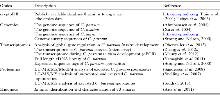
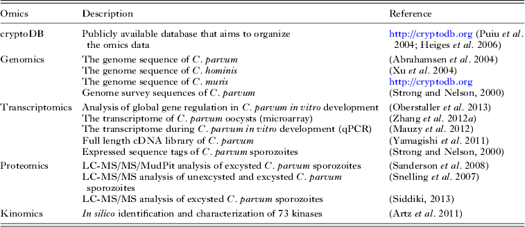
An overview of the omic literature can be found in Table 3.
Literature published on various omics studies on Cryptosporidium
CONCLUDING REMARKS
Cryptosporidium is one of the most resistant protozoan parasites. Few drugs (nitazoxanid, paromomycin, halofuginone) have been found to be effective against Cryptosporidium, and these have only partial effects. None of the common treatments against protozoan parasites are effective against Cryptosporidium. Moreover, the small oocysts are highly resistant against most disinfectants. The rigidity of the oocyst is due to the multilayered structure of the oocyst wall protecting the sporozoites from mechanical, environmental and chemical influences. Further insights into the structure and chemical composition of the oocyst might give us the opportunity to develop more efficient disinfectants. Upon infection of the host, the parasite expresses many molecules on the sporozoite surface that mediate attachment and probably invasion. However, no data are available about the signalling events taking place in the parasite. Calcium is probably one of the important second messengers that are responsible for signal transduction, but only a few kinases and transporters have been addressed so far. CDPKs might be responsible for microneme exocytosis as shown for Toxoplasma but there is no proof yet. The application of experimental results from other coccidian parasites to Cryptosporidium is not possible in many respects because of the substantial peculiarities of Cryptosporidium. Cryptosporidium is special in that it is embraced by the host cell instead of invading it, that it stays in an epicellular location and that it induces tremendous actin rearrangement in the cell. This shows that Cryptosporidium utilizes other infection strategies than, for example, Toxoplasma or Eimeria. This notion is supported by the analysis of the genomic data of C. parvum and C. hominis showing a highly streamlined metabolism that lacks many pathways found in other apicomplexans and depends on the import of essential nutrients from the host. Moreover, C. parvum expresses several potentially microneme-associated proteins of which several have no orthologues to proteins of other apicomplexans. Thus, Cryptosporidium probably releases unique molecules from the micronemes addressing different receptors/pathways in the target cell. Although we have some knowledge about the events within the target cell upon sporozoite attachment, many question marks remain. They include questions about the mediators released by the parasite, the receptors addressed, how nutrients are imported and what the molecular basis of egress is as well as questions concerning gametogenesis and oocyst formation. The publication of three Cryptosporidium genomes was a milestone giving insight into the organization and function of these parasites. However, the translation from in silico to in vitro/in vivo remains difficult. The lack of methods that allow full reproduction of the life cycle in vitro and a missing toolbox for genetic manipulation are major drawbacks. Moreover, it is not currently possible to store Cryptosporidium for a long period of time. Consequently, preservation of isolates or modified parasites relies on a constant passage of the parasites. Thus, the future challenges will be to improve in vitro systems and/or to establish alternative models such as Cryptosporidium baileyi or Cryptosporidium wrairi and to develop a transfection system for Cryptosporidium.