The coelenterate body plan of vertebrates consists of two protective barriers: the outer integumental (skin) and the inner mucosal (pulmonary, alimentary and urogenital) barriers. At these barriers, microbes of all sorts live in symbiotic and oftentimes commensal harmony with the host. A breach of the two barriers sets off a series of innate humoral and cellular responses beginning with blood clotting and culminating in an acute-phase response. When these immediate innate measures fail and microbes have gained entry past the barrier, cells of the innate immune system such as polymorphonuclear phagocytes (PMNs; neutrophils), macrophages and dendritic cells, which lie beneath the barrier or are recruited to the site of breach, deploy their defence mechanisms against the invader. These white blood cells (leukocytes) are professional scavengers that phagocytose (internalise) nonself (foreign) materials including microbes. They also alert other cells of the innate immune system [natural killer (NK) and natural killer T (NKT) cells] and cells of the adaptive immune system (T and B cells) of the entry of potentially harmful agents. The adaptive immune system allows a specific response against a pathogen and the development of immunological memory.
The property of macrophages and dendritic cells to respond rapidly to an invasion by foreign agents lies in their unique ability to recognise specific molecular patterns shared by large groups of microbes but absent from the host. Examples of such molecular patterns include lipoteichoic acid and lipopolysaccharide (LPS) of Gram-positive bacteria (e.g. Listeria, Staphylococcus and Streptococcus species) and Gram-negative bacteria (e.g. Escherichia coli and Salmonella), glycolipids of mycobacteria, mannans of yeast, and double-stranded RNA of viruses. These molecular patterns are recognised by specific receptors – the pattern-recognition receptors – expressed by the effector leukocytes, which include LPS-binding proteins, CD14, Toll-like receptors (TLRs), mannose-binding protein, surfactant protein A, and certain components of the complement cascade. Recognition of foreign patterns triggers a series of extracellular and/or intracellular mechanisms that eventually consume the infectious agent and hence limit the dissemination of the microbe at an early stage of infection. Activation of macrophages and immature dendritic cells by foreign patterns results in the differentiation of such cells and/or the elaboration of humoral factors. For example, signalling through the TLR on immature dendritic cells together with phagocytosis of the microbe triggers their differentiation to professional antigen-presenting cells (APCs). During this differentiation process they migrate from tissue sites of microbe entry (the homing site of immature dendritic cells) to secondary lymphoid organs such as the draining lymph nodes and spleen where mature dendritic cells are essential for the presentation of processed antigens to naive T cells.
Antigen processing and presentation are prerequisites for antigen recognition by T cells, and their activation and effector function. Internalisation of entire microbes (phagocytosis) or shed/secreted microbial products (pinocytosis or endocytosis) by macrophages and immature dendritic cells brings antigen to endosomal/lysosomal vesicles. These vesicles, also called class-II-enriched vesicles and the major histocompatibility (MHC) class II compartment (MIIC), are enriched in antigen-presenting molecules such as peptide-antigen-presenting MHC-encoded class II molecules as well as the lipid-antigen-presenting molecules collectively called CD1. Thus, the delivery of microbes or molecules shed or secreted by them to the MIIC followed by their processing provides a pool of derived products, some of which are presented to T cells by MHC class II and CD1 molecules.
César Milstein's group originally discovered CD1 as a thymocyte differentiation antigen that is recognised by a specific monoclonal antibody (Refs Reference Bradstock1, Reference McMichael2). Molecular cloning and nucleotide sequence analyses revealed that CD1 represents a family of molecules similar to MHC class I and class II molecules that comprises three groups: group I (CD1a, CD1b and CD1c), expressed by humans but not by mice; group II (CD1d), expressed by humans and mice; and group III (CD1e) expressed by humans but not by mice (Refs Reference Martin3, Reference Martin, Calabi and Milstein4). Subsequently, Michael Brenner's group discovered that human CD1b molecules restricted (regulated) the functions of a subset of Mycobacterium tuberculosis-specific T cells (Refs Reference Porcelli5, Reference Porcelli, Morita and Brenner6). They also discovered that CD1 restriction entailed the presentation of mycobacterial cell-wall lipids rather than peptides to reactive T cells (Refs Reference Beckman7, Reference Sieling8, Reference Moody9, Reference Moody10). Thus, CD1 molecules fundamentally differ from the closely related, peptide-antigen-presenting MHC proteins in that they present lipid antigens to T cells.
Soon thereafter, Albert Bendelac and Randy Brutkiewicz discovered that CD1d molecules restricted the functions of a unique subset of T cells: NKT cells (Ref. Reference Bendelac11). Mouse NKT cells were previously discovered as a subset of recent thymic emigrants that react to self-antigens, express an NK cell phenotype together with an invariant Vα14Jα18 T-cell receptor (TCR) α-chain, and have a predilection for IL-4 secretion upon activation. Paolo Dellabona, Steven Porcelli and their colleagues independently identified a homologous human T cell subset, which expressed an invariant Vα24Jα18 TCR α-chain (Refs Reference Dellabona12, Reference Porcelli13) and whose functions were restricted by human CD1d (Ref. Reference Exley14). Finally, Masaru Taniguchi, Yasuhiko Koezuka and their colleagues, and Mitchell Kronenberg and co-workers, discovered a potent, model CD1d-restricted iNKT cell antigen: α-galactosylceramide (αGalCer), a glycosphingolipid with anticancer activity derived from the marine sponge Agelas mauritianus (Refs Reference Cui15, Reference Kawano16, Reference Burdin17, Reference Brossay18). These major discoveries set the stage for our current understanding of CD1d and iNKT cell biology.
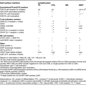
NKT cells are an unusual subset of thymus-derived lymphocytes. Their cell-surface phenotype resembles that of NK cells and T cells (Table 1) – hence their name. This article concentrates on the predominant and best-studied subset, called invariant NKT (iNKT) cells. iNKT cells express an invariant TCR α-chain [Vα14Jα18 (Vα14i) in mouse or Vα24Jα18 (Vα24i) in human] (Refs Reference Dellabona12, Reference Porcelli13, Reference Lantz and Bendelac22), which predominantly pairs with the Vβ8.2 (mouse) or Vβ11 (human) TCR β-chain to form a functional TCR (Refs Reference Dellabona12, Reference Porcelli13, Reference Lantz and Bendelac22). Some iNKT cells express CD4 or CD8 (the latter depending on species); others are double negative (DN) for CD4 and CD8 (Refs Reference Motsinger19, Reference Motsinger20, Reference Marks-Konczalik21). It should be noted that a small subset of NKT cells express the γδ TCR or diverse TCR α-chains, and can be restricted by MR1 (another MHC-like molecule). The introductory topics discussed above are reviewed excellently elsewhere (Refs Reference Bendelac, Savage and Teyton23, Reference Brigl and Brenner24, Reference Trinchieri and Sher25, Reference Villadangos and Schnorrer26, Reference Porcelli27).
Cell-surface phenotype of invariant natural killer T cells and comparison with other lymphocytesa

aBased on information in Refs Reference Van Kaer28, Reference Bendelac146, Reference Janeway147, Reference Joyce148 and Reference Wilson and Byrne149.
bA very small subset expresses γδ TCR.
c40–60% of mouse iNKT cells express CD4; the remainder express neither CD4 nor CD8 coreceptor. Human and monkey iNKT cells are either double negative for CD4 and CD8, or single positive for CD4 or CD8.
dExpressed upon T cell activation.
eExpression downregulated upon activation.
fA subset of iNKT cells expresses members of the Ly49 receptor family (e.g. a few express Ly49A or Ly49G2 and a large majority express Ly49C/I).
gThymocyte differentiation marker.
hA large subset but not all NKT cells express Ly6C.
Abbreviations: CD, cluster of differentiation; CTL, cytotoxic T lymphocyte; ICAM-1, intercellular adhesion molecule 1; IL, interleukin; iNKT, invariant natural killer T; LECAM, leukocyte endothelial cell adhesion molecule; LFA-1, lymphocyte function-associated antigen 1; NK, natural killer; TCR, T-cell receptor; Th, T helper.
Because iNKT cells are implicated in microbial and tumour immunity, vaccine adjuvants and regulation of autoimmunity (Refs Reference Bendelac, Savage and Teyton23, Reference Brigl and Brenner24, Reference Van Kaer28), this review focuses on CD1d-restricted antigen-presentation principles, the interaction of the iNKT cell receptor (mouse Vα14i/human Vα24i TCR) and antigen, and iNKT cell biology.
Antigen-presentation principles
CD1d-restricted antigens and distinct modes of iNKT cell activation
α-Anomeric glycosphingolipid antigens
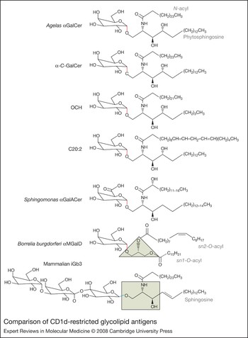
Since the original discovery that CD1d controls iNKT cell functions, no question has so captivated NKT cell watchers as to the nature of the natural iNKT cell antigen(s). Much of our understanding of iNKT cell biology is gleaned from in vitro and in vivo studies by using the synthetic form (KRN7000) of αGalCer (Fig. 1) as the probe (Refs Reference Bendelac, Savage and Teyton23, Reference Brigl and Brenner24). αGalCer is an α-anomeric glycolipid originally discovered as a marine-sponge-derived product with antimetastatic activities in mice (Ref. Reference Cui15). Its function depends on its presentation by CD1d and the specific activation of iNKT cells (Refs Reference Kawano16, Reference Burdin17, Reference Brossay18, Reference Spada, Koezuka and Porcelli29); thus αGalCer emerged as a powerful tool to track and probe the ontogeny and functions of iNKT cells in mice and humans (Refs Reference Bendelac, Savage and Teyton23, Reference Brigl and Brenner24, Reference Van Kaer28). Because most metazoan species synthesise β-anomeric glycolipids and sponges do not threaten mammalian life, αGalCer is variously thought of as an artificial iNKT cell antigen, a superantigen (dissimilar to bacterial superantigens that activate MHC-class-II-restricted T cells), or simply an immuno-pharmacological agent. However, certain bacterial species synthesise α-anomeric glycolipids that are similar to αGalCer (Fig. 1), some of which directly activate iNKT cells in a manner similar to αGalCer (Fig. 2) (Refs Reference Kinjo30, Reference Mattner31, Reference Sriram32). Moreover, many natural products isolated from marine sponges are derived from diverse bacterial symbionts that colonise them (Ref. Reference Haygood33). Hence, it is quite possible that αGalCer is a bacterial rather than a sponge-derived product.
Comparison of CD1d-restricted glycolipid antigens. (Legend; see previous page for figure.) The α-anomeric glycolipid α-galactosylceramide (αGalCer), derived from the marine sponge Agelas, and its synthetic variants α-C-GalCer, OCH and C20:2, have been used widely to probe invariant natural killer T (iNKT) cell function. These antigens elicit distinct cytokine responses from iNKT cells (see Table 2). Sphingomonas cell-wall-derived glycolipids, which contain α1′-1-O-linked galacturonic acid (αGalACer) (or glucuronic acid; not shown) resemble Agelas αGalCer. Borrelia burgdorferi cell-wall-derived α-monogalactosyldiacylglycerol (αMGalD) is a recently identified glycolipid antigen that weakly elicits an interferon γ (IFN-γ) response from iNKT cells; its hydrophobic O-acyl chains are predicted to form a structure similar to that adopted by phosphatidylcholine when bound to mouse CD1d (see structure on the right in Fig. 3d). Isoglobotrihexosylceramide (iGb3) is an endogenous self-antigen. The triangle and the box indicate differences in the glycerol and sphingosine backbone that make up αMGalD and αGalCer and its derivatives, respectively. The axial (red) versus equatorial (blue) disposition of the anomeric bond (hydroxyl group linked to carbon atom 1 in a hexose ring), which results in α versus β anomers, respectively, is highlighted in the structures shown. Other NKT cell antigens include the self-antigens sulphatide and phosphatidylcholine (see Fig. 3), phosphatidylinositol and phosphatidylethanolamine, as well as mycobacterial phosphatidylinositoltetramannoside (not shown).

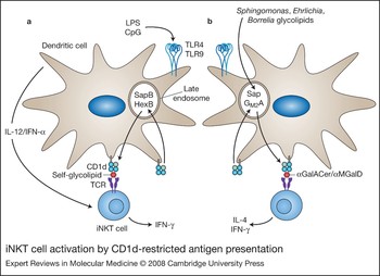
iNKT cell activation by CD1d-restricted antigen presentation. (a) Microbes containing Toll-like receptor (TLR) ligands such as LPS and CpG activate invariant natural killer T (iNKT) cells by inducing IL-12 or IFN-α production by dendritic cells, which amplifies weak responses elicited upon the recognition of CD1d–self-glycolipid complexes by the iNKT cell receptor. The generation of some self-antigens requires lysosomal glycosidases such as HexB, while binding of certain self-glycolipids to CD1d requires SapB (see Fig. 4). The end result is an IFN-γ response by iNKT cells. (b) α-Anomeric glycolipids from the cell walls of some microbes of the α-Proteobacteria class, such as Sphingomonas spp., can activate iNKT cells directly when presented by CD1d molecules. iNKT cells so activated secrete IL-4 and IFN-γ. As with the loading of CD1d with αGalCer, the loading of the bacterial glycolipids such as αGalACer and αMGalD (see Fig. 1) may require lysosomal lipid-transfer proteins GM2A and/or Sap. Abbreviations: CpG, oligonucleotides containing deoxycytidine-phospho-deoxyguanosine motif; GM2A, GM2 activator; HexB, hexosaminidase B; IFN, interferon; IL, interleukin; LPS, lipopolysaccharide; Sap, saposin.

The α-anomeric glycolipids similar to αGalCer that are synthesised by bacteria include the glycosphingolipids α-glucuronosylceramide and α-galacturonosylceramide (αGalACer) (Fig. 1) of LPS-negative and Gram-negative bacteria of the α-Proteobacteria class, to which Sphingomonas capsulata and Ehrlichia muris belong (Refs Reference Kinjo30, Reference Mattner31, Reference Sriram32). CD1d molecules bind both these glycolipids and present them to iNKT cells (Fig. 2). This presentation of Sphingomonas glycolipids appears to be important in bacterial-specific immunity because high-dose Sphingomonas infection of wild-type mice results in septic shock caused by inflammatory cytokines rapidly released by activated iNKT cells. Similar infection of mice that lack iNKT cells delays bacterial clearance (Refs Reference Kinjo30, Reference Mattner31). Thus, the direct recognition of α-anomeric glycolipids by iNKT cells has important immunological implications.
Diacylglycerolipid antigens
iNKT cells have a broad antigen specificity in that they recognise not only ceramide-based lipids but also diacylglycerol-based microbe-derived lipid antigens, such as α-monogalactosyldiacylglycerol (αMGalD; a nonphosphorylated diacylglycerol-based glycolipid) (Fig. 1) and phosphatidylinositoltetramannoside (PtdInoMan4) (Refs Reference Kinjo34, Reference Fischer35). These glycolipids are cell-wall components or their precursor synthesised by Borrelia burgdorferi (Refs Reference Ben-Menachem36, Reference Schroder37), the agent of Lyme disease, and M. tuberculosis (Ref. Reference Fischer35), respectively. Nevertheless, very little is known regarding the role of these unusual glycolipids in immunity to B. burgdorferi or M. tuberculosis. Notwithstanding, how ceramide-based α-anomeric glycosphingolipid and diacylglycerol-based glycolipids, which are structurally distinct antigens, are recognised by the iNKT cell receptor remains to be established.
Endogenous antigens
iNKT cells are autoreactive – that is, they react to self-antigens expressed by the host: freshly isolated thymic iNKT cells are activated by a variety of CD1d-positive cells including thymocytes and tumour cells without the addition of exogenous antigens of any sort (Ref. Reference Bendelac11). Likewise, iNKT-cell-derived hybridomas recognise CD1d-positive cells and cell lines (Refs Reference Bendelac11, Reference Brossay38). This presentation of self-antigen(s) in mice (Refs Reference Chiu39, Reference Chiu40, Reference De Silva41) but not in humans (Ref. Reference Exley14) requires that CD1d molecules traffic through the MIIC. Furthermore, neither cells deficient in β-glucosylceramide (βGlcCer) synthase nor cell-free CD1d–βGlcCer complexes activate mouse iNKT-cell-derived Vα14iTCR-positive hybridomas (Ref. Reference Stanic42). This finding provided the first clue that the natural mouse iNKT cell antigen is a cellular, βGlcCer-derived glycosphingolipid. One such CD1d-restricted self-antigen, which resides within the MIIC, was recently identified to be the cellular glycosphingolipid isoglobotrihexosylceramide (iGb3; Fig. 1) (Ref. Reference Zhou43). It is noteworthy that the terminal residue of iGb3 is an α-anomeric galactose, which forms the epitope for Vα14i and Vα24i TCR (Ref. Reference Zhou43), as does α-galactose and α-galacturonic acid in αGalCer and αGalACer, respectively.
The importance of self-antigen recognition was realised in studies where coculture of freshly isolated iNKT and dendritic cells with either the Gram-positive cocci Staphlococcus aureus or the Gram-negative Salmonella typhimurium resulted in human and mouse iNKT cell activation. In the case of S. typhimurium, iNKT cell activation resulted from bacterial LPS. Curiously, LPS does not directly activate iNKT cells, but instead activates dendritic cells through TLR4 (Ref. Reference Brigl44). Indeed, the activation of mouse iNKT cells by dendritic cells stimulated by heat-killed Salmonella required an enzyme (hexosaminidase B) that converts the precursor iGb4 to iGb3. These data were interpreted to mean that Gram-negative Salmonella activates mouse iNKT cells indirectly through the recognition of self-iGb3 presented by CD1d expressed on dendritic cells (Ref. Reference Mattner31).
Whether iGb3 is a bone fide endogenous antigen or the sole iNKT cell self-antigen has been contentious because this glycolipid was identified only in mouse dorsal root ganglion but not in human thymocytes or dendritic cells (Ref. Reference Speak45), which are known to directly activate iNKT cell hybridomas without the addition of exogenous lipid antigens. Nonetheless, contrary to a previous report, human thymocytes synthesise iGb4 (Ref. Reference Li46) and hexosaminidase B (Ref. Reference Zhou43), which can convert iGb4 to iGb3. Moreover, a recent study demonstrated that activation of dendritic cells through TLR9 ligation resulted in CD1d-restricted presentation of sialylated cellular glycolipids and the elaboration of interferon α (IFN-α). The CD1d-restricted sialylated antigen together with IFN-α resulted in iNKT cell activation (Ref. Reference Paget47). Thus, a number of distinct cellular lipids including iGb3 (Ref. Reference Zhou43), GD3 (Ref. Reference Wu48), phosphatidylinositol (PtdIno) (Ref. Reference Gumperz49) and phosphatidylethanolamine (PtdEtN) (Ref. Reference Rauch50) under different physiological and pathophysiological conditions could form endogenous iNKT cell antigens.
Summary of CD1d-restricted antigens
In summary, iNKT cells have evolved to sense bacterial infections either directly through recognition of exogenous glycolipid antigens, which are parts of cell walls, or indirectly through recognition of endogenous self-glycolipids upon activation of dendritic cells by cell-wall products such as LPS or microbial nucleic acid (Fig. 2). Because several CD1d-restricted antigens contain a shared α-anomeric epitope essential for iNKT cell recognition, the invariant TCR expressed by this T cell subset is thought to be a pattern-recognition receptor usually utilised by cells of the innate immune system to detect microbes. Furthermore, iNKT cells respond rapidly, within a few hours (discussed below under ‘Functional consequences’), to specific antigen, as do cells of the innate immune system. Therefore, iNKT cells, like NK cells, are thought to be innate lymphocytes.
Structures of CD1d–lipid-antigen complexes
Organisation and structure of CD1d
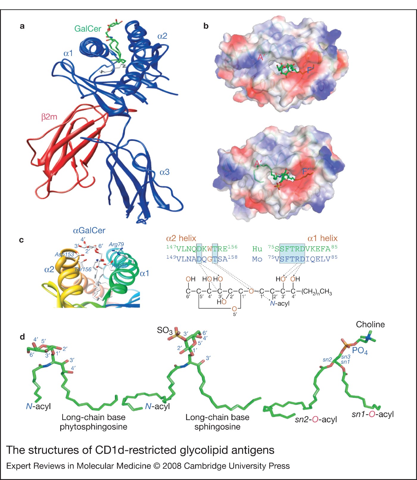
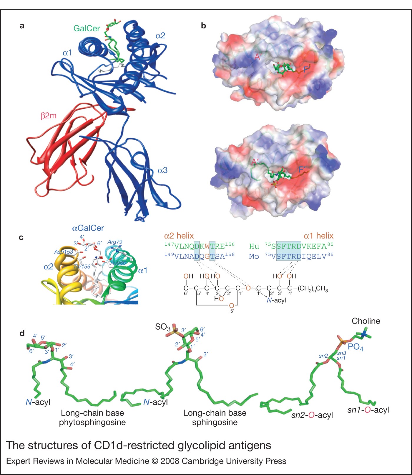
CD1d is a heterodimer consisting of a heavy chain that is noncovalently associated with the light-chain β2-microglobulin (Fig. 3a), which it shares with MHC class I molecules. The heavy chain folds into five domains: the extracellular α1, α2 and α3 domains (Fig. 3a) are membrane-anchored by the transmembrane region, which ends in a short cytoplasmic tail.
Solution of the three-dimensional structures of mouse and human CD1d molecules, which differ subtly (<5%) from each other, in complex with several distinct lipid antigens [αGalCer (Refs Reference Koch51, Reference Zajonc52), the acidic glycosphingolipid sulphatide (Ref. Reference Zajonc53), PtdInoMan4 (Ref. Reference Zajonc54) and phosphatidylcholine (PtdCho) (Ref. Reference Giabbai55)] revealed that the α1 and α2 domains of the heavy chain fold into a superdomain to form the antigen-binding groove (Fig. 3a and b). The antigen-binding groove is laterally confined by two antiparallel α-helices that are supported at the bottom by two β-sheets, each made up of four antiparallel β-strands. The membrane-proximal immunoglobulin-like α3 domain and the noncovalently associated light chain support the superdomain (Fig. 3a). Therefore, the topology of CD1d resembles peptide-antigen-presenting MHC molecules.
The structures of CD1d-restricted glycolipid antigens. (Legend; see previous page for figure.) (a) Lateral view of the three-dimensional structure of mouse CD1d bound to the potent invariant natural killer T (iNKT) cell antigen αGalCer. The CD1d molecule is made up of a heavy chain that is noncovalently associated with the light-chain β2-microglobulin (β2m). The membrane-distal α1 and α2 domains of the heavy chain fold in such a manner that they form an apical antigen-binding groove into which antigenic ligands bind. This antigen-binding domain is followed by the α3 domain, which adopts an immunoglobulin constant-domain-like fold. (b) Apical en face view of the antigen-binding domain shows the solvent-exposed sugars of αGalCer (top) and sulphatide (bottom). The antigen-binding groove contains two deep pockets – A′ and F′– into which the hydrocarbon tails of the N-acyl chain and the phytosphingosine (αGalCer)/sphingosine (sulphatide) base, respectively, bind. When so bound, the α-linked sugar in αGalCer and the β-linked sugar in sulphatide are differently disposed to solvent; the former lies parallel to the groove whereas the latter rises perpendicular out of the groove. (c) Close-up three-dimensional view of mouse CD1d–αGalCer structure (left) reveals critical amino acid side chains within hydrogen-bonding distance from atoms of the glycolipid antigen (right). Mouse (Mo) and human (Hu) sequences are shown for comparison, with arrows indicating hydrogen bonds. (d) The structures adopted by three different lipids – αGalCer (left), sulphatide (middle) and phosphatidylcholine (right) – when bound to mouse CD1d are similar but not identical. This allows a conserved hydrogen-bond network between polar atoms of lipids and that of the side chains of amino acids that line the entrance of the antigen-binding groove. Key carbons of the sugar and the phytosphingosine base are numbered. The sulphate (SO3) of sulphatide as well as phosphate (PO4) and choline in phosphatidylcholine are also marked. The three-dimensional structures were generated with Chimera (parts a, c and d) or PyMol (part b) software using published (Refs Reference Zajonc52, Reference Zajonc53, Reference Giabbai55) X-ray crystallographic coordinates (1Z5L, 2AKR and 1ZHN) deposited with the Protein Data Bank.
Display of αGalCer and related antigens
The arrangement of the amino acids that make up the antigen-binding groove is such that the narrow apical entrance leads into two deep-seated pockets (A′ and F′; Fig. 3a and b). The two pockets are lined predominantly by nonpolar and hydrophobic amino acid residues, and hence permit the binding of hydrocarbon tails of lipid molecules of varying lengths. The N-acyl chain tucks into the large A′ pocket while the long-chain phytosphingosine base fits into the F′ pocket (Fig. 3b and 3d). Similarly, the sn1- and sn2-O-acyl chains of diacylglycerol lipids (Fig. 3d) is predicted to bind the A′ and F′ pockets, respectively. This binding mode exposes the polar aspects of lipids out from the antigen-binding groove (Fig. 3a and b). Furthermore, the amino acids lining the entrance of the antigen-binding groove form a hydrogen-bond network with polar atoms of α-anomeric glycosphingolipid head groups of αGalCer (Fig. 3c) and αGalACer (Refs Reference Koch51, Reference Zajonc52, Reference Zajonc53, Reference Zajonc54, Reference Giabbai55, Reference Wu56). This hydrogen-bond network provides stability to the CD1d–lipid interaction. Thus, the physicochemical architecture of the antigen-binding groove dictates how stably a lipid ligand binds CD1d and exposes the polar epitope for recognition by the Vα14i/Vα24i TCR.
Display of other antigens
Molecular modelling experiments with αMGalD binding based on the CD1d–PtdInoMan4 structure suggests that the borrelial lipid might bind CD1d in a manner similar to αGalCer. However, despite similar binding modes, the polar galactose headgroup of αMGalD allows for fewer hydrogen-bond interactions with CD1d compared with those observed in the CD1d–αGalCer structure because the diacylglycerol backbone of αMGalD contributes to fewer hydrogen bonds (Ref. Reference Kinjo34). This perhaps results in poor binding and might explain the poor antigenic property of αMGalD.
Sulphatide binds CD1d in a distinct manner such that the 3′-sulphated galactose is solvent exposed and projects up and away from the antigen-binding groove as a result of its β linkage (Ref. Reference Zajonc53). This contrasts with the more intimate binding of the galactosyl headgroup of αGalCer to CD1d (Refs Reference Koch51, Reference Zajonc52) (Fig. 3b and d). However, despite differences in the binding mode, sulphatide engages CD1d in similar hydrogen-bond interactions to those formed by αGalCer (Refs Reference Koch51, Reference Zajonc52, Reference Zajonc53).
Akin to sulphatide, the first hexose of iGb3 – that is, glucose – is β-linked to ceramide and hence would be predicted to be solvent exposed in a manner similar to the sulphated galactose. This disposition of the first glucose of iGb3 would then result in an almost perpendicular exposition of the two terminal galactoses [Glc–β(1 → 4)Gal–α(1 → 3)Gal] of iGb3 out of the CD1d groove. Indeed, the recent structure of the mouse CD1d–iGb3 complex revealed that the terminal hexoses of this self-antigen were solvent exposed and displayed perpendicular to the CD1d groove (Ref. Reference Zajonc57). Nevertheless, the β-linked glucose, which unalike sulphatide lacks a 3′-sulphate and whose 4′-hydroxyl is equatorially disposed, perhaps results in poor binding of iGb3 to CD1d because the 3′-sulphate and the axial 4′-hydroxyl are involved in hydrogen bonding of sulphatide with CD1d.
Thus, the presentation principles for α- and β-linked glycolipids are distinct. How then the same Vα14i/Vα24i TCR of iNKT cells recognise these structurally distinct antigens remains to be elucidated.
Topological biochemistry of CD1d assembly and antigen presentation
CD1d molecules are expressed by APCs such as dendritic cells, macrophages and B cells, as well as CD4+CD8+ thymocytes, hepatocytes and intestinal epithelial cells (Refs Reference Amano58, Reference Bradbury59, Reference Brossay60, Reference Mandal61, Reference Roark62, Reference Dougan, Kaser and Blumberg63). The folding and assembly of CD1d occurs within the endoplasmic reticulum (ER) and is facilitated by the chaperones calnexin and calreticulin and the thiolreductase ERp57 (Fig. 4) (Ref. Reference Kang and Cresswell64). Because the antigen-binding groove of CD1d is largely nonpolar and hydrophobic (Fig. 3b) (Refs Reference Koch51, Reference Zajonc52, Reference Zajonc53, Reference Giabbai55, Reference Zeng65), it is thought to assemble with phospholipids indigenous to the ER (Refs Reference De Silva41, Reference Park66) perhaps by a process facilitated by the ER-resident lipid-transfer protein (LTP) microsomal triglyceride transfer protein (MTP; Fig. 4) (Refs Reference Brozovic67, Reference Dougan68, Reference Dougan69). Thence, by virtue of a short motif (Tyr-Gln-Gly-Val-Leu and Tyr-Gln-Asp-Ile-Arg in human and mouse CD1d, respectively) contained within the cytoplasmic tail, CD1d negotiates the plasma membrane, via the Golgi complex, en route to the MIIC where it assembles with iNKT cell antigens (Refs Reference Chiu39, Reference Chiu40, Reference De Silva41). Alternatively, CD1d can arrive directly at the MIIC by assembly with MHC-class-II-associated invariant chain (Ii), which contains an MIIC-targeting motif (Ref. Reference Jayawardena-Wolf70).
Topological biochemistry and the assembly of CD1d molecules with iNKT cell antigen, and its evasion. Being a type I integral membrane glycoprotein, the folding and assembly of CD1d occurs in the rough endoplasmic reticulum (ER). Here, several chaperones (calnexin and calreticulin) and the thiolreductase ERp57 assist in CD1d heavy chain (α) folding and β2-microglobulin (β2m) (light chain) binding. The α-chain–β2m complex forms a structure that is receptive to ER-resident lipids. Current evidence suggests that lipid-transfer proteins (LTPs) such as microsomal triglyceride-transfer protein (MTP), which facilitates the assembly of apolipoprotein B, might facilitate lipid binding to CD1d in the ER. Upon complete assembly, the CD1d–lipid complexes egress from the ER and negotiate the secretory pathway to the plasma membrane. By virtue of a late-endosome/lysosome-targeting motif within the cytoplasmic tail of CD1d, it recycles through the major histocompatibility complex (MHC)-class-II-enriched compartment (MIIC). Alternatively, CD1d can directly arrive at the MIIC by assembly with MHC-class-II-associated invariant chain (Ii), which contains a MIIC-targeting motif. Assembly with antigens involves the exchange of ER-derived lipid or Ii with glycosphingolipid antigens in the MIIC. The extraction of bound lipids from CD1d and the loading of antigenic glycolipids are facilitated by lysosomal LTPs such as saposin (Sap), GM2 activator (GM2A), and Niemann–Pick C-2 (not shown), which are essential for the enzymatic catabolism of glycolipids. Exogenous antigens such as those shed or secreted by microbes can be delivered to MIIC by binding to apolipoprotein-E-containing very-low-density lipoproteins (VLDLs) via low-density-lipoprotein receptor (LDLr)-mediated endocytosis. The arrival of self- and foreign lipids in the MIIC provides a pool of ligands that can bind to CD1d molecules during their transit through this intracellular compartment. Upon binding them, the CD1d–lipid complex egresses to the cell surface for the display of invariant natural killer T (iNKT) cell antigens. Pathogens can interfere with the CD1d-restricted antigen-presentation pathway by inhibiting key steps in intracellular CD1d trafficking. The modulator of immune recognition (MIR)-1 and MIR-2 proteins of Kaposi-sarcoma-associated herpesvirus (KHSV) ubiquitinylate the cytoplasmic tail of human CD1d, which triggers endocytosis of surface CD1d thereby reducing cell surface CD1d expression. The Nef (negative regulatory factor) protein of human immunodeficiency virus (HIV-1), which causes acquired immunodeficiency syndrome (AIDS), also reduces CD1d expression, perhaps by increased endocytosis of cell-surface CD1d molecules coupled with inhibition of CD1d transport back to the cell surface. Similarly, in herpes simplex virus (HSV)-1-infected cells, CD1d molecules accumulate in the MIIC, owing to a defect in recycling CD1d molecules back from endosomal compartments to the cell surface.

Assembly with antigens involves the exchange of ER-derived lipid or Ii-bound CD1d with glycosphingolipid antigens in the MIIC. These exchange and reassembly processes are facilitated by MIIC-resident LTPs such as saposins A, B, C and D, GM2 activator (Refs Reference Zhou71, Reference Kang and Cresswell72, Reference Yuan73) and Niemann–Pick type C2 protein (Ref. Reference Schrantz74), as well as cathepsin L and S (Refs Reference Honey75, Reference Riese76). Additionally, MTP appears to play an as yet undefined role in returning CD1d back to the cell surface upon negotiating the MIIC (Ref. Reference Sagiv77). These actions of various LTPs in CD1d assembly and antigen binding described above were deduced from cells derived from gene-deficient mice. However, it should be noted that the lack of proteins and enzymes involved in lipid metabolism also results in mistargeting of cellular lipids and hence data that have emerged from such studies should be interpreted with caution (Ref. Reference Gadola78).
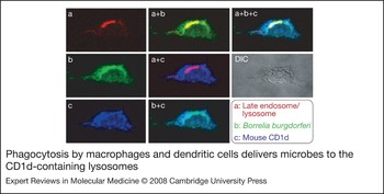
The presentation of bacterial cell-wall lipids by CD1d molecules depends on bringing them inside the APC (Fig. 5). This can occur by one of two ways: by phagocytosis of the bacterium itself; or by receptor-mediated or fluid-phase endocytosis (micro- or macropinocytosis) and eventual maturation of the phagosome or endosome to form MIIC. The internalised bacterium is then broken down by MIIC-resident hydrolases, thus making lipids available for CD1d-restricted presentation. In the case of shed or secreted bacterial lipids, soluble LTPs that regulate cellular lipid homeostasis such as apolipoproteins bind such microbial lipids, assemble into very-low-density lipoprotein (VLDL) complexes, and by LDL-receptor-mediated endocytosis arrive in the MIIC (Fig. 4) (Ref. Reference van den Elzen79). Thus, the topological distribution of cellular and microbial lipids and LTPs dictates the types of antigens that assemble with CD1d.
Phagocytosis by macrophages and dendritic cells delivers microbes to the CD1d-containing lysosomes. The differential interference contrast (DIC) picture of a macrophage that has phagocytosed a microbe observed under a light microscope shows the gross cellular outline; the prominent structure within this cell is its nucleus. Cellular organelles and their contents are observed by confocal fluorescence microscopy. In the micrographs shown: the lysosome is stained red (a) because it is marked with the fluorescent dye Lyso-tracker; the microbe, in this case Borrelia burgdorferi, is stained with the fluorescent dye PKH-II and is seen green (b); and CD1d, which is detected with a specific monoclonal antibody 1B1 tagged with the fluorescent dye allophycocyanin, is stained blue (c). B. burgdorferi bacteria colocalise with lysosomes, hence the yellow colour in the merged picture ‘a + b’. Similarly, the regions of the cell where lysosomes and CD1d colocalise appear pink in the merged picture ‘a + c’, while the regions where CD1d and microbe colocalise appear light blue in the merged picture ‘b + c’. The regions where lysosomes, B. burgdorferi and CD1d colocalise appear white in the merged picture ‘a + b + c’. New images produced based on published data described in Refs Reference Chiu39 and Reference Liu152.

Microbial subversion of CD1d-restricted antigen presentation
Because iNKT cells play a critical role in protective immune responses against a variety of microbes, it is not surprising that pathogens, especially viruses that establish latency, have devised ways to interfere with the CD1d-restricted antigen-presentation pathway (Fig. 4). Most accomplish this mainly by interfering with intracellular CD1d trafficking (Ref. Reference Van Kaer and Joyce80). For example, the modulator of immune recognition (MIR)-1 and MIR-2 proteins of Kaposi sarcoma-associated herpesvirus are ubiquitin ligases that ubiquitinylate the cytoplasmic tail of human CD1d, which triggers endocytosis of surface CD1d thereby reducing cell-surface CD1d expression (Refs Reference Sanchez, Gumperz and Ganem81, Reference Chen82). The Nef protein of human immunodeficiency virus also reduces CD1d expression perhaps by increased endocytosis of cell-surface CD1d molecules coupled with inhibition of CD1d transport to the cell surface (Ref. Reference Cho83). Similarly, in cells infected with herpes simplex virus 1, CD1d molecules accumulate in the MIIC owing to a defect in recycling CD1d molecules back from endosomal compartments to the cell surface (Ref. Reference Yuan, Dasgupta and Cresswell84). Vesicular stomatitis virus and vaccinia virus also impair antigen presentation by CD1d, perhaps by interfering with intracellular trafficking of CD1d molecules induced by mitogen-activated protein kinase signalling (Ref. Reference Renukaradhya85). Some bacteria have also devised strategies to evade CD1d-restricted antigen presentation. For example, infection of monocytes with M. tuberculosis also results in downmodulation of CD1d by reducing CD1d mRNA expression, implying regulation at the transcriptional level (Ref. Reference Roura-Mir86). Although these findings indicate that iNKT-cell-based immune recognition may play a significant role in viral and bacterial immunity, there is very little evidence yet for a role in viral immunity.
Recognition logic
The iNKT cell receptor consists of an invariant (mouse Vα14i or human Vα24i) TCR α-chain that pairs with highly diverse mouse Vβ8.2, Vβ7 and Vβ2 or human Vβ11 TCR β-chains (Refs Reference Dellabona12, Reference Porcelli13, Reference Lantz and Bendelac22). Akin to antigen recognition by the TCR of conventional T cells, gene-transfer and mutagenesis studies have demonstrated that the iNKT cell receptor α- and β-chains are sufficient for CD1d–lipid-antigen recognition (Refs Reference Gui87, Reference Scott-Browne88). Nonetheless, three lines of evidence indicate that the structure and/or organisation of the Vα14i TCR is distinct from the αβ TCR of conventional T cells (e.g. cytotoxic T lymphocytes; CTLs).
First, the FG loop within the Cβ domain is a large, evolutionarily conserved structure, which forms a wall at the region where Cβ and Vβ domains of the TCR β-chain join to form a cavity (Ref. Reference Wang89). Antibody mapping studies revealed that the FG loop is in close proximity to one of the CD3ɛ subunits (Ref. Reference Ghendler90). Transgenic mice expressing the TCR β-chain mutant lacking the FG loop have no gross deficiencies in the development and function of conventional CD4+ and CD8+ T cells (Ref. Reference Degermann, Sollami and Karjalainen91), implying that αβ TCR pairing and surface expression is not grossly impaired. However, a careful analysis in a single-TCR transgenic system revealed that thymocytes lacking the FG loop had impaired negative selection (Ref. Reference Sasada92) despite unhindered TCR αβ pairing and expression. By contrast, however, Vα14i TCR α-chain was found not to pair at all with the Vβ8.2-FG-loop mutant, and hence the mutant mice were impaired in iNKT cell development (Ref. Reference Degermann, Sollami and Karjalainen93). Second, interestingly, the fluorescently tagged anti-TCRβ antibody H57–597, which specifically binds the FG loop (Refs Reference Wang89, Reference Degermann, Sollami and Karjalainen91), exhibits strong FRET (fluorescence resonance energy transfer, which occurs between donor and acceptor fluorochromes within ~100 Å distance of each other) in conjunction with fluorescent CD1d1–αGalCer tetramer (Ref. Reference Stanic94). In striking contrast, MHC class I tetramers in conjunction with anti-TCR or anti-CD3ɛ antibody did not exhibit FRET (Ref. Reference Stanic94). Third, the high-affinity interaction between the Vα14i TCR and CD1d–αGalCer is independent of temperature and electrostatic forces (Ref. Reference Cantu95). This property is similar to the interactions between haptens and cognate antibodies that have undergone affinity maturation (Ref. Reference Wedemayer96), suggesting a rigid binding interface that is strikingly distinct from the flexible binding interface between the αβ TCR and peptide–MHC (Refs Reference Wu97, Reference Feng98). Taken together, these data strongly suggest that the structure and/or the organisation of the Vα14i TCR complex is distinct from αβ TCR of conventional T cells, which might potentially account for the co-operative engagement of glycolipid antigens (discussed below).
Indeed, the recently reported ternary structure of the Vα24iTCR–antigen cocomplex indicates that the receptor–antigen interface is distinct from that of the complex of TCR and peptide–MHC (Ref. Reference Borg99). Thus, we suggest that the Vα14i/Vα24i TCR has evolved a very sensitive mechanism for the recognition of high-affinity (αGalCer) (Refs Reference Stanic94, Reference Cantu95, Reference Sidobre100, Reference McCarthy101), medium-affinity (OCH) (Refs Reference Stanic94, Reference McCarthy101), and low-affinity (iGb3) (Refs Reference Zhou43, Reference Zajonc57) antigens (see section ′Kinetic parameters of Vα14iTCR–antigen interactions') that is perhaps key to responding to subtle changes in antigen structure (e.g. iGb3 versus αGalCer or αGalACer) and/or concentration (e.g. iGb3 under physiological versus pathophysiological conditions).
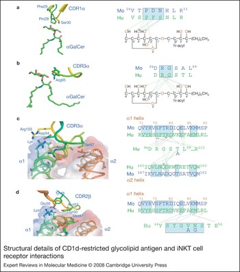
Gene-transfer and mutagenesis studies have suggested that the Vα14i TCR interfaces the combinatorial epitope formed by the CD1d–lipid complex such that it includes both the lipid head group and the two α-helices of CD1d (Refs Reference Gui87, Reference Scott-Browne88, Reference Grant102, Reference Melian103). This antigen-recognition mode is consistent with the recently solved three-dimensional structure of the ternary complex between the human iNKT cell receptor (Vα24i made of Vα24Jα18 and Vβ11 TCR chains) and its cognate antigen (human CD1d–αGalCer) (Ref. Reference Borg99). By contrast to TCR–peptide–MHC complexes in which the receptor docks diagonal on the antigen, the Vα24i TCR docks parallel on the CD1d antigen-binding groove (Ref. Reference Borg99). Additionally, the receptor docks on to the extreme C-terminal end of the CD1d antigen-binding groove using three of the six antigen-binding complementarity-determining regions (CDRs) – CDR1α, CDR3α and CDR2β – while almost excluding CDR2α, CDR1β and CDR3β from the interface (Fig. 6) (Ref. Reference Borg99). This docking mode enables a lock-and-key interaction with the α-linked galactose epitope on the antigen (Fig. 6) that was predicted from biophysical studies of Vα14iTCR–antigen binding (Refs Reference Borg99, Reference Cantu104). Furthermore, alanine-scanning mutagenesis of the mouse Vα14i TCR revealed that the mouse iNKT cell receptor interfaces mouse CD1d–αGalCer in a manner similar to the Vα24iTCR–antigen interaction (Ref. Reference Scott-Browne88).
Structural details of CD1d-restricted glycolipid antigen and iNKT cell receptor interactions. (a, b) αGalCer bound to human CD1d interacting with human invariant natural killer T (iNKT) cell receptors CDR1α and CDR3α, respectively; (c, d) three-dimensional views of human CD1d interacting with human iNKT cell receptors CDR3α and CDR2β. Close-up three-dimensional views are shown on the left, revealing critical amino acid side chains within hydrogen-bonding distance from atoms of the glycolipid antigen; and the details of the interactions observed in the three-dimensional structures are schematically rendered on the right. Blue arrows indicate hydrogen bonds; green dashed arrows indicate Van der Waals interactions; red arrows indicate salt bridges. Mo, mouse; Hu, human. Amino acid residues within the iNKT cell receptors are labelled in blue whereas those of CD1d are italicised. Mouse and human sequences are shown for comparison. The three-dimensional structures were generated using Chimera from published (Ref. Reference Borg99) co-ordinates 2PO6 deposited with the Protein Data Bank.

The above recognition logic raises the important question of how the mouse Vα14i and human Vα24i TCRs recognise structurally distinct antigens such as αMGalD, PtdInoMan4, iGb3, GD3, PtdIno and PtdEtN (see ‘Structures of CD1d–lipid antigens’). Alanine-scan mutants of Vα14i TCR revealed that this receptor recognises many α-linked glycolipids (αGalCer, OCH, αGalACer and iGb3, which contains an α-linked terminal galactose; see Fig. 1) by means of a ‘hot-spot’ of germline-encoded amino acids within CDR1α, CDR3α and CDR2β loops (Ref. Reference Scott-Browne88). This finding suggests that the interaction of iNKT cell receptor with structurally distinct α-linked antigens is accomplished by similar recognition logic. Whether the recognition of diacylglycerol lipids is accomplished by similar logic remains to be established.
Kinetic parameters of iNKT cell receptor–antigen interactions
The kinetic parameters of iNKT cell receptor–antigen interaction have been extensively studied. Surface plasmon resonance and tetramer binding studies have revealed high-affinity interaction between CD1d–αGalCer (or derived analogues) and Vα14i/Vα24i TCR: the relative avidity of this interaction is similar to that of high-affinity interactions between the TCR and peptide presented by MHC class I molecules (Refs Reference Stanic94, Reference Cantu95, Reference Sidobre100, Reference McCarthy101, Reference Gadola105, Reference Kjer-Nielsen106). Interestingly, the half-life of mouse CD1d–αGalCer–Vα14iTCR interaction was unusually long (Refs Reference Cantu95, Reference Sidobre100, Reference McCarthy101, Reference Gadola105, Reference Kjer-Nielsen106). How these kinetic parameters of lipid-antigen–TCR interactions relate to the rapid and robust iNKT cell response remains to be elucidated. In this regard, it is interesting to note that the αGalCer analogue OCH (Fig. 1), which has a shortened long-chain sphingosine base (C9 versus C18) and acyl chain (C24 versus C26) and interacts with the Vα14i and Vα24i TCR with lower relative affinity/avidity compared with αGalCer (Ref. Reference Stanic94), specifically elicits sustained IL-4 with very little IFN-γ response (Ref. Reference Miyamoto, Miyake and Yamamura107). A similar IL-4-biased response is elicited by a diunsaturated (C20-diene) N-acyl analogue of αGalCer (see Fig. 1) whose binding constant is similar to αGalCer but dissociation rate is similar to OCH (Refs Reference McCarthy101, Reference Yu108).
Consistent with the kinetic parameters of αGalCer and derived analogues, αGalCer and C20-diene efficiently elicit classic immune synapse formation by iNKT cells with about ten molecules of antigen/μm2. By contrast, OCH elicits immune synapse formation at tenfold higher antigen concentration (Ref. Reference McCarthy101). Thus, at equal antigen concentration, αGalCer and C20-diene induce very quick and sustained iCa2+ flux (a measure of very early T cell activation), and rapidly polarise secretory granules to the immune synapse when compared with OCH (Ref. Reference McCarthy101). Therefore, the kinetic parameters of iNKT cell receptor–antigen interactions dictate the qualitative and quantitative aspects of the iNKT cell response.
Co-operative antigen recognition
Some functions of iNKT cells depend on the recognition of endogenous self-antigens (discussed above). Therefore, self-antigen recognition must be finely tuned to prevent iNKT cell activation during physiological conditions, but to allow a rapid response to disturbances in intracellular physiology. In other words, iNKT cells need to be very sensitive to modest changes in antigen structure and/or concentration. In biological systems, such fine tuning is often achieved by employing co-operative ligand–receptor interactions (Ref. Reference Germain109). The possibility of co-operative engagement of cognate antigen by iNKT cells was investigated by calculating the Hill coefficient of Vα14i TCR binding to CD1d–αGalCer and comparing it with that of interaction of TCRs with peptide antigens (Ref. Reference Stanic94). The Hill coefficient of the interaction between the tetrameric antigen and the Vα14i TCR was ≥2; in stark contrast, all MHC class I-restricted TCRs had a calculated Hill coefficient of ~1, indicating a lack of co-operativity (Ref. Reference Stanic94). Moreover, the Hill coefficient for CD1d–OCH and CD1d–αGalCer for the same Vα14i TCR were very similar (Ref. Reference Stanic94). Thus, in contrast to conventional T cells, glycolipid antigen recognition by iNKT cells involves co-operativity. Co-operative glycolipid antigen recognition by iNKT cells might explain why these lymphocytes respond rapidly and robustly to small amounts of high- and low-affinity antigen in vivo and in vitro.
Functional consequences
iNKT cell homing and peripheral homeostasis
Mouse iNKT cells develop in postnatal thymus and then home to secondary lymphoid organs (blood, bone marrow, spleen and liver) (Refs Reference Bendelac, Savage and Teyton23, Reference Godfrey and Berzins110, Reference MacDonald and Mycko111). They account for about a million cells in each mouse lymphoid organ (Ref. Reference Ohteki and MacDonald112). Within the mouse spleen, iNKT cells are mainly dispersed in the T cell area and scattered in the B cell follicles, in sites where dendritic cells are located. They also congregate around the marginal zone where B cells and macrophages are present (Ref. Reference Stetson113). Moreover, mouse iNKT cells are recruited to the peritoneum following intraperitoneal delivery of pathogens such as Salmonella choleraesuis and Listeria monocytogenes (Refs Reference Matsuzaki114, Reference Naiki115). Thus, iNKT cells home to critical anatomical sites where they can promptly execute their immunoregulatory functions.
In striking contrast, human iNKT cells develop in utero within the fetal thymus. Their distribution within peripheral lymphoid organs is poorly defined. Curiously, however, iNKT cell frequency varies dramatically between individuals, and hence ranges from 0.01–1.00% of peripheral blood leukocytes (Refs Reference Motsinger19, Reference Lee116). The reason for this variation remains an enigma.
Following activation, iNKT cells rapidly downmodulate key surface markers that are used to identify them. Thus, within 2 h of αGalCer-induced activation, iNKT cells begin to downregulate their TCR expression, which is followed by CD161 downmodulation by 8–12 h postactivation. Shortly thereafter, TCR expression resumes, and iNKT cells undergo a period of proliferation, expansion, and CD161 re-expression. Within a week or two postactivation, most of the expanded cells die and the population reaches homeostatic levels (Refs Reference Van Kaer28, Reference Wilson117, Reference Crowe118, Reference Harada119). During this process of expansion and contraction, iNKT cells undergo functional reprogramming such that they are unresponsive to subsequent activation by αGalCer (Refs Reference Parekh120, Reference Bezbradica121). They neither downregulate their TCR upon restimulation nor secrete cytokines essential for transactivation of dendritic and NK cells (see section ‘Modulating the innate immune response’) but do produce low levels of IL-4 (Refs Reference Parekh120, Reference Bezbradica121). It is currently thought that repeated stimulation with αGalCer results in thymus-dependent repopulation of peripheral iNKT cells, which have increased expression of Ly49 family of MHC class I-specific receptors (Ref. Reference Hayakawa122). These inhibitory receptors, when engaged during recurrent αGalCer stimulation, are thought to prohibit iNKT cell activation (Refs Reference Van Kaer28, Reference Hayakawa122). These secondary events are perhaps responsible for iNKT cell unresponsiveness to second and subsequent encounters with glycolipid antigens.
Functions of iNKT cells
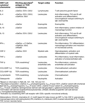
iNKT cells respond very rapidly, within the first several hours of antigen recognition, as do other cells of the innate immune system (e.g. PMNs, macrophages, dendritic cells and NK cells). Upon activation by antigen, iNKT cells elaborate copious amounts of effector cytokines and chemokines (Table 2). The quality of the cytokine response depends on the type of iNKT cell antigen that elicits the response. For example, the synthetic antigen αGalCer elicits a wide variety of cytokines within 30–90 min (Table 2). However, derivatives of αGalCer that are modified for lipid chain length predominantly elicit an IL-4 cytokine response from iNKT cells. By contrast, the αGalCer variant α-C-GalCer (Fig. 1) potently, and αMGalD (Fig. 1) weakly, elicit an IFN-γ-biased response from iNKT cells. The distinct iNKT cell cytokine response elicited by different antigens is currently being harnessed to tailor downstream immune responses that optimally benefit the host (reviewed in Ref. Reference Van Kaer28).
Properties of cytokines and chemokines elicited from invariant natural killer T cells by glycolipid antigensa

aBased on information in Refs Reference Van Kaer28, Reference Miyamoto, Miyake and Yamamura107, Reference Joyce148, Reference Wilson150 and Reference Yu151.
bGlycolipid composition: αGalCer, C18:0 phytosphingosine, C26:0N-acyl; OCH, αGalCer variant with shortened lipid tail (C9:0 phytosphingosine, C24:0N-acyl); C20:2, αGalCer N-acyl variant (C18 phytosphingosine, C20:2N-acyl); αGalACer, Sphingomonas-derived cell-wall glycolipid α-galacturonosylceramide; iGb3, mammalian-cell-derived isoglobotrihexosylceramide; αMGalD, Borellia burgdorferi cell-wall-derived α-monogalactosyldiacylglycerol.
cActivation by crosslinking iNKT cell receptor with CD3ɛ-specific monoclonal antibody.
Abbreviations: CCL3/4, chemokine (C–C motif) ligand 3/4 (also known as MIP-1α/1β); CSF-2, granulocyte–macrophage colony-stimulating factor); IFN, interferon; IL, interleukin; iNKT, invariant natural killer T; Th, T helper; TNF, tumour necrosis factor.
Human CD4+ and DN iNKT cells show functional dichotomy: the CD4+ iNKT cells secrete IL-4 upon antigen stimulation (Refs Reference Lee135, Reference Gumperz136), whereas DN iNKT cells secrete IFN-γ and TNF-α (Refs Reference Lee135, Reference Gumperz136). Both CD4+ and DN subsets upregulate perforin in the presence of inflammatory signals (Ref. Reference Gumperz136). However, DN iNKT cells also upregulate NKG2D expression (Ref. Reference Gumperz136), which, together with perforin, has the potential to bestow upon iNKT cells cytolytic activity against infected and cancer cells.
Functional differences in mouse iNKT cells are also evident. CD4+ iNKT cells have a suppressive role in the development of diabetes in nonobese diabetic mice. The DN iNKT cells in the liver have superior anticancer activities as compared with the liver-derived CD4+ subset and both the thymic and splenic CD4+ and DN subsets (Ref. Reference Crowe137). However, unalike human iNKT cell subsets, mouse iNKT cells that differ in CD4 expression do not show clear differences in cytokine responses (Ref. Reference Crowe137); therefore, why the two mouse iNKT cell subsets show functional dichotomy remains unclear.
Modulating the innate immune response
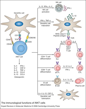
A key feature of iNKT cell activation by αGalCer is crosstalk with other cells of the immune system (Fig. 7). Dendritic cells, which are critical for the activation of naive conventional T cells, are essential for glycolipid antigen presentation and iNKT cell activation as well (Refs Reference Bezbradica121, Reference Fujii123, Reference Fujii124). Dendritic cells are the immediate target of this activation – that is, crosstalk between iNKT cells and dendritic cells results in the stimulation and maturation of dendritic cells through activated iNKT-cell-derived IFN-γ and interaction of CD154 (the CD40 ligand) on iNKT cells with CD40 on dendritic cells (Refs Reference Fujii124, Reference Vincent125). This elicits an IL-12 response from dendritic cells and the activation of NK cells to secrete copious amounts of IFN-γ. During this process, NK cells also attain cytolytic functions against infected or cancer cells. In fact, much of the IFN-γ detected in plasma upon activation of iNKT cells by αGalCer results from this indirect stimulation of NK cells (Ref. Reference Carnaud126). By contrast, a combination of IL-4, IL-13 and granulocyte–macrophage colony-stimulating factor (CSF-2) can induce tolerogenic dendritic cells, and hence suppress unwanted immune response to self-antigens as occurs in downregulating certain autoimmune diseases such as type I diabetes (Ref. Reference Naumov127). Moreover, IFN-γ and tumour necrosis factor (TNF)-α secreted by activated iNKT cells stimulate macrophages (Ref. Reference Nakagawa128), which attain microbicidal activity and promote delayed-type hypersensitivity. This source of TNF-α is also known to recruit dendritic cells that have captured antigens from the skin to the local draining lymph node where they subserve conventional T cell activation (Ref. Reference Fujii129). Osteopontin secreted by activated iNKT cells can activate PMNs (Ref. Reference Diao130). In this manner, αGalCer-activated iNKT cells can modulate the activities of a variety of innate immune system cells (Fig. 7).
The immunological functions of iNKT cells. (Legend; see previous page for figure.) The interactions between the invariant natural killer T (iNKT) cell receptor and its cognate antigen as well as interactions between costimulatory molecules CD28 and CD40 and their cognate ligands CD80/86 (B7.1/7.2) and CD40L, respectively, activate iNKT cells. Activated iNKT cells participate in crosstalk between members of the innate and the adaptive immune system by deploying cytokine/chemokine messengers. Upon activation in vivo, iNKT cells rapidly secrete a variety of cytokines/chemokines (see Table 2). These cytokines/chemokines influence the polarisation of CD4+ T cells toward T helper (Th)1 or Th2 cells as well as the differentiation of precursor CD8+ T cells (pCTLs) to effector cytotoxic T lymphocytes (CTLs), and B cells to antibody-secreting plasma cells. Some of these cytokines/chemokines facilitate the recruitment, activation and differentiation of macrophages and dendritic cells, which result in the production of interleukin (IL)-12 and possibly other factors. IL-12, in turn, stimulates NK cells to secrete interferon (IFN)-γ. IL-4, IL-13 and granuloyte–macrophage colony-stimulating factor (CSF-2) induce tolerogenic dendritic cells, which play an important role in suppressing pathogenic immune responses. Thus activated iNKT cells have the potential to potentiate as well as temper the immune response.

Modulating the adaptive immune response
iNKT cell activation also modulates adaptive immune responses mediated by lymphocytes (Fig. 7). For example, IFN-γ secreted by αGalCer-activated iNKT cells facilitates crosstalk with CTLs, which acquire the ability to secrete IFN-γ and to mediate cytotoxicity (Ref. Reference Fujii129). Such crosstalk rapidly elicits a specific CTL response and results in the eradication of cancerous growth (Ref. Reference Hermans131). Moreover, IL-4 elicited from iNKT cells activates B cells, which, within a week or so, secrete antigen-specific IgEs (Ref. Reference Singh132), a hallmark of Th2 immunity. Thus, αGalCer has adjuvant-like activity, which can be exploited in vaccine concoctions.
Medical implications of iNKT cell function
The ability to steer antigen-specific immune responses down the Th2 pathway has been exploited to prevent Th1-mediated autoimmune diseases in mouse models. Thus, activated iNKT cells protect mouse models from several autoimmune diseases, including type 1 diabetes, experimental allergic encephalomyelitis, systemic lupus erythematosus, collagen-induced arthritis, inflammatory colitis and Graves thyroiditis (Ref. Reference Van Kaer28). Although less clearly defined, several mechanisms might be responsible for the immunoregulatory protective role of iNKT cells in autoimmune diseases. Its ability to rapidly secrete IL-4 and IL-13 in a primary response to αGalCer and sustain IL-4 production upon chronic activation with this glycolipid in vivo suggests Th2 deviation of an otherwise Th1 response as a possible mechanism. This, in conjunction with IL-10, also secreted by activated iNKT cells, could potentially suppress autoreactive Th1 and B cell responses. In nonobese diabetic mice, a model for human type 1 diabetes, IL-4, IL-13 and CSF-2 released by αGalCer-activated iNKT cells induce tolerogenic dendritic cells, which in turn promote a nondestructive Th2 response while they suppress pathogenic Th1 immunity. Additional mechanisms, which include the activation and/or recruitment of regulatory T (Treg) cells such as IL-10-producing Treg-1, TGF-β-secreting Th3 or CD4+CD25+ Treg cells, might also be operative. Thus, αGalCer is a powerful pharmacological agent that holds promise as a therapeutic against autoimmune diseases (Ref. Reference Van Kaer28). Consequently, αGalCer analogues that elicit beneficial effects without the harmful ones are currently being sought (e.g. OCH and C20:2; Fig. 1) (Refs Reference Forestier133, Reference Ndonye134).
How iNKT cells function: a unified hypothesis
Dendritic cells have evolved at least two mechanisms to alert iNKT cells of in vivo bacterial infections (see section ‘CD1d-restricted antigens and distinct modes of iNKT cell activation’). One mechanism involves the direct presentation of α-anomeric glycolipids derived from bacteria that synthesise such products (e.g. S. capsulata-derived αGalACer) to iNKT cells (Refs Reference Kinjo30, Reference Mattner31, Reference Sriram32). Based on our current understanding of αGalCer presentation (Ref. Reference Bezbradica121), the direct presentation of such bacterial glycolipids and the ensuing robust iNKT cell response might critically depend on dendritic cells. Sphingomonas and Ehrlichia are extracellular pathogens. Whether dendritic cells internalise the whole bacterium, their shed cell walls or free glycolipids for presentation is currently unknown. Although αGalCer can directly bind cell-surface CD1d, the internalisation of αGalCer is essential for efficient activation of iNKT cells. We predict that the efficient presentation of bacterial α-anomeric glycolipids also follows the same rules (Refs Reference Sriram32, Reference Long138). If free bacterial α-anomeric glycolipids are present, B cells might be able to present them, as it does αGalCer, and elicit an IL-4-biased response from iNKT cells (Ref. Reference Bezbradica121). Whether such a response is sufficient to clear the infections is not known.
The second mechanism by which dendritic cells activate iNKT cells is exemplified by the iNKT cell response to Salmonella and other bacteria such as S. aureus that involves the activation of the APC by pathogen-derived LPS through TLRs (Refs Reference Mattner31, Reference Brigl44). LPS-mediated dendritic cell activation results in IL-12 secretion (Ref. Reference Brigl44), which in conjunction with a self-glycolipid antigen presented by CD1d activates iNKT cells (Ref. Reference Mattner31). Dendritic cell activation through TLRs perhaps induces/increases the self-glycolipid(s) antigen expression (Refs Reference Paget47, Reference Salio139). B cells do not elicit an autoreactive response from iNKT cells (Ref. Reference Park, Roark and Bendelac140) perhaps because the former are not known to express IL-12 in response to LPS-induced activation that is required for self-antigen-induced iNKT cell activation. These data suggest that indirect iNKT cell activation requires the recognition of a self-glycolipid whose presentation depends on dendritic cells. Thus, dendritic cells play a critical role in the activation of iNKT cells in response to infections.
Cellular lipid content is stringently regulated. Activation of dendritic cells with bacteria (S. aureus, E. coli, Bacillus subtilis, or M. bovis) or by their cell-wall products (LPS, lipoteicoic acid, the lipopeptide Pam3Cys or nucleic acid) induces cellular glycolipid syntheses, some of which are presented to self-reactive, group I CD1-restricted T cells (Ref. Reference De Libero141). Membrane biogenesis is part of the ER-stress-induced unfolded protein response and hence requires de novo lipid biosynthesis (Refs Reference Nunnari and Walter142, Reference Cox, Chapman and Walter143, Reference Chapman, Sidrauski and Walter144, Reference Brewer and Hendershot145). Therefore, bacterial and viral infections, either by dendritic cell activation through TLR ligation or by ER stress induced by overexpression of virus-derived membrane glycoproteins, have the potential to alter cellular lipid content, both in variety and in concentration. Such changes in cellular self-lipid content can alter the quality and quantity of the ligands presented by CD1d. Because Vα14i TCR exhibits co-operative ligand binding (Ref. Reference Stanic94), the iNKT cell receptor has the potential to recognise subtle qualitative and quantitative changes reflected in CD1d-associated lipid content. We hypothesise that sensitive antigen recognition at very early stages of infection, which is initiated by very few infectious particles, is key to iNKT cell function in vivo.
Thus, in our model, CD1d functions as a sensor, to detect alterations in cellular lipid content by virtue of its inherent affinity for such ligands. The presentation of foreign α-anomeric glicolipids by dendritic cells or, alternatively, the presentation of an induced self- or a neoself-lipid by dendritic cells upon stimulation by pathogen-derived TLR ligands, activates iNKT cells. In vivo activation of iNKT cells results in the rapid release of pro- and anti-inflammatory cytokines and chemokines, which have the potential to ‘jump start’ the immune system (Fig. 7). In this regard, iNKT cells, akin to an enzyme, perhaps function by lowering the ‘energy of activation’ required for the initiation of an immune response. In doing so, the immune system is alerted by the entry of only a few intruders as occurs in natural infections.
Concluding remarks
The nature of CD1d-restricted antigens coupled with the innate-like pattern-recognition logic and the unique effector functions of iNKT cells suggest an innate immunoregulatory role for this T cell subset. Their ability to respond fast and our ability to steer iNKT cell cytokine response to altered lipid antigens make them an important target for vaccine design and immunotherapies against autoimmune diseases.
Acknowledgements and funding
We thank Dr D.B. Lacy (Vanderbilt University) for instructions in analyses of X-ray crystallographic co-ordinates using PyMol and Chimera programmes. We also thank the three anonymous reviewers of this manuscript, Dr L. Van Kaer (Vanderbilt University), D. Zhou (MD Anderson), and past and current members of the Joyce laboratory for critical reading of the manuscript and helpful discussions. Supported by grants from the NIH (AI042284, AI061721 and AI040079) to S.J.