Approximately 450 cardiac transplants are performed in children with end-stage heart disease secondary to cardiomyopathies or congenital heart disease each year.Reference Kirk, Dipchand and Edwards 1 Although the short-term survival has improved significantly over the last several decades, the medium- and long-term survival remains suboptimal, with 40% of patients not surviving to 10 years post transplant.Reference Kirk, Dipchand and Edwards 1 , Reference Morales, Dreyer and Denfield 2 Many risk factors presenting before transplantation have been identified to affect 1- and 5-year survival, including underlying congenital heart disease, increased pre-transplant support with mechanical ventilation or dialysis, and elevated panel reactive antibody levels.Reference Kirk, Dipchand and Edwards 1 , Reference Rossano, Morales and Zafar 3 Following cardiac transplantation, the primary determinants of graft survival include rejection episodes and allograft vasculopathy, with infection also playing a role predominantly in the first year when immunosuppression is at its highest levels.Reference Kirk, Dipchand and Edwards 1 , Reference Morales, Dreyer and Denfield 2 , Reference Groetzner, Reichart and Roemer 4 , Reference Ross, Kouretas and Gamberg 5
On assessing for infectious aetiologies associated with mortality in children who have undergone cardiac transplant, the presence of viral genome in the cardiac allograft was determined to be associated with early graft loss.Reference Shirali, Ni and Chinnock 6 , Reference Moulik, Breinholt and Dreyer 7 The mechanism for graft loss in these patients is unknown, but appears to be associated with the early development of transplant coronary artery disease.Reference Shirali, Ni and Chinnock 6 – Reference Breinholt, Moulik and Dreyer 8 It has been speculated that prolonged viral genome exposure promotes an ongoing immune response with expression of inflammatory mediators that may provoke the development of accelerated coronary arteriopathy.Reference Shirali, Ni and Chinnock 6
Thus, we sought to test the hypothesis that children who have undergone cardiac transplant and in whom a virus was identified in the myocardium in the first year post transplant would have increased inflammation and worse haemodynamics compared with patients who were virus free.
Materials and methods
This study was approved by the Baylor College of Medicine Institutional Review Board.
Study population
All patients younger than 18 years of age undergoing orthotopic cardiac transplantation between January 2004 and May 2008 at a single centre (Texas Children's Hospital) were identified and included in a retrospective analysis.
Data collection
Medical records were reviewed to identify patient demographics, clinical course, endomyocardial biopsy pathology, and viral polymerase chain reaction results, as well as cardiac catheterisation haemodynamic data, at 1 year post transplant. Patients underwent serial cardiac catheterisations as per institutional protocol to evaluate for rejection and the presence of virus by polymerase chain reaction.Reference Kim, Dreyer and O'Brian 9 The standard immunosuppressive regimen used included triple-drug therapy with cyclosporine or tacrolimus, prednisone, and mycophenolate mofetil. No induction therapy was provided. Before transplant surgery, patients were given mycophenolate mofetil, with steroid administration in the operative room. Following surgery, the ongoing steroid administration and mycophenolate mofetil was continued, and the patients were given an addition of calcineurin inhibitor once they were tolerant towards enteral nutrition, predominantly tacrolimus. Data were also included if rejection was suspected clinically at other times and the patient underwent catheterisation.
Catheterisation data recorded at the time of biopsy 1 year post transplant included cardiac index – determined by both thermodilution and Fick calculation – pulmonary capillary wedge pressure, pulmonary vascular resistance index, right ventricular end-diastolic pressure, and coronary angiography. Approximately six endomyocardial biopsy samples were obtained from the right ventricle using the standard percutaneous transvenous approach. Each biopsy sample was screened for a panel of viral genomes including adenovirus, cytomegalovirus, Epstein–Barr virus, enterovirus, and parvovirus B19. The method of viral polymerase chain reaction analysis has been detailed in previous studies and was performed by technicians at the John Welsh Cardiovascular Diagnostic Laboratory who had no knowledge of the clinical or serologic data.Reference Shirali, Ni and Chinnock 6 , Reference Breinholt, Moulik and Dreyer 8 , Reference Martin, Webber and Fricker 10 , Reference Griffin, Kearney and Ni 11 The degree of inflammation and rejection was graded on the Texas Heart Institute-McAllister scaleReference McAllister 12 – a 10-point scale with higher scores indicating more inflammation – by a cardiac pathologist blinded to the polymerase chain reaction results. Examples of histologic grading according to both Texas Heart Institute numerical scale and the International Society of Heart and Lung Transplant rejection scoring are outlined in Figure 1 for comparison assessment. Treatment for acute cellular rejection was considered if the grade was >4 – equivalent of an International Society of Heart and Lung Transplant rejection score of 2R.
Histologic grading of inflammation and rejection according to Texas Heart Institute assessment (0–10 numeric scale) and International Society of Heart and Lung Transplant rejection score. ( a ) Focal mild rejection with focal perivascular lymphocytes – Texas Heart Institute scale 1 or International Society of Heart and Lung Transplant score 1R. ( b ) Focal myocyte degeneration – Texas Heart Institute scale 4 or International Society of Heart and Lung Transplant score 1R. ( c ) Multifocal moderate rejection involving <50% of biopsy sample – Texas Heart Institute scale 4 or International Society of Heart and Lung Transplant score 2R.

Patients with virus identified at any time during the first year post transplant were compared at 1 year post transplant with virus-free patients. This time point was chosen because all transplant patients routinely underwent annual evaluation with cardiac catheterisation and their immunosuppression profile had generally been reduced to similar levels as per institutional guidelines. Owing to death or moving out of area, five patients did not have an annual catheterisation. Data from these patients were assessed using their last available cardiac catheterisation.
Outcome measures
The primary outcome measures were the haemodynamic data obtained at cardiac catheterisation at 1 year post transplantation and the degree of inflammation present on the biopsy specimen from that catheterisation. Patients who tested positive for viral endomyocardial infection by polymerase chain reaction at any time during the first year post transplant were compared with patients who remained virus free on endomyocardial biopsy. The incidence of rejection between the two groups was also assessed.
Statistical analysis
We examined the data using standard descriptive statistics, including median and interquartile range. Continuous variables were compared using the Mann–Whitney U test, as the data were not normally distributed. Fisher's exact test was used to assess categorical data. Statistical significance was achieved for p < 0.05. The SPSS 17 software (SPSS Inc, Chicago, Illinois, United States of America) was used for statistical analysis.
Results
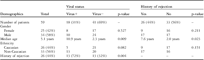
During the study period, there were 59 patients who underwent cardiac transplant. The baseline characteristics for the patient population are outlined in Table 1. This included 34 male patients (58%). The median age at transplant was 5.1 years (interquartile range: 1.2–12.2 years). The majority of the patients were Caucasian (44%), followed by African American (31%) and Hispanic (22%) ethnicities, with a small Asian (3%) population. The indication for cardiac transplant predominantly included congenital heart disease (44%) and dilated cardiomyopathy (34%), with the remaining cases due to other cardiomyopathies (19%) or transplant coronary artery disease (3%). In all, 26 patients (44%) experienced rejection episodes during their first year post transplant, with acute cellular rejection being the most common in 20 patients. A total of 40 episodes of acute cellular rejection – Texas Heart Institute score >4; equivalent to International Society of Heart and Lung Transplant score of 2R and greater – were treated with increased immunosuppression during the study time period, with 13 patients requiring treatment for more than one episode. Within the first year post transplant, three patients died from rejection, one of whom had a history of multiple endomyocardial biopsy specimens with parvovirus B19. Of these patients, two died from antibody-mediated rejection at 9 and 11 months post transplant, with the latter patient having a history of virus-positive endomyocardial biopsy specimens. Cellular rejection was present in one patient who died 4 months after transplant.
Baseline characteristics of the study population.

A total of 469 endomyocardial biopsy specimens from the 59 patients were analysed for the presence of viral genome by polymerase chain reaction (range: 2–12 per patient; median: 9 samples per patient). Viral genomes were isolated in 18 patients (31%) from 70 specimens (15%). Parvovirus B19 was the most common viral genome detected (89%), and was found in 68 specimens from 16 patients. In two additional patients, two specimens were positive for the Epstein–Barr virus. No specimens were positive for adenovirus, cytomegalovirus, or enterovirus in this patient population. The majority of the patients with parvovirus B19-positive endomyocardial specimens had evidence of persistent infection (n = 12, 75%), with up to eight biopsy samples showing ongoing evidence of parvovirus B19 genome.
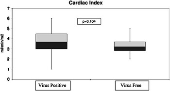
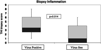
When analysing whether certain demographic characteristics, such as age at the time of transplant, gender, or ethnicity, were associated with a history of virus-positive endomyocardial biopsy specimen, only age was found to be significantly associated, with older individuals more likely to have a positive specimen (median: 10.9 versus 2.3 years, p = 0.009). Patients with virus-positive specimens were also more likely to have experienced an episode of rejection (1 episode of rejection versus 0 episodes, p = 0.004). Catheterisation haemodynamics between the two groups was comparable with no statistical difference appreciated (Fig 2). The virus-positive group had a median cardiac index of 3.7 ml/min/m2 (interquartile range: 3.0–4.5), median pulmonary capillary wedge pressure of 10 mmHg (interquartile range: 7–11), and median pulmonary vascular resistance index of 1.7 U m2 (interquartile range: 1.1–2.4) compared with the virus-free group, with a median cardiac index of 3.2 ml/min/m2 (interquartile range: 2.8–3.7), median pulmonary capillary wedge pressure of 8 mmHg (interquartile range: 7–11), and median pulmonary vascular resistance index of 1.7 U m2 (interquartile range: 1.2–2.1). The virus-positive group had an increased degree of inflammation on endomyocardial biopsy at 1 year post transplant, with a median score of 4 (interquartile range: 1–4), compared with the virus-free group, with a median score of 1 (interquartile range 0 to 4; p = 0.026) (Fig 3).
Cardiac index haemodynamic information in the virus-positive and virus-free endomyocardial biopsy population at 1 year post transplant. The vertical lines represent the full range of values with the 50th quartile demonstrated by the middle black horizontal line. 25th–50th interquartile range is shown in purple, and 50th–75th interquartile range is shown in yellow.

Comparison of inflammation on biopsy samples obtained 1 year post transplant in patients with a history of virus-positive endomyocardial biopsy versus virus-free samples. Quantitative inflammation score is based on the Texas Heart Institute biopsy score.

Statistical analysis of demographic characteristics and history of rejection only showed significance with age at transplant, with older patients having a higher likelihood of rejection (median ages: 7.6 versus 2.0 years; p = 0.023). In comparing the haemodynamic profiles at 1 year between patients with a history of rejection, only the cardiac index determined using the Fick equation showed significance, with lower values for those with a prior history of rejection (median: 3.9 versus 4.5 ml/min/m2; p = 0.016); however, both values remained within normal limits. The cardiac index determined by thermodilution showed a similar trend; however, it was not significant (p = 0.07). In addition, the biopsy score at the annual catheterisation was significantly higher in patients with a previous history of rejection on prior biopsy specimens (median: 4 versus 1; p = 0.001).
In all, four patients (7%) had evidence of transplant coronary artery disease on the basis of angiography on their catheterisation 1 year post transplant, with one additional patient having evidence of mild disease on an earlier catheterisation performed for the assessment of acute cellular rejection that the individual subsequently died from within 4 months of transplantation. Of the four patients, three had a previous history of rejection, and two of these patients had a history of parvovirus B19-positive endomyocardial biopsy specimen.
Discussion
Although much has been elucidated about the role of viral myocardial infection in patients with dilated cardiomyopathy and myocarditis, less information is known about the role that these viruses play in immunocompromised children following cardiac transplant.Reference Moulik, Breinholt and Dreyer 7 , Reference Breinholt, Moulik and Dreyer 8 , Reference Friman, Wesslen, Fohlman, Karjalainen and Rolf 13 – Reference Eid, Brown, Patel and Razonable 18 Viral genome has been detected in the cardiac allograft after transplantation and has been associated with an increased risk for rejection and a greater than sixfold increase in graft loss.Reference Shirali, Ni and Chinnock 6 , Reference Schowengerdt, Ni and Denfield 19 A more recent study does not find the same correlation with cellular rejection and the presence of PVB19 genome in the myocardium; however, early development of advanced coronary artery disease was seen in those children with persistent infection.Reference Breinholt, Moulik and Dreyer 8 These studies have led to speculation regarding the true pathogenesis of these viral genomes leading to early graft loss. Their presence may cause increased inflammation and a heightened immune response, which subsequently leads to increased episodes of rejection or endothelial damage and the development of premature transplant coronary artery disease. Our study shows a significant association between the presence of viral genome in the endomyocardium and evidence of rejection, as well as increased levels of inflammation at 1 year post transplant. Using the data in this study, we were not able to clearly distinguish the effect of virus infection from episodes of acute rejection in leading to this increased level of inflammation. The degree of inflammation at 1 year did not affect medical management, as both scores of 1 and 4 meet International Society of Heart and Lung Transplant rejection score IR criteria and no change in management was necessary. However, we speculate that if this subtle degree of inflammation persisted, it may account for the association of worse long-term graft survival seen in other studies with presence of viral genome due to chronic low levels of rejection.Reference Shirali, Ni and Chinnock 6 – Reference Breinholt, Moulik and Dreyer 8
The question regarding transplant coronary artery disease is difficult to fully assess in such a limited time period of only 1 year, especially because the majority of patients do not develop angiographic evidence of vasculopathy until later. Our study does highlight an increased degree of inflammation in patients with viral-positive endomyocardial biopsy specimens, which over the long term could promote early graft loss and development of coronary vasculopathy. However, longer-term follow-up is needed to verify whether this increased inflammation persists over time. No significant haemodynamic changes were seen with this elevated level of inflammation, although these variables may change over time with longer persistence of inflammation. Additional data and longer-term follow-up are needed to clarify this.
The viruses present in this study population are almost exclusively parvovirus B19, which mirrors the trend seen in many other studies. Viral pathogens have changed over time, with parvovirus B19 becoming the prevalent organism found in myocardial tissue samples in the most recent decade.Reference Breinholt, Moulik and Dreyer 8 , Reference Pankuweit, Moll, Baandrup, Portig, Hufnagel and Maisch 20 – Reference Donoso, Nitsche and Meyer 22 Parvovirus infection is ubiquitous in humans with IgG antibodies detectable in ∼50% of the population by age 15 years.Reference Young and Brown 23 This finding may help explain why older individuals in our study were more likely to have virus-positive endomyocardial biopsy specimens; we speculate that they would come into contact with more people in their peer group with history of exposure to parvovirus compared with the younger age set. In addition, persistence of parvovirus B19 is associated with long-term diastolic dysfunction,Reference Kuhl, Pauschinger and Seeberg 15 , Reference Tschope, Bock and Kasner 21 which may be related to its direct effect on the coronary vasculature and subsequent endothelial dysfunction.Reference Kuhl, Pauschinger and Bock 24 , Reference Bultmann, Klingel and Sotlar 25 This could have long-term consequences for transplant patients who already have a limited immune response to clear the virus from their myocardium.
Limitations
This study is a retrospective, single-centre review and has limitations intrinsic to such an analysis. The series consisted of a small patient group and therefore may not be powered to detect important differences in biopsy inflammation or haemodynamic parameters. Owing to the fact that this was a 1-year review, limited assertions can be made regarding long-term outcomes. Furthermore, without multiple viral types represented, it is difficult to determine whether these results reflect how all viral types affect patients following cardiac transplantation or rather a unique effect seen among those with parvovirus B19.
Conclusions
Children who have undergone cardiac transplant and who have develop viral endomyocardial infections have evidence of increased allograft inflammation and rejection compared with virus-free patients during the first year post transplant. However, the haemodynamic profile is similar between the groups. The role of ongoing subclinical inflammation in early and long-term graft loss requires further study.
Acknowledgements
The authors acknowledge the contribution of E. O'Brian Smith, PhD, for his assistance with the statistical analysis.
Financial support
None.
Conflict of interest
The authors have no conflicts of interest or additional funding resources to disclose.
Author contributions
Kimberly M. Molina, MD, concept/design, data collection, data analysis, drafting the article; Susan W. Denfield, MD, critical revision of the article; Yuxin Fan, MD, PhD, data collection, critical revision of the article; Mousumi Moulik, MD, data collection; Jeffrey A. Towbin, MD, critical revision of the article; William J. Dreyer, MD, critical revision of the article; Joseph W. Rossano, MD, concept/design, data analysis, critical revision of the article.
Ethical Standards
The authors assert that all procedures contributing to this work comply with the ethical standards of the relevant national guidelines on human experimentation and with the Helsinki Declaration of 1975, as revised in 2008, and has been approved by the Baylor College of Medicine institutional review board.