




No CrossRef data available.
Introduction to immune cell signalling
Published online by Cambridge University Press: 11 November 2005
Abstract
The dynamic interaction of cells of the immune system with other cells, antigens and secreted factors determines the nature of an immune response. The response of individual cells is governed by the sequence of intracellular signalling events triggered following the association of cell surface molecules during cell-cell contact or the detection of soluble molecules of host or pathogen origin. In this review we will first outline the general principles of intracellular signal transduction. We will then describe the signalling pathways triggered following the recognition of antigen, as well as the detection of cytokines, and discuss how the signalling pathways activated regulate the effector response.
Information
- Type
- Research Article
- Information
- Copyright
- © 2005 Cambridge University Press
BASIC PRINCIPLES OF CELL SIGNALLING
Cells sense, respond to and integrate a multiplicity of signals from their environment. In the context of the immune system, these include signals resulting from interactions with neighbouring cells (cell-cell contact), and the detection of soluble factors such as cytokines, which may originate at distant sites and be transported via the blood, as well as the recognition of pathogens and their products. Following ligation of cell surface receptors, intracellular signal transduction cascades are initiated, resulting in the activation of transcription factors and other proteins that regulate processes such as gene induction, phagocytosis, apoptosis, proliferation and secretion (Harnett, 1999a,b).
Signal transduction cassettes comprise specific cell-surface membrane receptors, effector signalling elements and regulatory proteins. These signalling cassettes serve to detect, amplify and integrate diverse external signals to generate the appropriate cellular response. In this review, we first discuss how cell-surface receptors sense and transduce signals by transmembrane coupling to effector enzyme systems, generating low-molecular-weight molecules termed second messengers and activating a range of key protein kinases with distinct substrates that control gene induction and other cellular responses (Harnett, 1999a,b). We will then describe the signalling cascades underlying some of the key responses of cells of the immune system to environmental stimuli of host as well as pathogen origin.
Phosphorylation is an important mechanism of control of protein recruitment and activation; kinases catalyse the transfer of a phosphate group from ATP onto specific amino acids, a process that can be reversed by the action of phosphatases. Individual kinases have distinct substrate specificities e.g. tyrosine kinases, dual specificity serine/threonine or tyrosine/threonine kinases (Bromann, Korkaya & Courtneidge, 2004; Cannons & Schwartzberg, 2004; Gambacorti-Passerini, 2004; Piiper & Zeuzem, 2004). Lipid kinases, such as phosphoinositide-3-kinase (PI3K), catalyse the phosphorylation of lipids (Cantrell, 2001, 2002, 2003b).
Recruitment of kinases and other enzymes, such as phospholipases, to the activated receptor following ligation triggers a cascade of enzymatic activation and the generation of second messengers such as cyclic AMP (cAMP) (Houslay, 1998; Houslay & Adams, 2003; Houslay & Baillie, 2003), inositol trisphosphate (IP3) (Irvine, 2003a,b; 2004), diacylglycerol (DAG) (Powner & Wakelam, 2002; McDermott, Wakelam & Morris, 2004) and calcium ions (Ca2+) (Berridge, Bootman & Roderick, 2003; Berridge, 2004), which results in propagation and amplification of the signal. Hydrolysis of phosphatidylcholine, which comprises about 40% of the total cellular phospholipid, by various phospholipases results in the generation of a range of second messengers, including arachidonic acid (phospholipase A2, PLA2), DAG (phospholipase C, PLC) and phosphatidic acid (phospholipase D, PLD), depending on the site of action of the enzyme (Wakelam & Harnett, 1998; Powner & Wakelam, 2002; McDermott et al. 2004). Arachidonic acid is a key lipid second messenger involved in the regulation of signalling enzymes, such as PLC-γ and –δ, and the α, β and γ isoforms of the protein kinase C (PKC) family of serine/threonine kinases. DAG is a cofactor required for the activation of PKC isoforms; it can also be metabolised to generate arachidonic acid by the action of DAG lipase. Furthermore, phosphatidic acid can be interconverted to DAG by the action of phosphatidic acid phosphohydrolase (Wakelam & Harnett, 1998; Powner & Wakelam, 2002; McDermott et al. 2004).
One of the key outcomes of intracellular signalling cascades is the activation of transcription factors, such as NF-κB, NFAT, Fos, Jun and Oct. Transcription requires the binding of RNA polymerase to the promoter region situated 5′ of the transcription start site. Transcription factors, which bind to specific regulatory DNA sequences, co-operate to permit or deny RNA polymerase access to the promoter, and thereby regulate gene expression (Clevenger, 2004; Coffer & Burgering, 2004; Eggert et al. 2004; Smith & Sigvardsson, 2004).
ANTIGEN RECEPTOR SIGNALLING
Recognition of specific antigens by B cells is achieved by surface-bound immunoglobulin (sIg). However, sIg is unable to transduce intracellular signals due to its short cytoplasmic tail, and hence recruits two Ig-α/β heterodimers, which possess Intracellular Tyrosine-based Activatory Motifs (ITAMs; D/E-X7-D/E-XX-YXXL-X7-YXXL/I) in their cytoplasmic tails. The earliest detectable B cell receptor (BCR) signal is tyrosine phosphorylation of proteins including the Ig-α/β ITAMs; mutations in either of the conserved tyrosines disrupts signalling. ITAM phosphorylation results in the initiation of a signalling scaffold around the active BCR (see Fig. 1). The tyrosine kinases Lyn and Syk bind to the phosphorylated ITAMs via their SH2 domains (Dal Porto et al. 2004; Gauld & Cambier, 2004; Harnett, Katz & Ford, 2005). Further recruitment of adaptors/signalling proteins occurs via SH2, SH3 (binds proline-rich sequences) and PH (binds phosphoinositides) domain interactions. The tyrosine phosphatase SHP-1 also associates with the BCR to prevent aberrant activation (Kurosaki, 2002; Cannons & Schwartzberg, 2004; Simeoni et al. 2004).

Fig. 1. B cell receptor (BCR) signalling. Surface-bound immunoglobulin (sIg) alone cannot transduce intracellular signals following recognition of specific antigen. Instead signalling is initiated by tyrosine phosphorylation of Ig-α/β dimers, which associate with sIg to form the BCR complex. A signalling scaffold forms around the activated receptor complex with recruitment of adaptors and other signalling proteins, which trigger the activation of various cascades including the PI3K, PLC-γ and MAP kinase pathways, ultimately resulting in the promotion of gene induction, B cell survival and proliferation.
The phosphoinositide-3-kinase (PI3K) pathway is a key pathway promoting survival, gene induction and cell cycle progression following B cell activation (Fruman, 2004a,b). PI3K catalyses the conversion of phosphatidylinositol-4,5-bisphosphate (PIP2) to phosphatidylinositol-3,4,5-trisphosphate (PIP3). This enables the recruitment and activation of the serine/threonine kinase protein kinase B (PKB), also known as Akt, which prevents apoptosis and promotes B cell survival, as well as the serine/threonine kinase p70 S6 kinase, which is required for the entry of cells into S phase of the cell cycle. Akt phosphorylates the pro-apoptotic protein Bad, which is then sequestered by association with the 14-3-3 protein, thereby preventing the release of cytochrome c and activation of the caspase cascade. Thus PIP3 promotes B cell survival and proliferation (Cantrell, 2001, 2002, 2003b; Fruman, 2004a).
PIP3 can also regulate gene induction via the activation of atypical isoforms of PKC (ε and ζ), which activate transcription factors such as AP-1, NFAT and NF-κB (Hao, Kurosaki & August, 2003; Guo, Su & Rawlings, 2004). Furthermore, PIP3 can recruit PLC-γ, which hydrolyses PIP2 to generate IP3 and DAG, resulting in the release of Ca2+ ions from intracellular stores and the activation of classical and novel isoforms of PKC. Ca2+ ion binding is required for the serine/threonine kinase activity of the Ca2+/calmodulin (CaM)-dependent protein kinase (CaMKII) and enables the CaM protein phosphatase calcineurin to dephosphorylate and activate NFAT. DAG is necessary for the activation of various PKC isoforms, which may also require Ca2+ binding, resulting in the activation of transcription factors such as AP-1, NFAT, NF-κB and Oct (Hao et al. 2003; Guo et al. 2004).
Another key event following ligation of the BCR is the activation of the mitogen-activated protein kinase (MAP kinase) pathways. There are three major subfamilies of MAP kinases – the extracellular regulated kinases (Erk1/2), the p38 MAP kinases and the c-Jun kinases/stress-activated protein kinases (JNK/SAPK) (Adams et al. 2004; Roux & Blenis, 2004; Rubinfeld & Seger, 2004; Harnett et al. 2005). Recruitment of a GTPase to the activated receptor complex is required for the activation of a kinase cascade, ultimately leading to activation of MAP kinases, which target transcription factors to regulate gene induction. For example, BCR coupling to the Erk cascade is achieved via the GTPase Ras (see Fig. 1). Inactive Ras (Ras-GDP) is anchored at the plasma membrane and is recruited by the Grb2-Sos complex. Sos, a guanine nucleotide exchange factor (GEF), promotes the exchange of GDP for GTP. Active Ras (Ras-GTP) activates Raf (a MAP kinase kinase kinase) by binding to its N-terminal regulatory domain. Raf then phosphorylates MEK-1 (a MAP kinase kinase), which in turn phosphorylates Erk1/2. The Erk MAP kinase pathway promotes the proliferation and differentiation of B cells by targeting transcription factors such as c-Myc, Sap and Elk-1 (Cantrell, 2003a,b, Dal Porto et al. 2004; Harnett et al. 2005).
CYTOKINE RECEPTOR SIGNALLING
Cytokines and their receptors can be classified according to their structure. Most cytokine receptors belong to the class I cytokine receptor family; they are multichain receptor complexes comprising separate chains for specific cytokine recognition and signal transduction. Signalling chains may be shared by several cytokines within a subfamily. For example, IL-3, IL-5 and GM-CSF, which are structurally similar, share a common β chain, while IL-2, IL-4, IL-7, IL-9 and IL-15 share a common γ chain. gp130 is the signalling subunit of the IL-6 and IL-11 receptors, and the gp130-related IL-12Rβ1 chain is utilised by IL-12 and IL-23 (Hofmann et al. 2002; O'Shea, 2004; O'Shea et al. 2004).
Most cytokines signal through PLC, PI3K, and Ras MAPK pathways, as well as JAK-STAT pathways (reviewed in Leonard & Lin, 2000; Hofmann et al. 2002; Agnello et al. 2003; O'Shea, 2004; O'Shea et al. 2004). Binding of cytokines to their receptors triggers the recruitment of receptor-associated tyrosine kinases of the Janus kinase (JAK) family (JAK1-3, Tyk2). Activated JAKs phosphorylate tyrosine residues in the β and γ signalling receptor chains, enabling the recruitment of other proteins via their SH2 domains, in particular the cytosolic STAT proteins (signal transducers and activators of transcription; STAT1-6). STATs are then phosphorylated and released from the receptor. Phosphorylated STAT proteins homodimerise or heterodimerise via pTyr-SH2 domain interactions, and rapidly translocate into the nucleus where they act as transcription factors by binding to GAS motifs (see Fig. 2).

Fig. 2. Cytokine receptor signalling via JAK-STAT pathways.Following cytokine receptor ligation, JAKs are recruited and phosphorylate tyrosine residues in the β and γ signalling receptor chains, enabling STAT recruitment and phosphorylation. pSTAT proteins form dimers, which rapidly translocate to the nucleus where they act as transcription factors by binding to GAS elements.
Different combinations of JAKs and STATs can be utilised to achieve specificity. For example, the JAK-STAT pathways play key roles in the control of polarisation of CD4+ helper T (Th) cell responses (Murphy et al. 1999, 2000; Murphy & Reiner, 2002; Agnello et al. 2003). Cytokine signals received by Th precursors determine their differentiation towards a Th1 or Th2 phenotype. Antigen recognition by the TCR in the presence of IL-12 and IFN-γ leads to the development of Th1 responses, while a Th2 bias is achieved by exposure to IL-4. Polarisation is achieved via the action of two transcription factors, T-bet and GATA-3 (Zheng & Flavell, 1997; Szabo et al. 2000; Murphy & Reiner, 2002; see Fig. 3). IL-12 and IFN-γ signal through their receptors on the surface of precursor Th cells, leading to the activation of STAT4 and STAT1 respectively, both of which induce T-bet expression. T-bet promotes IFN-γ expression by chromatin remodelling and increases IL-12Rβ2 chain expression, thereby further enhancing the IL-12 and IFN-γ signals, in part by sustaining its own expression.

Fig. 3. Signalling pathways underlying Th1/Th2 polarisation. JAK-STAT pathways underlie the polarisation of CD4+ helper T (Th) cell responses. IL-12 and IFN-γ signal via STAT4 and STAT1 respectively to induce expression of the transcription factor T-bet, which promotes Th1 polarisation via enhanced IFN-γ and IL-12Rβ2 expression. IL-4 induces activation of STAT6, leading to production of GATA-3, which promotes transcription of the Th2 cytokines IL-4, IL-5 and IL-13. Positive and negative feedback mechanisms reinforce the Th1/Th2 bias.
Conversely, recognition of IL-4 by its receptor triggers the activation and nuclear translocation of STAT6, which rapidly induces GATA-3 expression (Zheng & Flavell, 1997; Zhou & Ouyang, 2003). GATA-3 regulates the expression of Th2 cytokines by co-ordinate regulation of the Th2 locus, which contains the genes encoding IL-4, IL-5 and IL-13, by not only inducing the expression of IL-5 and IL-13, but also by promoting chromatin remodelling to enable IL-4 transcription (Murphy et al. 1999, 2000; Lee et al. 2000; Ouyang et al. 2000; Murphy & Reiner, 2002; Agnello et al. 2003). GATA-3 thereby augments its own expression by positive feedback autoregulation. Furthermore, negative feedback mechanisms reinforce the fate decision. GATA-3 inhibits Th1 development by suppressing T-bet expression, at least in part by downregulating STAT4, and by increasing IL-4 production, thereby suppressing IL-12Rβ2 chain expression (reviewed in Ho & Glimcher, 2002). Similarly, T-bet may also inhibit GATA-3 expression and IL-4 production (Murphy et al. 1999, 2000; Murphy & Reiner, 2002; Agnello et al. 2003).
TOLL-LIKE RECEPTOR SIGNALLING
Detection of pathogen products by cells of the innate immune system is mediated by pattern recognition receptors (PRR), with or without the aid of soluble host factors such as complement. PRRs include scavenger receptors, complement receptors and the recently identified family of Toll-like receptors (TLRs). TLRs, which were originally identified by their homology to the Drosophila anti-fungal protein Toll, have been demonstrated to be crucial for the recognition of a variety of pathogens and their products, including proteins, carbohydrates and nucleotides derived from bacteria, viruses, fungi, protozoa and helminth parasites (reviewed in Takeda, Kaisho & Akira, 2003).
TLRs, which are members of the Toll/IL-1R (TIR) superfamily, comprise extracellular leucine-rich repeats (LRRs), a transmembrane region and an intracellular region containing a conserved intracellular TIR domain. TLRs signal via the recruitment of adaptor proteins, such as MyD88, TIR-containing adaptor protein/MyD88 adaptor-like (TIRAP/Mal), TIR-containing adaptor inducing IFN-β/TIR-containing adaptor molecule-1 (TRIF/TICAM-1), TRIF-related adaptor molecule (TRAM) and sterile α and HEAT-Armadillo motifs (SARM) (reviewed in McGettrick & O'Neill, 2004). Specificity is achieved by homo- or heterodimerisation of TLRs and differential adaptor recruitment. Different downstream events are triggered by the association of various adaptors with the TLRs e.g. MyD88-dependent NF-κB and MAP kinase activation, and TRIF-dependent activation of the transcription factor IRF-3.
The signalling pathways downstream of TLR4, the receptor required for the recognition of and response to bacterial lipopolysaccharide (LPS), are the most thoroughly investigated so far (reviewed in Takeda & Akira, 2004; see Fig. 4). A MyD88-dependent pathway is required for LPS-triggered induction of IL-12 and pro-inflammatory cytokines such as IL-6 and TNF-α. Following LPS recognition, IL-1 receptor-associated kinase (IRAK)-1, IRAK-4 and TNF receptor-associated factor (TRAF)-6 are recruited to the receptor complex. IRAK-1 associates with MyD88 via the interaction of their death domains, and is phosphorylated by IRAK-4. Phosphorylated IRAK-1 interacts with and phosphorylates TRAF6, and they dissociate from the receptor to form a complex with TGFβ-activated kinase (TAK)1, TAK1-binding protein (TAB)1 and TAB2. Activation of TAK1 results in phosphorylation of the IKK complex, which consists of the I-κB kinases IKKα, IKKβ and NEMO/IKKγ. Phosphorylation of I-κB by the IKK complex leads to its ubiquitination and degradation, releasing the transcription factor NF-κB to enter the nucleus and promote gene induction. TAK1 is also responsible for the activation of MAP kinase pathways, leading to gene induction via activation of transcription factors such as Elk, AP-1 and ATF-2.

Fig. 4. Toll-like receptor 4 (TLR4) signalling. Following LPS binding to its receptor complex, signalling is initiated via TLR4. Adaptor proteins, including MyD88 and TRIF, are recruited, as well as IRAK-1, IRAK-4 and TRAF6. Following phosphorylation of IRAK-1 by IRAK-4, and TRAF6 by IRAK-1, IRAK-1 and TRAF6 dissociate from the receptor and form a complex with TAK1, TAB1 and TAB2. TAK1 activates the MAP kinase and NF-κB pathways, resulting in transcription of pro-inflammatory cytokines. A MyD88-independent, TRIF-dependent pathway via TBK-1 results in phosphorylation of the transcription factor IRF-3, which induces IFN-β production.
In contrast, knockout studies indicated that MyD88 is not required for the induction of IFN-inducible genes, such as IP-10 and GARG16, following LPS stimulation (Kawai et al. 2001). These genes are induced indirectly via MyD88-independent production of IFN-β, which activates STAT1 (Toshchakov et al. 2002). The transcription factor IRF-3, which is activated by signalling via the TRIF adaptor, regulates IFN-β production. This occurs via activation of the IKK isoform TANK-binding kinase 1 (TBK1), leading to IRF-3 phosphorylation and nuclear translocation (Fitzgerald et al. 2003; Sharma et al. 2003; Hemmi et al. 2004).
CONCLUSIONS
Activation of intracellular signalling cascades, which integrate and amplify signals received following receptorligation enables the controlled response of cells to multiple environmental stimuli. These highly co-ordinated processes are vital for the generation of appropriate effector responses in the immune system, to achieve effective removal of pathogens while limiting host damage. However, these communication pathways are also key targets utilised by pathogens to achieve modulation of host immune responses to promote their survival and propagation.
References
REFERENCES
ADAMS, C. L.,
GRIERSON, A. M.,
MOWAT, A. M.,
HARNETT, M. M. &
GARSIDE, P. (
2004).
Differences in the kinetics, amplitude, and localization of ERK activation in energy and priming revealed at the level of individual primary T cells by laser scanning cytometry.
Journal of Immunology
173,
1579–1586.CrossRefGoogle Scholar
AGNELLO, D.,
LANKFORD, C. S.,
BREAM, J.,
MORINOBU, A.,
GADINA, M.,
O'SHEA, J. J. &
FRUCHT, D. M. (
2003).
Cytokines and transcription factors that regulate T helper cell differentiation: new players and new insights.
Journal of Clinical Immunology
23,
147–161.CrossRefGoogle Scholar
BERRIDGE, M. (
2004).
Conformational coupling: a physiological calcium entry mechanism.
Sci STKE
2004,
pe33.CrossRefGoogle Scholar
BERRIDGE, M. J.,
BOOTMAN, M. D. &
RODERICK, H. L. (
2003).
Calcium signalling: dynamics, homeostasis and remodelling.
Nature Reviews Molecular and Cell Biology
4,
517–529.CrossRefGoogle Scholar
BROMANN, P. A.,
KORKAYA, H. &
COURTNEIDGE, S. A. (
2004).
The interplay between Src family kinases and receptor tyrosine kinases.
Oncogene
23,
7957–7968.CrossRefGoogle Scholar
CANNONS, J. L. &
SCHWARTZBERG, P. L. (
2004).
Fine-tuning lymphocyte regulation: what's new with tyrosine kinases and phosphatases?
Current Opinion in Immunology
16,
296–303.Google Scholar
CANTRELL, D. A. (
2001).
Phosphoinositide 3-kinase signalling pathways.
Journal of Cell Science
114,
1439–1445.Google Scholar
CANTRELL, D. (
2002).
Protein kinase B (Akt) regulation and function in T lymphocytes.
Seminars in Immunology
14,
19–26.CrossRefGoogle Scholar
CANTRELL, D. A. (
2003 a).
GTPases and T cell activation.
Immunology Reviews
192,
122–130.Google Scholar
CANTRELL, D. A. (
2003 b).
Regulation and function of serine kinase networks in lymphocytes.
Current Opinion in Immunology
15,
294–298.Google Scholar
CLEVENGER, C. V. (
2004).
Roles and regulation of stat family transcription factors in human breast cancer.
American Journal of Pathology
165,
1449–1460.CrossRefGoogle Scholar
COFFER, P. J. &
BURGERING, B. M. (
2004).
Forkhead-box transcription factors and their role in the immune system.
Nature Reviews Immunology
4,
889–899.CrossRefGoogle Scholar
DAL PORTO, J. M.,
GAULD, S. B.,
MERRELL, K. T.,
MILLS, D.,
PUGH-BERNARD, A. E. &
CAMBIER, J. (
2004).
B cell antigen receptor signaling 101.
Molecular Immunology
41,
599–613.Google Scholar
EGGERT, M.,
KLUTER, A.,
ZETTL, U. K. &
NEECK, G. (
2004).
Transcription factors in autoimmune diseases.
Current Pharmaceutical Design
10,
2787–2796.CrossRefGoogle Scholar
FITZGERALD, K. A.,
McWHIRTER, S. M.,
FAIA, K. L.,
ROWE, D. C.,
LATZ, E.,
GOLENBOCK, D. T.,
COYLE, A. J.,
LIAO, S. M. &
MANIATIS, T. (
2003).
IKKepsilon and TBK1 are essential components of the IRF3 signaling pathway.
Nature Immunology
4,
491–496.CrossRefGoogle Scholar
FRUMAN, D. A. (
2004 a).
Phosphoinositide 3-kinase and its targets in B-cell and T-cell signaling.
Current Opinion in Immunology
16,
314–320.Google Scholar
FRUMAN, D. A. (
2004 b).
Towards an understanding of isoform specificity in phosphoinositide 3-kinase signalling in lymphocytes.
Biochemical Society Transactions
32,
315–319.Google Scholar
GAMBACORTI-PASSERINI, C. (
2004).
Oncogenic tyrosine kinases.
Cellular and Molecular Life Sciences
61,
2895–2896.CrossRefGoogle Scholar
GAULD, S. B. &
CAMBIER, J. C. (
2004).
Src-family kinases in B-cell development and signaling.
Oncogene
23,
8001–8006.CrossRefGoogle Scholar
GUO, B.,
SU, T. T. &
RAWLINGS, D. J. (
2004).
Protein kinase C family functions in B-cell activation.
Current Opinion in Immunology
16,
367–373.CrossRefGoogle Scholar
HAO, S.,
KUROSAKI, T. &
AUGUST, A. (
2003).
Differential regulation of NFAT and SRF by the B cell receptor via a PLCgamma-Ca(2+)-dependent pathway.
EMBO Journal
22,
4166–4177.CrossRefGoogle Scholar
HARNETT, M. M. (
1999 a).
Cell growth, Differentiation & Cancer. In
Medical Biochemistry (
Eds, Baynes, J. and Dominiczak, M. H.)
Mosby,
London.
HARNETT, M. M. (
1999 b)
Membrane Receptors and Signal Transduction. In
Medical Biochemistry (
Eds, Baynes, J. and Dominiczak, M. H.)
Mosby,
London.
HARNETT, M. M.,
KATZ, E. &
FORD, C. A. (
2005).
Differential signalling during B cell maturation.
Immunology Letters,
98,
33–44.CrossRefGoogle Scholar
HEMMI, H.,
TAKEUCHI, O.,
SATO, S.,
YAMAMOTO, M.,
KAISHO, T.,
SANJO, H.,
KAWAI, T.,
HOSHINO, K.,
TAKEDA, K. &
AKIRA, S. (
2004).
The roles of two IkappaB kinase-related kinases in lipopolysaccharide and double stranded RNA signaling and viral infection.
Journal of Experimental Medicine
199,
1641–1650.CrossRefGoogle Scholar
HO, I. C. &
GLIMCHER, L. H. (
2002).
Transcription: tantalizing times for T cells.
Cell
109 (Suppl.)
S109–120.CrossRefGoogle Scholar
HOFMANN, S. R.,
ETTINGER, R.,
ZHOU, Y. J.,
GADINA, M.,
LIPSKY, P.,
SIEGEL, R.,
CANDOTTI, F. &
O'SHEA, J. J. (
2002).
Cytokines and their role in lymphoid development, differentiation and homeostasis.
Current Opinion in Allergy and Clinical Immunology
2,
495–506.CrossRefGoogle Scholar
HOUSLAY, M. D. (
1998).
Adaptation in cyclic AMP signalling processes: a central role for cyclic AMP phosphodiesterases.
Seminars in Cell and Developmental Biology
9,
161–167.CrossRefGoogle Scholar
HOUSLAY, M. D. &
ADAMS, D. R. (
2003).
PDE4 cAMP phosphodiesterases: modular enzymes that orchestrate signalling cross-talk, desensitization and compartmentalization.
Biochemical Journal
370,
1–18.CrossRefGoogle Scholar
HOUSLAY, M. D. &
BAILLIE, G. S. (
2003).
The role of ERK2 docking and phosphorylation of PDE4 cAMP phosphodiesterase isoforms in mediating cross-talk between the cAMP and ERK signalling pathways.
Biochemical Society Transactions
31,
1186–1190.CrossRefGoogle Scholar
IRVINE, R. (
2004).
Inositol lipids: to PHix or not to PHix?
Current Biology
14,
R308–310.Google Scholar
IRVINE, R. F. (
2003 a)
20 years of Ins(1,4,5)P3, and 40 years before.
Nature Reviews Molecular and Cell Biology
4,
586–590.Google Scholar
IRVINE, R. F. (
2003 b)
Nuclear lipid signalling.
Nature Reviews Molecular and Cell Biology
4,
349–360.Google Scholar
KAWAI, T.,
TAKEUCHI, O.,
FUJITA, T.,
INOUE, J.,
MUHLRADT, P. F.,
SATO, S.,
HOSHINO, K. &
AKIRA, S. (
2001).
Lipopolysaccharide stimulates the MyD88-independent pathway and results in activation of IFN-regulatory factor 3 and the expression of a subset of lipopolysaccharide-inducible genes.
Journal of Immunology
167,
5887–5894.CrossRefGoogle Scholar
KUROSAKI, T. (
2002).
Regulation of B cell fates by BCR signaling components.
Current Opinion in Immunology
14,
341–347.CrossRefGoogle Scholar
LEE, H. J.,
TAKEMOTO, N.,
KURATA, H.,
KAMOGAWA, Y.,
MIYATAKE, S.,
O'GARRA, A. &
ARAI, N. (
2000).
GATA-3 induces T helper cell type 2 (Th2) cytokine expression and chromatin remodeling in committed Th1 cells.
Journal of Experimental Medicine
192,
105–115.CrossRefGoogle Scholar
LEONARD, W. J. &
LIN, J. X. (
2000).
Cytokine receptor signaling pathways.
Journal of Allergy and Clinical Immunology
105,
877–888.CrossRefGoogle Scholar
McDERMOTT, M.,
WAKELAM, M. J. &
MORRIS, A. J. (
2004).
Phospholipase D.
Biochemistry and Cell Biology
82,
225–253.CrossRefGoogle Scholar
McGETTRICK, A. F. &
O'NEILL, L. A. (
2004).
The expanding family of MyD88-like adaptors in Toll-like receptor signal transduction.
Molecular Immunology
41,
577–582.CrossRefGoogle Scholar
MURPHY, K. M.,
OUYANG, W.,
FARRAR, J. D.,
YANG, J.,
RANGANATH, S.,
ASNAGLI, H.,
AFKARIAN, M. &
MURPHY, T. L. (
2000).
Signaling and transcription in T helper development.
Annual Reviews in Immunology
18,
451–494.CrossRefGoogle Scholar
MURPHY, K. M.,
OUYANG, W.,
RANGANATH, S. &
MURPHY, T. L. (
1999).
Bi-stable transcriptional circuitry and GATA-3 auto-activation in Th2 commitment.
Cold Spring Harbor Symposia on Quantitative Biology
64,
585–588.CrossRefGoogle Scholar
MURPHY, K. M. &
REINER, S. L. (
2002).
The lineage decisions of helper T cells.
Nature Reviews Immunology
2,
933–944.CrossRefGoogle Scholar
O'SHEA, J. J. (
2004).
Targeting the Jak/STAT pathway for immunosuppression.
Annals of the Rheumatic Diseases
63 (Suppl 2),
ii67–ii71.CrossRefGoogle Scholar
O'SHEA, J. J.,
HUSA, M.,
LI, D.,
HOFMANN, S. R.,
WATFORD, W.,
ROBERTS, J. L.,
BUCKLEY, R. H.,
CHANGELIAN, P. &
CANDOTTI, F. (
2004).
Jak3 and the pathogenesis of severe combined immunodeficiency.
Molecular Immunology
41,
727–737.CrossRefGoogle Scholar
OUYANG, W.,
LOHNING, M.,
GAO, Z.,
ASSENMACHER, M.,
RANGANATH, S.,
RADBRUCH, A. &
MURPHY, K. M. (
2000).
Stat6-independent GATA-3 autoactivation directs IL-4-independent Th2 development and commitment.
Immunity
12,
27–37.CrossRefGoogle Scholar
PIIPER, A. &
ZEUZEM, S. (
2004).
Receptor tyrosine kinases are signaling intermediates of G protein-coupled receptors.
Current Pharmaceutical Design
10,
3539–3545.CrossRefGoogle Scholar
POWNER, D. J. &
WAKELAM, M. J. (
2002).
The regulation of phospholipase D by inositol phospholipids and small GTPases.
FEBS Letters
531,
62–64.CrossRefGoogle Scholar
ROUX, P. P. &
BLENIS, J. (
2004).
ERK and p38 MAPK-activated protein kinases: a family of protein kinases with diverse biological functions.
Microbiology and Molecular Biology Reviews
68,
320–344.CrossRefGoogle Scholar
RUBINFELD, H. &
SEGER, R. (
2004).
The ERK cascade as a prototype of MAPK signaling pathways.
Methods in Molecular Biology
250,
1–28.CrossRefGoogle Scholar
SHARMA, S.,
TENOEVER, B. R.,
GRANDVAUX, N.,
ZHOU, G. P.,
LIN, R. &
HISCOTT, J. (
2003).
Triggering the interferon antiviral response through an IKK-related pathway.
Science
300,
1148–1151.CrossRefGoogle Scholar
SIMEONI, L.,
KLICHE, S.,
LINDQUIST, J. &
SCHRAVEN, B. (
2004).
Adaptors and linkers in T and B cells.
Current Opinion in Immunology
16,
304–313.CrossRefGoogle Scholar
SMITH, E. &
SIGVARDSSON, M. (
2004).
The roles of transcription factors in B lymphocyte commitment, development, and transformation.
Journal of Leukocyte Biology
75,
973–981.CrossRefGoogle Scholar
SZABO, S. J.,
KIM, S. T.,
COSTA, G. L.,
ZHANG, X.,
FATHMAN, C. G. &
GLIMCHER, L. H. (
2000).
A novel transcription factor, T-bet, directs Th1 lineage commitment.
Cell
100,
655–669.CrossRefGoogle Scholar
TAKEDA, K. &
AKIRA, S. (
2004).
TLR signaling pathways.
Seminars in Immunology
16,
3–9.CrossRefGoogle Scholar
TAKEDA, K.,
KAISHO, T. &
AKIRA, S. (
2003).
Toll-like receptors.
Annual Reviews in Immunology
21,
335–376.CrossRefGoogle Scholar
TOSHCHAKOV, V.,
JONES, B. W.,
PERERA, P. Y.,
THOMAS, K.,
CODY, M. J.,
ZHANG, S.,
WILLIAMS, B. R.,
MAJOR, J.,
HAMILTON, T. A.,
FENTON, M. J. &
VOGEL, S. N. (
2002).
TLR4, but not TLR2, mediates IFN-beta-induced STAT1alpha/beta-dependent gene expression in macrophages.
Nature Immunology
3,
392–398.CrossRefGoogle Scholar
WAKELAM, M. J. &
HARNETT, M. M. (
1998).
Phospholipase A2 (EC 3.1.1.4) and D (EC 3.1.4.4) signalling in lymphocytes.
Proceedings of the Nutrition Society
57,
551–554.Google Scholar
ZHENG, W. &
FLAVELL, R. A. (
1997).
The transcription factor GATA-3 is necessary and sufficient for Th2 cytokine gene expression in CD4 T cells.
Cell
89,
587–596.CrossRefGoogle Scholar
ZHOU, M. &
OUYANG, W. (
2003).
The function role of GATA-3 in Th1 and Th2 differentiation.
Immunology Research
28,
25–37.CrossRefGoogle Scholar

Fig. 1. B cell receptor (BCR) signalling. Surface-bound immunoglobulin (sIg) alone cannot transduce intracellular signals following recognition of specific antigen. Instead signalling is initiated by tyrosine phosphorylation of Ig-α/β dimers, which associate with sIg to form the BCR complex. A signalling scaffold forms around the activated receptor complex with recruitment of adaptors and other signalling proteins, which trigger the activation of various cascades including the PI3K, PLC-γ and MAP kinase pathways, ultimately resulting in the promotion of gene induction, B cell survival and proliferation.
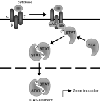
Fig. 2. Cytokine receptor signalling via JAK-STAT pathways.Following cytokine receptor ligation, JAKs are recruited and phosphorylate tyrosine residues in the β and γ signalling receptor chains, enabling STAT recruitment and phosphorylation. pSTAT proteins form dimers, which rapidly translocate to the nucleus where they act as transcription factors by binding to GAS elements.
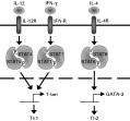
Fig. 3. Signalling pathways underlying Th1/Th2 polarisation. JAK-STAT pathways underlie the polarisation of CD4+ helper T (Th) cell responses. IL-12 and IFN-γ signal via STAT4 and STAT1 respectively to induce expression of the transcription factor T-bet, which promotes Th1 polarisation via enhanced IFN-γ and IL-12Rβ2 expression. IL-4 induces activation of STAT6, leading to production of GATA-3, which promotes transcription of the Th2 cytokines IL-4, IL-5 and IL-13. Positive and negative feedback mechanisms reinforce the Th1/Th2 bias.
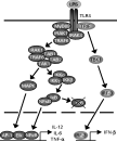
Fig. 4. Toll-like receptor 4 (TLR4) signalling. Following LPS binding to its receptor complex, signalling is initiated via TLR4. Adaptor proteins, including MyD88 and TRIF, are recruited, as well as IRAK-1, IRAK-4 and TRAF6. Following phosphorylation of IRAK-1 by IRAK-4, and TRAF6 by IRAK-1, IRAK-1 and TRAF6 dissociate from the receptor and form a complex with TAK1, TAB1 and TAB2. TAK1 activates the MAP kinase and NF-κB pathways, resulting in transcription of pro-inflammatory cytokines. A MyD88-independent, TRIF-dependent pathway via TBK-1 results in phosphorylation of the transcription factor IRF-3, which induces IFN-β production.
 You have
Access
- 13
- Cited by