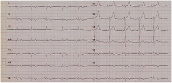
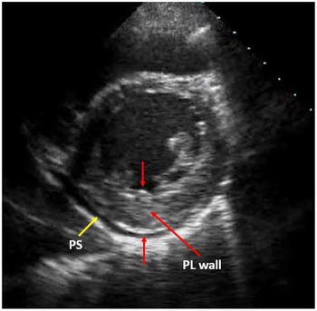
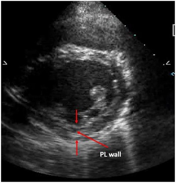
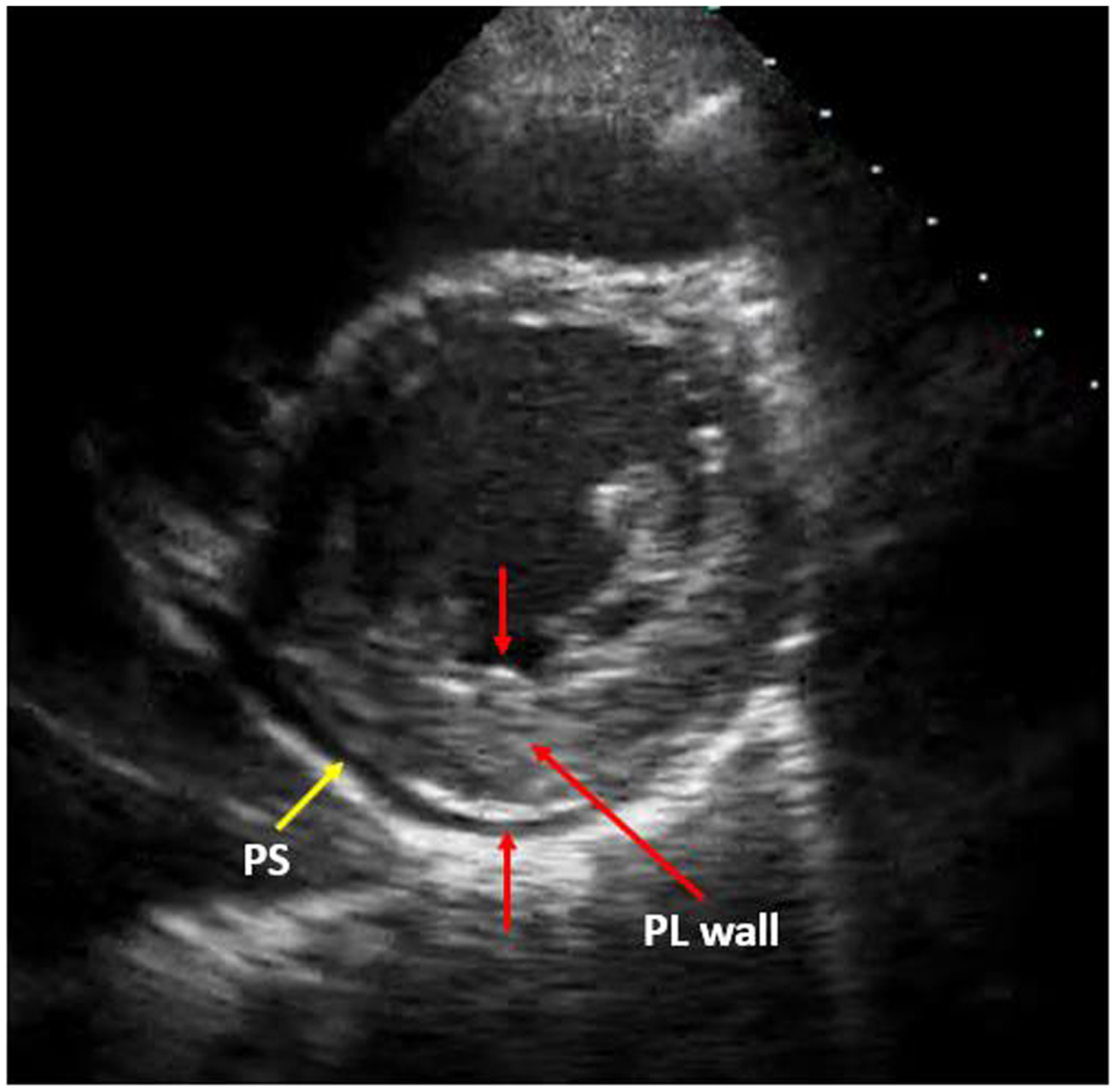
A 17-year-old boy complained of pleuritic chest pain exacerbated by movement and deep inspiration, 1 week after having an upper respiratory tract infection. On physical exam, he was noted to have a friction rub over the precordium. His ECG showed T wave inversion in leads I, II, AVF, V5, V6, and high peaked T waves in leads V1–V3 (Fig 1). Blood tests were ordered and showed elevated troponin T (0.1 ng/ml, normal upper limit 0.03 ng/ml) and CRP (40 mg/L) levels. Echocardiography was performed and showed thickening (1.7 mm) and hypokinesis of the posterolateral wall with a mild circumferential pericardial effusion (Fig 2). His left ventricular ejection fraction remained preserved at >50%. The patient was diagnosed to have myopericarditis and treated with indomethacin and colchicine. His symptoms resolved after 15 days. Repeat echocardiography done 2 months later showed normal posterolateral wall thickness and function (Fig 3).
ECG on presentation showing T wave inversion in leads I, II, AVF, V5, V6, and high peaked T waves in leads V1–V3.

Echocardiographic image on presentation of the left ventricular base in the parasternal short-axis plane showing thickening of the posterolateral (PL) wall (red arrows). PS = Pericardial space.
Echocardiographic image after 2 months of treatment showing resolution of the myocardial oedema with a normal thickness of the posterolateral (PL) wall (red arrows).
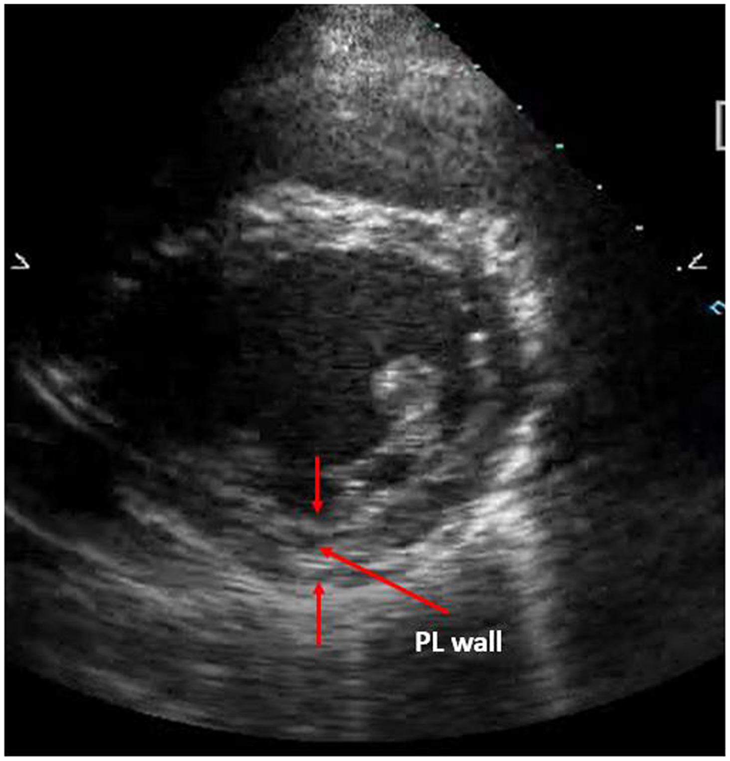
Acute myocarditis is associated with myocardial oedema secondary to inflammation which usually requires MRI for detection. Reference Cooper1–Reference Tschöpe, Ammirati, Bozkurt, Caforio, Cooper and Felix3 This case is unique in that the inflammation and myocardial oedema were very prominent causing significant ventricular wall thickening that was apparent on echocardiography and which resolved after treatment with non-steroidal anti-inflammatory drugs.
Acknowledgements
None.
Financial support
This research received no specific grant from any funding agency, commercial, or not-for-profit sectors.
Conflicts of interest
None.
Consent
The authors confirm that written consent for submission and publication of this case report including images and associated text has been obtained from the patient in line with COPE guidance.