Introduction
The early detection of Alzheimer's disease (AD) requires neuropsychological tools that can reveal the subtle cognitive impairments present prior to clinical onset (Sperling et al., Reference Sperling2011). Neuropsychological tasks should be sufficiently sensitive and specific to act as cognitive markers for AD pathology. Performance on cognitive tasks can often be affected by healthy aging (Balota et al., Reference Balota, Dolan, Duchek, Tulving and Craik2000; Brockmole and Logie, Reference Brockmole and Logie2013; Sperling et al., Reference Sperling, Karlawish and Johnson2013; Wakefield et al., Reference Wakefield, McGeown, Shanks and Venneri2014) making them unsatisfactory as screening tools for detecting dementia (Dubois et al., Reference Dubois2007; Parra et al., Reference Parra, Abrahams, Logie, Méndez, Lopera and Della Sala2010b). It is therefore important to investigate whether a neuropsychological task that could serve as a cognitive marker of AD is affected by age.
The current study investigated the effects of aging on two tasks devised to minimize the effects of age and that have been recently proposed as cognitive markers for AD: the Free and Cued Selective Reminding Task with Immediate Recall (FCSRT-IR) and a paradigm assessing temporary visual memory binding (TMB).
The FCSRT-IR was developed to distinguish memory impairments associated with amnesia from those associated with healthy aging (Grober and Buschke, Reference Grober and Buschke1987; Grober et al., Reference Grober, Buschke, Crystal, Bang and Dresner1988). The task exploits the encoding specificity principle (Tulving and Thomson, Reference Tulving and Thomson1973), wherein an individual's memory for items is enhanced by reference to the context in which they were learned. Specifically, in the FCSRT-IR, participants are asked to encode a list of items together with their semantic categories. At test, participants attempt to remember these items under free recall followed by selective reminding for unrecalled items using semantic categories as cues. Performance on this task is measured by free and cued recall scores, the sum of which produces a total recall score.
TMB is responsible for the integration of disparate visual information – such as shape and color – to produce a visual object which can be retained as a whole. Critically, the features of objects are retained along with the information of what features are attributed to which object. This information is necessary when detecting whether an object's features have changed. In the visual memory literature, this is typically assessed in a change detection paradigm whereby participants decide whether features between two visual objects have swapped between study and test arrays (Wheeler and Treisman, Reference Wheeler and Treisman2002). Performance on this task would represent a participants’ memory for bindings (e.g. colored shapes), which would then be compared to their memory for single features (e.g. black shapes or blocks of color) to reveal a “cost” of binding.
Both tasks are reported to be sensitive to clinical and preclinical AD. For instance, poor free recall on the FCSRT-IR has been observed prior to a diagnosis of AD (Grober and Kawas, Reference Grober and Kawas1997; Grober et al., Reference Grober, Hall, Lipton, Zonderman, Resnick and Kawas2008b, but see Papp et al., Reference Papp2015). Comparatively, poor TMB is associated with sporadic AD (Parra et al., Reference Parra, Abrahams, Fabi, Logie, Luzzi and Della Sala2009) and preclinical familial AD (Parra et al., Reference Parra, Abrahams, Logie, Méndez, Lopera and Della Sala2010b). Furthermore, both tasks are reported to be sensitive and specific to AD, potentially aiding differential diagnosis from other patient groups that are also associated with memory impairment. In the case of the FCSRT-IR, total recall is argued to be an index of AD pathology in contrast to frontotemporal dementia (FTD) (Lemos et al., Reference Lemos, Duro, Simões and Santana2014). Poor TMB is reported in AD but absent in FTD, vascular dementia (VaD), dementia associated with Parkinson's disease, and dementia with Lewy bodies (Della Sala et al., Reference Della Sala, Parra, Fabi, Luzzi and Abrahams2012) as well as depression (Parra et al., Reference Parra, Abrahams, Logie and Della Sala2010a). Thus, both tasks are promising aids to the differential diagnosis, follow-up, and early screening of AD.
However, these tasks rely on different cognitive systems, suggesting that they are not revealing the same memory impairments in AD. Crucially, the FCSRT-IR is an assessment of associative (or relational) long-term memory, whereas binding is an assessment of conjunctive short-term memory (Parra et al., Reference Parra, Fabi, Luzzi, Cubelli, Hernandez Valdez and Della Sala2013). Specifically, items in the FCSRT-IR are successfully retrieved if the association between the item, its category, and its encoding context are retained. Items in the TMB task are successfully remembered if their constituent parts are correctly bound and such bindings are accurately held in short-term memory. Recent studies have revealed that relational and conjunctive binding are two memory functions that dissociate not only across memory systems but within short-term (Piekema et al., Reference Piekema, Rijpkema, Fernandez and Kessels2010; Parra et al., Reference Parra, Fabi, Luzzi, Cubelli, Hernandez Valdez and Della Sala2013) and long-term memory (Bastin and Van der Linden, Reference Bastin and Van der Linden2005). Moreover, the two tasks rely on different retrieval functions with the FCSRT-IR dependent on recall and the TMB task on recognition.
Accordingly, these two forms of memory – as representing different cognitive systems – are also thought to depend on different structures and networks in the brain (Moses and Ryan, Reference Moses and Ryan2006; Mayes et al., Reference Mayes, Montaldi and Migo2007). Variation in free recall in the FCSRT-IR has been shown to be associated with hippocampal volume and metabolic rate (Zimmerman et al., Reference Zimmerman2008; Sarazin et al., Reference Sarazin2010). In AD patients, it has been posited that poor performance on the FCSRT-IR may be due to a disconnection between hippocampal and frontal areas necessary for checking the context in which items are learned (Lekeu et al., Reference Lekeu2003). Both findings are in line with the argument that the hippocampus is central to the formation of relational memories that link item and context information together (Davachi, Reference Davachi2006). In contrast, lesion and imaging data suggest that the hippocampus proper is not necessary for successful TMB (Baddeley et al., Reference Baddeley, Allen and Vargha-Khadem2010; Piekema et al., Reference Piekema, Rijpkema, Fernandez and Kessels2010; Parra et al., Reference Parra, Della Sala, Logie and Morcom2014). A dissociation between the binding paradigm and other hippocampal tasks has been observed, as performance on a shape-color binding paradigm is not correlated with performance on the Paired Associates Task (Parra et al., Reference Parra, Della Sala, Abrahams, Logie, Méndez and Lopera2011). Moreover, diffusion tensor imaging evidence suggests that, in familial AD patients, the impaired white matter structures associated with poor Paired Associates Task performance (hippocampal part of cingulum bundle) dissociate from the structures implicated in poor performance on the shape-color binding paradigm (Parra et al., Reference Parra2015). Instead, the maintenance of bindings is dependent on the ventral visual stream (Staresina and Davachi, Reference Staresina and Davachi2010; Parra et al., Reference Parra, Della Sala, Logie and Morcom2014) and may bear on the functioning of the perirhinal cortex (Staresina and Davachi, Reference Staresina and Davachi2010; Tyler et al., Reference Tyler2013; Watson and Lee, Reference Watson and Lee2013; Clarke and Tyler, Reference Clarke and Tyler2014).
The evidence above complements the hypothesis that parahippocampal functions are affected earlier in the progression of AD than hippocampal functions (Didic et al., Reference Didic2011), which suggests that the FCSRT-IR may be sensitive to memory impairments that occur relatively later than those detected by the TMB (see Papp et al., Reference Papp2015). Additionally, the hippocampus is also affected by healthy cognitive aging (Balota et al., Reference Balota, Dolan, Duchek, Tulving and Craik2000; Mitchell et al., Reference Mitchell, Johnson, Raye and D'Esposito2000), which implies that tasks that assess its function are likely to reveal impairments in healthy elderly controls and AD patients (for example, performance on a relational (i.e. hippocampal) binding task declines with healthy aging and worsens with disease progression, van Geldorp et al., Reference van Geldorp, Heringa, van den Berg, Olde Rikkert, Biessels and Kessels2015). Therefore, performance on the FCSRT-IR is likely to be affected by both disease and healthy aging.
Indeed, free recall as assessed by the FCSRT-IR is seen to decline over time in a healthy population (Grober et al., Reference Grober2008a). This effect is further confounded by an individual's level of education and their gender (Ivnik et al., Reference Ivnik, Smith, Lucas, Tangalos, Kokmen and Petersen1997; Grober et al., Reference Grober, Lipton, Katz and Sliwinski1998; Frasson et al., Reference Frasson2011), necessitating the collection and use of norms across populations for the accurate identification of a genuine AD memory impairment (Ivnik et al., Reference Ivnik, Smith, Lucas, Tangalos, Kokmen and Petersen1997; Peña-Casanova et al., Reference Peña-Casanova2009).
Previous research suggests that performance on the TMB is less likely to be affected by healthy cognitive aging. Brockmole and Logie (Reference Brockmole and Logie2013) showed that although overall visual short-term memory capacity declined with age, this did not necessarily affect the ability to bind; the percentage of correctly remembered bound objects in 75-year-olds (82%) was comparable to that seen in 20-year-olds (85%). Similarly, healthy older participants do not show a greater binding cost in comparison to healthy young participants (Brockmole et al., Reference Brockmole, Parra, Della Sala and Logie2008), and their performance is as affected by the presence of a dual task as it is in young participants (Brown and Brockmole, Reference Brown and Brockmole2010). Thus, binding appears resistant to the effect of age, as confirmed with a recent replication (Isella et al., Reference Isella, Molteni, Mapelli and Ferrarese2015).
In sum, there is evidence to suggest that free recall and binding may be differentially affected by age, potentially due to their sensitivity to different cognitive systems supported by different neuronal networks. However, the two tasks have never been directly compared in the same study and as such it is not clear if this apparent differential sensitivity to aging is the result of different sampled populations between studies. Thus, the primary aim of the present study was to directly compare the effect of age on these two tasks in a sample drawn from one population. To this end, we observed performance on the FCSRT-IR and binding paradigm in healthy young and older participants, controlling for differences in gender distribution and years of education between these two groups.
Methods
Participants
Two groups, one of 24 healthy older participants (M = 70.6, SD = 6.41, range = 61–85) from the University of Edinburgh Volunteer Panel (14 females) and 24 healthy undergraduate and postgraduate students (M = 20.5, SD = 2.15, range = 18–25) from the University of Edinburgh (18 females) participated in this experiment. All reported good health and had normal or corrected to normal vision; none had premorbid neurological or psychiatric history. There was no significant difference between the years of education of the older (M = 15.8, SD = 3.23, range 11–20) and young (M = 16.0, SD = 2.31, range 12–20) participants (t(46) = −0.36, p = 0.72). All healthy older participants scored above the conservative cut-off of 88 points for the ACE-III (M = 97, SD = 2.75, range 90–100). All the participants signed a consent form prior to participation. The study was approved by the University of Edinburgh Psychology Research Ethics Committee (Ref. 36–1314/2).
Assessment
General assessment
English Version A of the ACE-III was used to screen for possible cognitive impairment associated with dementia in the healthy older group (Hsieh et al., Reference Hsieh, Schubert, Hoon, Mioshi and Hodges2013).
FCSRT-IR
The word version of Form A of the FCSRT-IR was selected for this experiment based on the recommendation that the word version is sensitive to healthy participants at risk of AD (Auriacombe et al., Reference Auriacombe, Helmer, Amieva, Berr, Dubois and Dartigues2010), and that all forms of the FCSRT-IR are psychometrically equivalent (Grober et al., Reference Grober, Ocepek-Welikson and Teresi2009); our results are therefore unlikely to be driven by idiosyncrasies associated with this particular form.
The FCSRT-IR was administered according to the instructions provided by the Albert Einstein Medical College. The task is composed of an acquisition and test phase. In the acquisition phase, participants were presented with one of four A4 cards, on which were four printed words, one in each quadrant. Participants were asked to point to and give the name of an item that belonged to a category cue (e.g. “What is the furniture?”). Each item belonged to its own, unique category. Once participants had correctly pointed to and named all four items on the card, the card was taken away, and immediate cued recall was assessed for items (e.g. “What was the furniture?”) in the same order they were encoded. Where participants failed to recall the item given a cue, the item and its cue were presented again (“The furniture was the desk.”) and assessed again to ensure the association was learned. This procedure was repeated for the remaining three cards. Once the final set of four items was identified, the test phase began. This was composed of three trials. At the start of each trial, participants were instructed to count backwards from a number by decrements of 3 for 20 sec as a form of articulatory suppression. Participants were then given up to 2 min to recall as many of the words as they could in any order (free recall). After free recall was completed, participants were then presented with the categories of items that they did not remember (cued recall). The sum of a participant's free and cued recall represented their total recall. Participants completed two more trials of the test phase.
We analyzed the sum of a participants’ free recall, which was calculated as the sum of all words correctly recalled under free recall across all three trials (0–48 words). Similarly, we analyzed the sum of a participants’ total recall across all three trials, which was calculated as the sum of all words correctly recalled under free recall and cued recall across all three trials (0–48 words). Additionally, a participants’ sensitivity to cueing was calculated to demonstrate the degree to which they benefited from cueing (0–100%). This was calculated according to the formula presented by Tounsi et al. (Reference Tounsi1999):
 $$\begin{eqnarray*}
&&{\rm{Sensitivity}}\,{\rm{to}}\,{\rm{cueing}} = 100\, \\
&&\quad*\,\frac{{{\rm{Sum}}\,{\rm{of}}\,{\rm{total}}\,{\rm{recall}} - {\rm{Sum}}\,{\rm{of}}\,{\rm{free}}\,{\rm{recall}}}}{{48 - {\rm{Sum}}\,{\rm{of}}\,{\rm{free}}\,{\rm{recall}}}}
\end{eqnarray*}$$
TMB
The TMB paradigm used here was the three-item version of the paradigm reported by Parra et al. (Reference Parra, Abrahams, Logie and Della Sala2010a; Reference Parra, Abrahams, Logie, Méndez, Lopera and Della Sala2010b) and Parra et al. (Reference Parra, Abrahams, Logie and Della Sala2010a). This paradigm has been used to identify preclinical AD and also differentiate AD from healthy cognitive aging. The use of a three-item paradigm was to better reveal the effect of age, as the two-item paradigm is associated with ceiling effects in single feature conditions in healthy older groups (Parra et al., Reference Parra, Abrahams, Logie and Della Sala2010a; Della Sala et al., Reference Della Sala, Kozlova, Stamate and Parra2016)
Items consisted of eight 6-sided abstract shapes that were either black or colored with non-primary colors (see Parra et al. (Reference Parra, Abrahams, Logie and Della Sala2010a; Reference Parra, Abrahams, Logie, Méndez, Lopera and Della Sala2010b)). The task was written and presented using E-Prime and E-Run 2.0, respectively (Psychology Software Tools, 2012). All visual information was presented through a 15.1″ LCD monitor, and viewing distance was not restrained. In all cases, participants responded with the mouse, using the left mouse button to indicate “different” and the right to indicate “same.”
Participants first completed a perceptual screen to ensure that any subsequent difficulties on the task were not due to perceptual problems. In each trial, participants are presented with three colored shapes in the upper and lower half of a screen, as separated by a horizontal line. In half of the trials, the color and shape of the objects above the line was identical to those below the line. In the remaining half, two of the shapes had swapped colors. Participants completed ten trials, and had to respond to at least eight correctly before continuing to the TMB task.
The paradigm contained two conditions: shape only and shape-color binding. Each condition contained 32 trials. In the shape only condition, participants were presented with black, 6-sided shapes. In the binding condition, these shapes were colored with non-primary colors. The stimuli used in both conditions are described in more detail in the Method section of Experiment 2, Brockmole et al. (Reference Brockmole, Parra, Della Sala and Logie2008). In all condition trials, participants were presented with three objects in a study array at 2,000 ms. After this, they were removed for a 900 ms retention interval and the test array was presented indefinitely until participants make their response. The next trial would then be initiated by the participant by pressing any mouse key.
In the shape only condition, half of the trials displayed the same shapes at test that were present at study. In the remaining half of these trials, two new shapes were presented in the test array. These two types of trials (same shapes, new shapes) were presented randomly within the shape only condition. In the shape-color binding condition, half of the trials displayed the same combinations of shape and color in the study and test arrays. In the remaining half, two of the shapes from the study array swapped colors when shown in the test array. The presentation of these types of trials was also random. In all conditions, the position of the objects between study and test arrays was always random, making location an uninformative feature.
Participants’ accuracy was calculated as a percentage of trials where they gave a correct response (hits and correct rejections). Furthermore, a participant's cost of binding was calculated as their accuracy in the binding condition subtracted from their accuracy in the shape condition.
Procedure and design
The order of the presentation of the FCSRT-IR and the TMB was counterbalanced, as was the presentation of the shape and binding condition within the binding paradigm. The experiment employed a mixed, fully counterbalanced design. Older participants completed the ACE-III at the end of the testing session. The assessment lasted 45 min.
Analysis
Variables from the FCSRT-IR and binding paradigm were analyzed separately.
Initially, the relationship between total recall and sensitivity to cueing was analyzed using paired correlations within each age group, as these variables have been seen to almost completely overlap (Grober et al., Reference Grober, Sanders, Hall and Lipton2010). Subsequently, differences between groups on the measures of free recall, total recall, and sensitivity to cueing were analyzed with unpaired t-tests.
Binding variables were assessed using a mixed, 2-way ANOVA over the between-subjects variable of age group and the within-subjects variable of condition (shape only, shape-color binding). Binding cost was analyzed between the two age groups using an unpaired t-test.
The association between age and performance on the TMB and FCSRT-IR variables was also analyzed with paired Pearson's correlations (for the older group only).
Results
FCSRT-IR
Total recall was high in both the young (M = 47.92, SD = 0.3, range = 47–48) and older (M = 47.63, SD = 0.8, range = 45–48) participants (sensitivity to cueing was observed to be high in young (M = 99.2, SD = 2.7) and older (M = 97.77, SD = 4.76) participants). A Mann–Whitney–Wilcoxon test revealed no statistically significant difference in total recall performance between these groups (W = 238, p = 0.1). A one-sample t-test revealed that young participants’ summed total recall was not significantly different from ceiling (t(23) = −1.45, p = 0.16). However, the same analyses revealed that older participants’ performance was significantly different from ceiling (t(23) = −2.39, p = 0.02), although the absolute difference here was 0.37 words.
Free recall
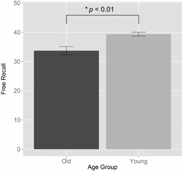
Participants' free recall is represented in Figure 1. Younger participants recalled significantly more words in free recall than older participants (M = 39.33, SD = 3.22, range = 32–45; M = 33.66, SD = 6.84, range = 14–44, respectively, t(32.67) = −3.67, p = 0.001).
Mean number of words recalled by young and older participants under free recall. Error bars represent standard error.

TMB task
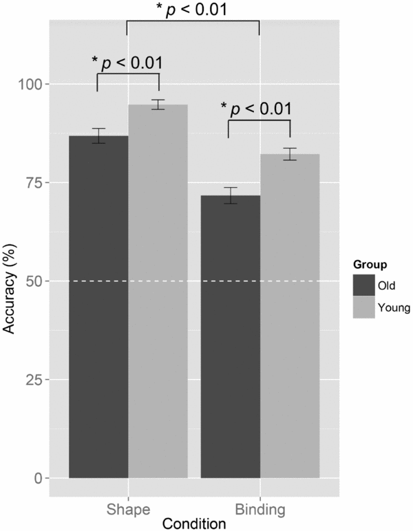
Participants’ performance on the TMB task is represented in Figure 2. Older participants were less accurate than the young participants overall, but both groups of participants were more accurate in the shape condition compared to the shape-color binding condition. A mixed, two-way ANOVA undertaken on accuracy revealed a significant effect of age (F(1, 46) = 21.09, p < 0.01, ηG2 = 0.24 (this represents general η 2, which is a preferred estimate of effect size for repeated measures ANOVAs.)) and condition (F(1, 46) = 112.28, p < 0.01, ηG2 = 0.42), but no significant interaction (F(1, 46) = 0.94, ηG2 = 0.006). Similarly, although a cost of binding was seen for young (M = 12.58, SD = 8.11, range = −3 to 28) and older participants (M = 15.13, SD = 9.92, range = 0–35), this difference was not statistically significant (t(44.29) = 0.972, p = 0.34). Hence, both young and older participants were less accurate in the binding condition, but there was no evidence to suggest that this difficulty was exacerbated by age.
Accuracy for young and older participants in the TMB paradigm. Error bars represent standard error. Dotted white line represents chance performance (50%).

The lack of a significant interaction between age and condition may be argued as representing limited power. Here, post-hoc analyses conducted using G*Power 3.1.9.2 revealed that the interaction effect of age and condition on accuracy would require at least 132 participants to become statistically significant with power at 90%. Similarly, at the same level of power, the difference in cost reported here would require 219 participants in each age group to become statistically significant. By contrast, the main effects of age and condition on accuracy were revealed with 99% and 100% power, respectively. Furthermore, the effect of age on free recall in the FCSRT-IR was revealed with 98% power. Thus, the lack of a statistically significant interaction effect suggests that any differential effect of age on binding memory is negligible in comparison to the effect of age on free recall, shape, and binding memory.
While the lack of an interaction effect suggests that there is no significant effect of age on binding cost, an effect was nonetheless observed (d = 0.155), but crucially smaller than what was observed for free recall (d = 1.061).
Correlational analysis
In the older group, a Pearson's correlation confirmed a significant relationship between age and free recall ability (r = −0.45, p = 0.027). There was a trend towards a significant negative correlation between age and shape memory (r = −0.37, p = 0.079) and age significantly correlated with binding memory (r = −0.41, p = 0.047), but no relationship was observed between cost and age (r = 0.074, p = 0.73).
Discussion
This study investigated the effect of age on the FCSRT-IR and the TMB. To our knowledge, this is the first study to directly compare performance of these two cognitive markers for AD in a single older cohort. In contrast to young participants, older participants demonstrated significantly poorer free recall on the FCSRT-IR, but did not significantly differ in their relative memory for binding. The comparable levels of education and gender distributions between the experimental groups suggest that this represents a differential effect of age on one task but not the other, which in turn may reflect their sensitivity to different memory systems. Specifically, the TMB paradigm appears less affected by memory systems that are also affected by healthy cognitive aging (i.e. hippocampal functions).
Both the FCSRT-IR and TMB paradigm have been used to distinguish patients with AD from healthy older controls and other patient groups, and have therefore been proposed as sensitive and specific cognitive markers. However, the literature associated with these tasks and the results from the current experiment suggest that these tasks may be sensitive to different memory systems.
Specifically, our findings comply with the idea of two separate forms of memory – recall and recognition – that in turn are differentially affected by age (Danckert and Craik, Reference Danckert and Craik2013) and distinctly associated with separate parts of the MTL, with recognition bearing on the entorhinal cortex, and recall the hippocampus (Yonelinas et al., Reference Yonelinas, Widaman, Mungas, Reed, Weiner and Chui2007). The clinical relevance of this dissociation is outlined in Didic et al.’s (Reference Didic2011) model of progressive memory impairments in AD pathology, wherein pathology occurs in sub-hippocampal areas prior to the hippocampus proper. Crucially, the authors review evidence to argue that sub-hippocampal areas (e.g. perirhinal and entorhinal cortices) are associated with context-free object memory, and thus impairments of this type of memory are observed before the context-dependent episodic memory impairment classically associated with AD. Of note, regions proposed to be part of the sub-hippocampal network declines in AD earlier than the hippocampus (Juottonen et al., Reference Juottonen1998) and seem to remain unaffected across the life span (Insausti et al., Reference Insausti1998). Overall, our results – in concert with previous findings regarding the TMB – conform to a framework that proposes two forms of memory (context free recognition vs. context dependent recall) that are dependent on different parts of the MTL and, as such, are differentially affected by healthy aging and impaired at different stages of AD pathology.
We have outlined fundamental differences between these tasks that may be responsible for this differential effect of age, with the key distinction being that one task assesses relational long-term memory and the other conjunctive short-term memory.
However, there is an additional difference in the use of recognition and recall, in that the former (as assessed in the binding paradigm) is less affected than the latter in healthy aging (Perlmutter, Reference Perlmutter1979; Danckert and Craik, Reference Danckert and Craik2013). This discrepancy has been interpreted as a need for more cognitive resources in recall tasks compared to recognition tasks (Craik and McDowd, Reference Craik and McDowd1987; Danckert and Craik, Reference Danckert and Craik2013). Indeed, it has been posited that free recall in the FCSRT-IR necessitates retrieval strategies (Lekeu et al., Reference Lekeu2003) that are affected by age-associated changes in anterior brain regions (Buckner, Reference Buckner2004). By contrast, the formation and memory for bindings has been argued to be automatic but fragile (Allen et al., Reference Allen, Baddeley and Hitch2006), and thus not as resource-demanding or as dependent on effective strategies. This position is supported by evidence that patients with dysexecutive symptoms (e.g. VaD patients, Román, Reference Román2003) are poorer than controls on the FCSRT-IR (Traykov et al., Reference Traykov2005) yet comparable in TMB (Della Sala et al., Reference Della Sala, Parra, Fabi, Luzzi and Abrahams2012). This in turn highlights the utility of the TMB impairment in differential diagnosis, as its high specificity and sensitivity to AD reflects that it is not driven by impairments associated with other dementias (Della Sala et al., Reference Della Sala, Parra, Fabi, Luzzi and Abrahams2012), aging, or depression (Parra et al., Reference Parra, Abrahams, Logie and Della Sala2010a).
It may be argued that the difference reported here is an experimental artifact given the use of words on the FCSRT-IR and visual information in the binding paradigm and the picture superiority effect in older individuals (Winograd et al., Reference Winograd, Smith and Simon1982). Specifically, older participants may rely only on verbal codes in the former task, but both verbal and visual codes in the latter. Although possible, this is unlikely, as the materials used in the binding paradigm were abstract shapes and low frequency colors that discourage verbal encoding. Furthermore, an effect of age on immediate and delayed free recall has also been demonstrated in a picture version of the FCSRT-IR (Frasson et al., Reference Frasson2011). Ultimately, it is more likely that the differential effect of age on free recall reflects the nature of the FCSRT-IR rather than its chosen presentation format.
Age effects shown by our results are usually addressed in clinical neuropsychology with the use of age-adjusted normative data. Although this is a necessary practice in some neuropsychological assessments, there are particular problems when this is done with free recall for the purposes of identifying AD (as opposed to comparison to peers for the purposes of ranking). Critically, Sliwinski et al. (Reference Sliwinski, Buschke, Stewart, Masur and Lipton1997) demonstrated with the Selective Reminding Task that a variable's sensitivity to dementia was reduced when confounding yet predictive factors (i.e. age) were controlled for. Moreover, adjusted, standardized scores can lead to a loss of discrimination between health and AD in very old populations (Bondi et al., Reference Bondi2003), and these forms of control are also seen to weaken the association between test performance and brain integrity (Mungas et al., Reference Mungas, Reed, Farias and DeCarli2009). Thus, although robust normative data are necessary for interpreting neuropsychological performance (see Mitrushina et al., Reference Mitrushina, Boone, Razani and D'Elia2005; Slick, Reference Slick, Strauss, Sherman and Spreen2006), this practice when using free recall as an indicator of AD must be approached with caution (Carlesimo et al. Reference Carlesimo, Perri and Caltagirone2011; Gainotti et al., Reference Gainotti, Quaranta, Vita and Marra2014).
Although we assessed a healthy elderly and young population's free recall, it should be noted that the FCSRT-IR was initially intended to be an assessment of episodic memory in older adults with reference to total recall (Grober et al., Reference Grober, Buschke, Crystal, Bang and Dresner1988). However, we revealed that total recall is also a limited variable, as we replicated the finding that healthy elderly participants were virtually at ceiling on measures of cue efficiency – total recall and sensitivity to cueing (Frasson et al., Reference Frasson2011). The ceiling effect seen in total recall introduces an increased risk of diagnostic false alarms; one isolated error would be misconstrued as abnormal. The high prevalence of this effect in the 40–94-year-old Frasson et al. (Reference Frasson2011) cohort suggests that this issue is evident throughout the lifespan. Other tasks following the same general principles, such as the RI48 (Ivanoiu et al., Reference Ivanoiu2005) capture a participants’ use of semantic cues without the ceiling effects associated with the FCSRT-IR, and thus may be more appropriate for assessing impairments in sensitivity to cueing in a healthy population.
Our results may be somewhat limited in that we have only observed the effect of age in a population with a high average level of education. However, previous research suggests that our findings may hold across a wider population. Specifically, TMB performance is not affected by differences in education between experimental groups (Parra et al., Reference Parra, Della Sala, Abrahams, Logie, Méndez and Lopera2011) or study cohorts (Parra et al., Reference Parra, Abrahams, Logie and Della Sala2010a; Reference Parra, Abrahams, Logie, Méndez, Lopera and Della Sala2010b), but free recall is negatively affected by education (Grober et al., Reference Grober, Lipton, Katz and Sliwinski1998). Thus, it may be that the differential age effect observed here is relatively conservative, and may be more striking in a low-education population.
In sum, we revealed that two tests that are promising tools in assisting the early detection of AD have differential sensitivity to the effect of age in a healthy older population. This suggests that the tasks may be sensitive to different memory systems that are differentially affected by age. The resilience of the binding task to aging combined with its sensitivity to AD indicates this may provide a more suitable baseline from which to detect cognitive deterioration due to pathological neurodegeneration of AD. Future research may confirm this possibility by directly comparing the tasks’ diagnostic utility in a clinical population.
Conflict of interest declaration
None.
Description of authors’ roles
LK designed the study, conducted data collection, data analysis, and wrote the manuscript. SA and SDS gave supervision on the design of the study and writing. MAP gave supervision on analysis and writing.
Acknowledgments
The FCSRT-IR is copyrighted by the Albert Einstein College of Medicine (AECOM) and is made freely available from the AECOM for teaching and academic research purposes. This work was supported by the John C Marshall Studentship via the School of Philosophy, Psychology and Language Sciences at the University of Edinburgh and the Medical Research Council [LK]; the Alzheimer's Society [AS-R42303 and AS-SF-14-008 to MAP].