Introduction
Early-stage glottic squamous cell carcinoma (SCC) has some exceptional characteristics which distinguish it from other mucosal head and neck malignancies, namely: it rarely metastasises to the neck lymph nodes; it almost never metastasises distantly; several treatment methods are equally effective in achieving a cure; and treatment does not cause significant functional disability and has a very good prognosis.Reference Spector, Sessions, Chao, Haughey, Hanson and Simpson1–Reference Sjögren, van Rossum, Langeveld, Voerman, van de Kamp and Friebel3
However, there are currently no published prospective, randomised, double-blind studies comparing different treatment modalities for tumour stage 1 (T1) glottic cancers. Many studies report results for a single modality treatment, and at many institutions one treatment modality has become established as standard practice.Reference Cellai, Frata, Magrini, Paiar, Barca and Fondelli4–Reference Franchin, Minatel, Gobitti, Talamini, Vaccher and Sartor6 However, since there are several treatment alternatives with similar oncological outcomes, other studies have sought to determine the advantages of individual modalities compared with other therapies.Reference Sjögren, van Rossum, Langeveld, Voerman, van de Kamp and Friebel3, Reference Karatzanis, Psychogios, Zenk, Waldfahrer, Hornung and Velegrakis7, Reference Peeters, van Gogh, Goor, Verdonck de Leeuw, Langendijk and Mahieu8 Non-randomised studies have many potential defects, selection bias being the most important. However, despite flaws, in the absence of higher quality evidence, non-randomised data are important as they offer the only scientific basis for treatment decision-making.
Although definitive treatment guidelines are not currently available to aid the selection of T1 glottic SCC treatment modality, patient and tumour characteristics are important determinants in the decision-making process. At our institution, patients are not routinely directed towards one specific treatment modality. Rather, each patient's age, co-morbidities, pulmonary status, tumour extent, and potential functional results for each treatment option are evaluated individually by our tumour review board. We believe that radiotherapy (RT), endolaryngeal laser surgery and open partial laryngectomy may have different relative benefits in different patients.
In this report, we aim to present and discuss the results of our T1 glottic SCC treatment algorithm.
Materials and methods
Setting
A retrospective clinical study was conducted at a university hospital serving as a tertiary referral centre.
Ethical approval was obtained from the institutional ethical board before beginning the study.
The medical records of patients managed for T1 laryngeal SCC between 2001 and 2011 were reviewed, and demographic, clinical and follow-up data were recorded. All pathology reports were re-evaluated in our pathology department. The American Joint Committee on Cancer tumour–node–metastasis (TNM) staging system was used for tumour staging.Reference Edge, Byrd, Compton, Fritz, Greene and Trotti9 Since the TNM staging system for T1 glottic tumours did not change over the study period, tumour stage classification was consistent for all the patients assessed. Laryngeal cancer was staged using endoscopic examination (in the out-patient department), imaging modalities (e.g. computed tomography (CT) and magnetic resonance imaging (MRI)) and direct laryngoscopic examination (under general anaesthesia). All lesions were confirmed histopathologically as SCC before any treatment was commenced.
Patients
Only patients with a T1 glottic SCC and at least 12 months' follow up were included in the study. We excluded any patients with in situ carcinoma, a previous history of head and neck cancer, or previous treatment for laryngeal cancer.
There were 102 consecutive patients treated between 2001 and 2011. Seven of these patients (6.9 per cent) were lost to follow up. One of these seven patients was treated with endolaryngeal laser surgery, another with open partial laryngectomy (comprising frontolateral laryngectomy) and the remaining five with RT. Thus, we evaluated follow-up data for 95 patients with T1 glottic SCC treated with either RT or surgery (i.e. endolaryngeal laser surgery or open partial laryngectomy).
Treatment
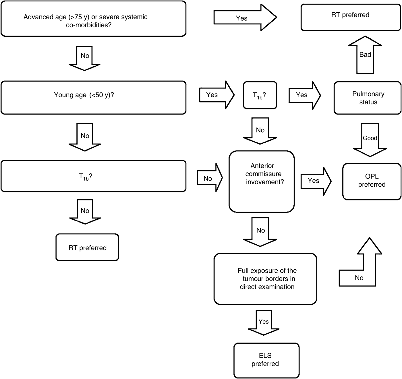
All possible treatment options, together with their advantages and disadvantages, were explained to each patient at the beginning of management. Patients were evaluated individually and the best possible treatment method was advised, following the treatment algorithm presented in Figure 1; however, the final treatment decision was made by the patient themselves. Treatment options were open partial laryngectomy, endolaryngeal laser surgery and external RT. However, only selected T1a tumours were treated with endolaryngeal laser surgery. If complete exposure of the tumour edges could not be achieved during direct laryngoscopy examination, or if anterior commissure involvement was present, endolaryngeal laser surgery was not considered.
Treatment algorithm for tumour stage 1 (T1) glottic squamous cell carcinoma. RT = radiotherapy; OPL = open partial laryngectomy; ELS = endoscopic laser surgery

Radiotherapy was delivered via the external route in all patients. One patient received a total dose of 50 Gy. All other patients in the RT group received a 1.8–2.0 Gy dose per fraction, one fraction per day, five days per week, with a total dose of 63 to 70 Gy over a 6 to 7 week time period. Since the risk of nodal metastasis is quite low for T1 lesions, the neck lymph nodes were not included in the treatment field.
Endolaryngeal laser surgery was performed using a CO2 laser source mounted on an operating microscope. Continuous mode, 5–10 W output power and a 0.3 mm spot size were used during laser surgery. For patients undergoing open partial laryngectomy, a tracheotomy was routinely performed before the main procedure. For both endolaryngeal laser surgery and open partial laryngectomy, a 2 mm surgical margin was considered safe during resection, and frozen section biopsies were examined during the operation.
Chemotherapy was never used for the initial treatment of T1 glottic cancer.
In the presence of a suspicious lymph node on clinical examination, a selective neck dissection (levels II–IV) was performed contemporaneously with the main procedure. All neck dissections and regional recurrences were recorded. The need for additional treatment, such as adjuvant RT or re-resection for positive surgical margins, was also recorded.
Follow-up examinations were performed in the out-patient department every two months in the first year, every three months in the second year, every four months in the third year, every six months in the fourth and fifth years, and yearly after the fifth year. Direct laryngoscopy examinations and biopsies of suspicious lesions were performed during the follow-up period.
Statistical analysis
The SPSS version 17.0 software program (SPSS Inc, Chicago, Illinois, USA) was used for statistical analysis.
Overall survival, disease-specific survival, laryngeal preservation, local recurrence and regional recurrence rates were the end-points of the study. Overall survival, disease-specific survival, laryngeal preservation, local recurrence and regional recurrence calculations were performed by the Kaplan–Meier method. The overall survival rate was defined as the proportion of patients still alive at the end of a given time period after diagnosis. The disease-specific survival rate was similarly determined, but taking into account only glottic cancer related deaths. The laryngeal preservation rate was defined as the proportion of patients with an intact larynx at the end of a given time period after diagnosis. The local recurrence rate was defined as the proportion of patients with local recurrence at the end of a given time period after diagnosis. The regional recurrence was similarly determined, but noting only recurrences in the neck.
The association between laryngeal preservation and anterior commissure involvement, treatment modality (RT or surgery) and T classification (T1a or T1b) was analysed using the chi-square test. The t-test was used to study the strength of the association between age and laryngeal preservation. A p value of 0.05 or less was considered significant.
Results
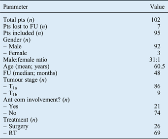
There were 95 patients (92 men and 3 women; male to female ratio, 31:1) who fulfilled the inclusion criteria and had sufficient follow-up data. An additional seven patients were lost to follow up. The mean patient age was 60.5 years (standard deviation, 9.7 years; range, 33–86 years). The median follow-up time was 48 months (range, 12–136 months).
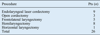
Twenty-six patients received surgery and 69 received definitive RT as initial treatment. Patient characteristics are listed in Table I and the distribution of primary procedures is shown in Table II.
Kaplan–Meier analysis of overall survival probability by treatment modality. RT = radiotherapy
Kaplan–Meier analysis of laryngeal preservation probability by treatment modality. RT = radiotherapy
Patient characteristics
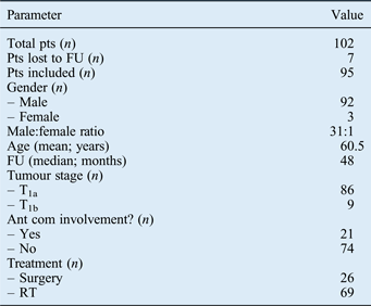
Pts = patients; FU = follow up; T = tumour stage; Ant com = anterior commissure; RT = radiotherapy
Primary procedures
Pts = patients
Ten patients in the surgical treatment group had a synchronous, ipsilateral, selective neck dissection (levels II–IV). Two patients in the surgery group had adjuvant RT for positive surgical margins. None of the patients had pathologically confirmed metastatic neck disease at presentation. No major post-operative complications occurred in the surgery group.
Recurrence and laryngeal preservation
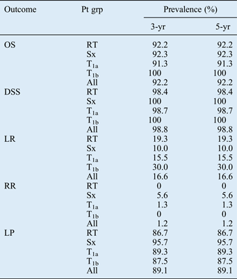
For all patients, over 5 years, the overall survival rate was 92.2 per cent, disease-specific survival was 98.8 per cent, local recurrence was 16.6 per cent, regional recurrence was 1.2 per cent and laryngeal preservation was 89.1 per cent (see Table III and Figures 2 and 3). The patient who received low-dose RT did not develop recurrence. There was no statistically significant difference between recurrence rates in the RT and surgery groups (19.3 per cent vs 10.0 per cent, respectively; p = 0.220, Fisher's exact test). Five-year local recurrence rates for patients with T1a and T1b disease were 15.5 and 30.0 per cent, respectively (p = 0.618, Fisher's exact test). The median time from primary treatment initiation to recurrence was 12 months.
Patient outcomes
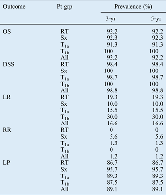
Pt grp = patient group; yr = year; OS = overall survival; RT = radiotherapy group; Sx = surgery group; T1a=tumour stage 1a; T1b = tumour stage 1b; DSS = disease-specific survival; LR = local recurrence; RR = regional recurrence; LP = laryngeal preservation
Laryngeal preservation was achieved in 4 of the 16 patients with local recurrence (25.0 per cent). Three of these patients were treated with vertical hemilaryngectomy and the fourth with horizontal glottic laryngectomy. The tumour was cured and functional laryngeal preservation achieved in all four patients.
In another 12 patients, total laryngectomy was performed. One patient developed a stomal recurrence four months after total laryngectomy and died three months later.
Only one patient developed a regional recurrence, 24 months after initial treatment, and this was successfully cured with a combination of modified radical neck dissection and concomitant chemo-radiotherapy.
There was no statistically significant difference in 5-year laryngeal preservation rate between the RT and surgery groups (86.7 vs 95.7 per cent, respectively; p = 0.220, Fisher's exact test). The 5-year laryngeal preservation rates for patients with T1a and T1b stage disease were 89.3 and 87.5 per cent, respectively (p = 0.266, Fisher's exact test). Age and anterior commissure involvement showed no association with laryngeal preservation (p = 0.779, t-test, and p = 0.081, chi-square test, respectively).
Second primary tumours and systemic causes of death
Five of the patients developed second primary cancers (four lung cancer cases and one thyroid cancer case); apart from the thyroid cancer patient, all of these patients died of their second primary cancer. Second lung cancers were synchronous in one patient and metachronous in three patients (developing in the third, seventh and eighth year of follow up, variously).
Four patients died of coronary artery disease and one patient died of renal failure within the follow-up period.
Discussion
Laryngeal cancer and oral cancer are the two most common mucosal head and neck cancers. Due to the early appearance of symptoms and the low risk of neck metastasis, glottic cancer is diagnosed at an early stage in the majority of cases. Tumour stage 1 vocal fold SCCs are limited to the vocal fold, with normal mobility, and can be treated with RT or surgery with equally high success rates.Reference Spector, Sessions, Chao, Haughey, Hanson and Simpson1 Since the 5-year disease-specific survival rate for T1 glottic cancer is over 95 per cent, the main goal of treatment is to preserve laryngeal function without compromising cure rates.Reference Cellai, Frata, Magrini, Paiar, Barca and Fondelli4, Reference Karatzanis, Psychogios, Zenk, Waldfahrer, Hornung and Velegrakis7
With the development and popularisation of endolaryngeal laser surgery over the last two decades, the role of open partial laryngectomy has dramatically reduced, and endolaryngeal laser surgery has become the mainstay of surgical treatment.Reference Steiner, Ambrosch, Rödel and Kron5, Reference Karatzanis, Psychogios, Zenk, Waldfahrer, Hornung and Velegrakis7 It has also been shown that endolaryngeal surgery can be safely performed using ‘cold steel’ instruments in selected patients.Reference Kasapoglu, Erisen, Coskun and Basut10
Endolaryngeal laser surgery has several advantages over open partial laryngectomy and RT. It is a minimally invasive procedure which can be performed without a tracheotomy. It offers similar oncological results to RT and open partial laryngectomy, but with the advantages of shorter hospitalisation period, reduced operative morbidity and mortality, and relatively lower cost.Reference Karatzanis, Psychogios, Zenk, Waldfahrer, Hornung and Velegrakis7, Reference Goor, Peeters, Mahieu, Langendijk, Leemans and Verdonck-de Leeuw11, Reference Schrijvers, van Riel, Langendijk, Dikkers, Schuuring and van der Wal12 In selected cases, especially for mid-fold or superficial lesions, voice outcome after endolaryngeal laser surgery has been shown to be equal to that for RT.Reference Sjögren, van Rossum, Langeveld, Voerman, van de Kamp and Friebel3, Reference Goor, Peeters, Mahieu, Langendijk, Leemans and Verdonck-de Leeuw11
On the other hand, endolaryngeal laser surgery may not be a perfect treatment choice in every situation. Chone et al. reported that, when the anterior commissure was involved, the 5-year local control rate for endolaryngeal laser surgery was 17 per cent lower, although this result was not statistically significant.Reference Chone, Yonehara, Martins, Altemani and Crespo13 Steiner et al. reported that anterior commissure involvement decreased local control rates by 6 and 19 per cent in cases of T1a and T1b glottic tumours, respectively.Reference Steiner, Ambrosch, Rödel and Kron5 Sachse et al. reported 5-year local control rates as low as 42 per cent in cases with anterior commissure involvement, compared with 87 per cent in anterior commissure negative cases; by comparison, patients receiving open partial laryngectomy had a 5-year local control rate of 82 per cent if anterior commissure positive and 93 per cent if anterior commissure negative.Reference Sachse, Stoll and Rudack14 Rödel et al. reported that 5-year local control rates for T1a and T1b tumours decreased by 16 and 18 per cent, respectively, in cases with anterior commissure involvement.Reference Rödel, Steiner, Müller, Kron and Matthias15
Although forming a robust conclusion is not possible in the absence of prospective, randomised studies, these results indicate that anterior commissure involvement may have a negative impact on local control in patients undergoing endolaryngeal laser surgery, and that open partial laryngectomy may be a better alternative. This may be due to inadequate exposure of the tumour or under-staging in some cases. A T3 or T4a tumour can be easily under-staged as T1, especially if imaging is not routinely used. The American Joint Committee on Cancer TNM staging manual (seventh edition) suggests the following staging principle: if there is uncertainty in assigning a T, N, or M classification, a stage modifying factor, or an anatomical stage or prognostic group, then one should default to the lower (lesser) of the two categories in the uncertain range.Reference Edge, Byrd, Compton, Fritz, Greene and Trotti16 Even in cases of routine evaluation by CT or MRI, correct staging may not be possible. The sensitivity and specificity of CT in detecting laryngeal cartilage involvement are 64–72 per cent and 86–94 per cent, respectively.Reference Connor17 Magnetic resonance imaging is not perfect, having an 89–95 per cent sensitivity and a 74–84 per cent specificity for laryngeal cartilage involvement.Reference Connor17 In our practice, we do not recommend endolaryngeal laser surgery for T1 glottic cancer patients with extensive anterior commissure involvement, for these reasons. In addition to cases with extensive anterior commissure involvement, an open partial laryngectomy approach should be considered for patients in whom individual anatomical and tumour-related factors do not permit complete tumour exposure during microlaryngoscopy.
It should be well known that adjuvant RT is not needed in the treatment of T1 glottic cancer; single modality treatment is sufficient.
Although RT and surgery have similarly high local control rates, RT is traditionally seen as superior to surgery due to its better voice outcomes. For this reason RT is preferred, especially in the presence of T1b and more extensive tumours. In many countries and clinics, RT is still the preferred and most popular method for the treatment of early-stage glottic cancers.Reference Nomiya, Nemoto, Wada, Takai and Yamada2, Reference Cellai, Frata, Magrini, Paiar, Barca and Fondelli4, Reference Franchin, Minatel, Gobitti, Talamini, Vaccher and Sartor6, Reference Gowda, Henk, Mais, Sykes, Swindell and Slevin18 As it is non-invasive and has very good cure rates and voice outcomes, RT would appear to be the ideal treatment method for T1 glottic cancer.
However, RT has some adverse effects and disadvantages. In the acute phase, severe mucositis and weight loss may occur. Radiotherapy is also more expensive, requires a very long treatment duration, and cannot be applied repeatedly. For young patients, considering the potential need to use RT to treat second primary head and neck cancers, and the risk of RT-induced cancer, we suggest that surgical alternatives should be used where possible, and RT reserved for future use. However, we note that this is not a well studied issue.
In our series, 4 of 16 local recurrences (25 per cent) were treated with partial laryngectomy, resulting in successful salvage in all four cases. This is similar to the results of Holsinger et al. who reported that 31 per cent of patients experiencing RT failure were treated using larynx-conserving surgery, with a final laryngeal preservation rate of approximately 25 per cent.Reference Holsinger, Funk, Roberts and Diaz19 However, lower and higher rates have been reported. Ganly et al. reported a 44 per cent laryngeal preservation rate, following recurrence of early glottic cancer, although McLaughlin et al. reported successful functional laryngeal preservation in only 8 per cent of their series.Reference Ganly, Patel, Matsuo, Singh, Kraus and Boyle20, Reference McLaughlin, Parsons, Fein, Stringer, Cassisi and Mendenhall21 In our series, the total 5-year overall survival and disease-specific survival rates were 92.2 and 98.8 per cent, respectively. The 5-year overall survival and disease-specific survival rates of the RT and surgery groups were almost equal. The 5-year laryngeal preservation rates were 95.7 per cent in the surgery group, 86.7 per cent in the RT group and 89.1 per cent overall.
Despite comparable survival results, the reduced laryngeal preservation rate in our RT patients may be related to the careful selection process for patients receiving surgery. Similar results have been previously reported. Schrijvers et al. reported that, although local control and overall survival rates did not differ significantly, the laryngeal preservation rate was better in patients treated by endolaryngeal laser surgery (95 per cent) compared with those receiving RT (77 per cent).Reference Schrijvers, van Riel, Langendijk, Dikkers, Schuuring and van der Wal12 Sjögren et al. reported laryngeal preservation rates of 100 per cent in patients treated by endolaryngeal laser surgery, versus 83 per cent in those receiving RT; they concluded that strategies should be developed to improve treatment results for patients who have larger lesions and are not suitable for endolaryngeal laser surgery.Reference Sjögren, Langeveld and Baatenburg de Jong22
There are two main oncological principles in the treatment of head and neck cancers. The first is that, in the presence of different available treatment methods, the method with the highest chance of cure should be used. The second principle is that, in the presence of different treatment modalities with similar results, the method offering better quality of life, better function preservation and/or better cosmetic results should be preferred. These two principles apply to the treatment of T1 glottic cancers; however, the treatment modality which best meets both criteria will differ in different patients. Both tumour-related and patient-related factors should guide treatment selection. Patient-related factors include occupation, age, general health status, systemic co-morbidity, accessibility of radiotherapy centres, compliance with follow up and expectations. Important tumour-related factors include anterior commissure involvement, tumour exposure during microlaryngoscopy, and tumour extent (e.g. one or both vocal folds). When making treatment decisions, patients should be informed about the different treatment options in detail, and the patient's choices should always be considered.
• Tumour stage 1 (T1) glottic carcinoma can be treated by different modalities
• Many studies report results of a single modality, and in many centres one modality is preferred
• There is little evidence supporting this
• An individualised treatment algorithm is presented, based on tumour- and patient-related factors
The present study findings indicate that, in cases of T1 glottic carcinoma, an individualised treatment strategy can be used while still ensuring favourable disease-specific survival, laryngeal preservation and recurrence rates. Our oncological results are consistent with the previous literature. At present, we do not have enough scientific data to support a preference for any one T1 glottic carcinoma treatment modality; furthermore, in recent years there have been advances in both surgical and RT techniques. However, we believe that patient and tumour characteristics should be important determinants of treatment selection, especially for patients in the same stage of the disease.