Introduction
Methamphetamine use disorder (MUD) has emerged as the most prevalent drug of abuse in China (55.2% of drug users, 1.186 million), leading to high morbidity and mortality (China National Narcotics Control Committee, 2020). MUD is associated with the altered dopaminergic transmission, and impaired prefrontal cortical network functions (Volkow, Wise, & Baler, Reference Volkow, Wise and Baler2017). These alterations are often accompanied by cognitive deficits that include impaired executive function, decision-making, inhibitory control (Everitt & Robbins, Reference Everitt and Robbins2016), and working memory (WM) deficits (Chang et al., Reference Chang, Ernst, Speck, Patel, DeSilva, Leonido-Yee and Miller2002; Gonzalez, Bechara, & Martin, Reference Gonzalez, Bechara and Martin2007; Simões et al., Reference Simões, Silva, Pereira, Marques, Grade, Milhazes and Macedo2007). WM is a fundamental component of executive function, which is relevant to impulsivity (Finn, Reference Finn2002). Impulsivity, in turn, is linked to compulsivity and repetitive drug use (de Wit, Reference de Wit2009). Compromised WM systems lead to higher impulsivity, poor self-regulated behavior, inability to hold long-term goals (e.g. abstinence) when challenged with craving cues. Enhancing WM function may accordingly help to decrease addiction symptoms and reduce the relapse risk, which is further supported by evidence that WM training decreases delay discounting (measurement indicating impulsivity) (Bickel, Yi, Landes, Hill, & Baxter, Reference Bickel, Yi, Landes, Hill and Baxter2011), improves control of impulsivity and self-regulation (Brooks et al., Reference Brooks, Wiemerslage, Burch, Maiorana, Cocolas, Schiöth and Stein2017) and reduces substance abuse (Houben, Wiers, & Jansen, Reference Houben, Wiers and Jansen2011). Therefore, examining methods to improve WM performance and understanding any neurophysiological changes associated with such WM improvement may lead to improved treatments and better clinical outcomes for methamphetamine (MA) dependent patients.
Repetitive transcranial magnetic stimulation (rTMS) has been investigated as a treatment to change cognitive functions (Rossi & Rossini, Reference Rossi and Rossini2004), to treat schizophrenia (Dougall, Maayan, Soares-Weiser, McDermott, & McIntosh, Reference Dougall, Maayan, Soares-Weiser, McDermott and McIntosh2015), and to treat cognitive deficits in schizophrenia (Barr et al., Reference Barr, Farzan, Rajji, Voineskos, Blumberger, Arenovich and Daskalakis2013). Most notably, rTMS has also been approved for the treatment of depression by the US Food and Drug Administration. A recent meta-analysis has suggested that excitatory rTMS (10 Hz) is a promising treatment for craving in cocaine and MA (Zhang, Fong, Ouyang, Siu, & Kranz, Reference Zhang, Fong, Ouyang, Siu and Kranz2019). Intermittent theta-burst stimulation (iTBS) is a brief form of rTMS and shown to be as effective as 10 Hz rTMS for depression (Blumberger et al., Reference Blumberger, Vila-Rodriguez, Thorpe, Feffer, Noda, Giacobbe and Downar2018). iTBS allows the accelerated application of several sessions in a day, which might greatly facilitate clinical treatment practices (e.g. financial and time efforts) (Baeken, Duprat, Wu, De Raedt, & van Heeringen, Reference Baeken, Duprat, Wu, De Raedt and van Heeringen2017; Caeyenberghs et al., Reference Caeyenberghs, Duprat, Leemans, Hosseini, Wilson, Klooster and Baeken2019). Multiple iTBS sessions are tolerable and effective in reducing craving for psychostimulant patients (Steele, Maxwell, Ross, Stein, & Salmeron, Reference Steele, Maxwell, Ross, Stein and Salmeron2019). Previous studies have also reported that a single session of iTBS could enhance WM performance in healthy subjects (Chung et al., Reference Chung, Sullivan, Rogasch, Hoy, Bailey, Cash and Fitzgerald2019; Hoy et al., Reference Hoy, Bailey, Michael, Fitzgibbon, Rogasch, Saeki and Fitzgerald2016). Our primary study aimed to understand the safety and efficiency of twice-daily iTBS on craving for MA patients. The present study is a secondary analysis of this primary study to examine if iTBS restores WM function in MA dependents. The dorsolateral prefrontal cortex (DLPFC) was selected as the stimulating target because neuronal activity in the DLPFC of primates was first recorded to represent WM information (Fuster & Alexander, Reference Fuster and Alexander1971) and DLPFC was then regarded as the neural correlates of WM (Curtis & D'Esposito, Reference Curtis and D'Esposito2003; Goldman-Rakic, Reference Goldman-Rakic1995). In addition, DLPFC had been proved to filter distraction and control the accesses to WM (McNab & Klingberg, Reference McNab and Klingberg2008), and TMS over DLPFC suppressed remote posterior areas representing the distractors and increased activity in regions representing the current memory target, suggesting DLPFC is vital in selective processing of target-specific information (Feredoes, Heinen, Weiskopf, Ruff, & Driver, Reference Feredoes, Heinen, Weiskopf, Ruff and Driver2011). In terms of the left-DLPFC (l-DLPFC), previous studies suggested that it mainly correlated with spatial WM (Barbey, Koenigs, & Grafman, Reference Barbey, Koenigs and Grafman2013; Rottschy et al., Reference Rottschy, Langner, Dogan, Reetz, Laird, Schulz and Eickhoff2012; Sandrini, Rossini, & Miniussi, Reference Sandrini, Rossini and Miniussi2008). A coordinate-based meta-analysis indicated that the n-back task, which poses stronger demands on manipulation, is attributed to the dorsolateral portions of the left-PFC (Rottschy et al., Reference Rottschy, Langner, Dogan, Reetz, Laird, Schulz and Eickhoff2012). Therefore, we purposed that apply iTBS over l-DLPFC could not only affect craving but also improve spatial WM performance.
Electroencephalography (EEG) provides neurophysiological assessments for cortical dynamics and plasticity with high temporal resolution. Investigating the EEG power alterations accompanying responses to rTMS treatment could provide clinical biomarkers for prognosis as well as a mechanistic understanding that underlies rTMS induced neuroplasticity in cortical networks. For instance, EEG power changes (e.g. increased alpha power at frontal and parieto-occipital regions) have been found to be associated with treatment responses in depression patients (Rolle et al., Reference Rolle, Fonzo, Wu, Toll, Jha, Cooper and Etkin2020). Substance abuse has been associated with decreased alpha power, increased delta and beta power in the resting EEG spectrum (King, Herning, Gorelick, & Cadet, Reference King, Herning, Gorelick and Cadet2000; Newton et al., Reference Newton, Cook, Kalechstein, Duran, Monroy, Ling and Leuchter2003; Rass, Ahn, & O'Donnell, Reference Rass, Ahn and O'Donnell2016; Son et al., Reference Son, Choi, Lee, Park, Lim, Lee and Kwon2015). Specifically, Newton et al. (Reference Newton, Cook, Kalechstein, Duran, Monroy, Ling and Leuchter2003) indicate that MUD subjects with 4 days of abstinence had increased EEG power in the delta and theta bands while the alpha and beta bands remain unchanged. It was previously reported that rTMS over the DLPFC elicited both global connectivity and local excitability changes (Eshel et al., Reference Eshel, Keller, Wu, Jiang, Mills-Finnerty, Huemer and Etkin2020), and machine learning with resting EEG band coherence was linked to treatment response prediction in depression (Zandvakili et al., Reference Zandvakili, Philip, Jones, Tyrka, Greenberg and Carpenter2019).
Here we aimed to characterize the response pattern for WM performance of MUD subjects receiving twice-daily iTBS or sham treatment at left DLPFC for 10 days and to determine potential EEG signatures associated with behavioral improvements. We hypothesized that DLPFC-iTBS would enhance WM performance and be associated with local EEG band power changes.
Method
Participants
We performed analysis on clinical data from the trial (ChiCTR1900024405), in which twice-daily iTBS or sham intervention was applied to L-DLPFC on male MUD subjects (aged 18–50 years old) for 10 days (total 20 sessions). All subjects met diagnostic for stimulant use disorder in accordance with the fifth edition diagnostic and statistical manual of mental disorders (DSM-5), had a positive urine sample for MA upon admission to drug addiction rehabilitation program. The subjects in this study maintained abstinence from the entrance to discharge. Exclusion criteria included major medical illness (high blood pressure, stroke, diabetes cardiovascular complications, such as heart failure, heart attack, and Aneurysm, etc.), history of mental disorders, neurological disorders (epilepsy, acute spinal cord injury, brain tumor, etc.) and other TMS contraindications (implemented artificial cochlea, intracranial metallic or magnetic pieces, pacemakers, and other implantable medical devices). In the rehabilitation center, all participants received standardized rehabilitation, including daily physical exercise and supportive therapy, but no pharmacological medications. Ethics approval was obtained from the research ethics boards of Shanghai mental health center and local safety monitoring board. All participants signed informed consent for the study.
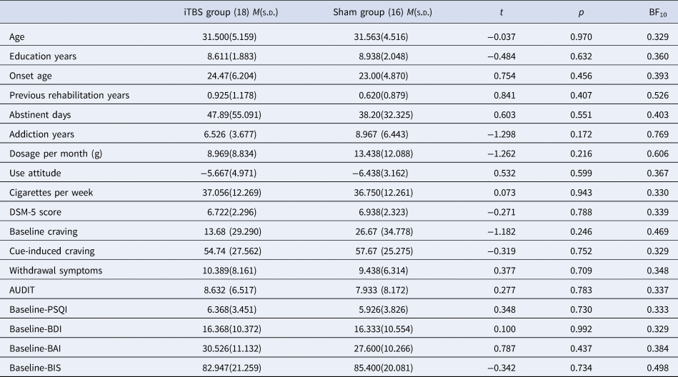
A total of 40 MUD subjects took part in the present study and were randomly assigned (with computer-generated number sequence) into the iTBS group (n = 20) and sham group (n = 20). Six subjects were transferred to a different rehabilitation center during the intervention and 14 subjects were transferred during the follow-up, separately. As we only analyzed the pre- and post-intervention effect, so the six subjects were not included in the analysis but the 14 subjects were kept. In total, 34 subjects completed 20 sessions of treatment and were included in the analysis (16 in the sham intervention, 18 in the active intervention). There were no detectable differences in age, addiction years, current abstinent duration, monthly dosage, number of cigarettes smoked per week, alcohol use severity, baseline craving, sleep quality, depression, anxiety, and impulsivity among the two groups, respectively (Table 1).
Demographic characteristics of participants

AUDIT: Alcohol Use Disorders Identification Test; DSM-5 score: The score of stimulant use disorder in The Diagnostic and Statistical Manual of Mental Disorders, Fifth Edition; PSQI: Pittsburgh Sleep Quality Index; BDI: Beck Depression Inventory; BAI: Beck Anxiety Inventory; BIS: Barratt Impulsiveness Scale; Use attitude: a visual analog scale to assess the impact of drug use on themselves is negative or positive, −10 represents the very negative impact on themself, 0 represents no impact on themselves, 10 represents a very positive impact on themselves; M: mean; s.d.: standard deviation; t: t test; p: p value.
According to the results of the Bayesian independent sample t test, the BF10 values did not well support the null hypothesis that there were no differences between the two groups at baseline, especially in addiction years (BF10 = 0.769) and dosage per month (BF10 = 0.606). Besides, we could not rule out the possibility of some imbalances for behavioral variables at baseline (baseline 1-back ACC: BF10 = 0.351; 2-back ACC: BF10 = 0.350; 3-back ACC: BF10 = 0.431). Therefore, to remove the potential effect of differences in baseline task differences, years of addiction, and dosage per month between two groups, the variables were included in the behavioral task analysis as covariates.
Behavioral measurements
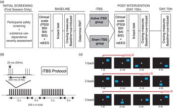
WM tasks (n-back) were only assessed before the treatment session and after 20 sessions. Pittsburgh Sleep Quality Index (PSQI), Beck Depression Inventory (BDI), Beck Anxiety Inventory (BAI), and Barratt Impulsiveness Scale (BIS), were assessed before the treatment session, after 20 sessions, and at 2-month follow up study (Fig. 1).
Procedure of the study. (A) flowchart of the experiment, (B) iTBS protocol, (C) N-back paradigm. PSQI: Pittsburgh Sleep Quality Index; BDI: Beck Depression Inventory; BAI: Beck Anxiety Inventory; BIS: Barratt Impulsiveness Scale; RMT: Resting motor threshold; rsEEG: resting-state electroencephalogram.

For the n-back task, a blue square as the stimulus presented in 3 × 3 matrix in 44, 46, 48 trials for 1-back, 2-back, and 3-back tests, respectively. The subjects were required to indicate whether each blue square was in the same location as the dot ‘n-back’ (either 1-back, 2-back, or 3-back, depending on task instructions), if the square was in the same location, then press ‘A’ button. Each n-back level had the same proportion of ‘matching’ responses (40%) and ‘non-matches’ (60%). Subjects were asked to respond as accurately and quickly as possible. A crosshair was displayed for 1000 ms, followed by a 500 ms stimulus, and then a 1000 ms window for response time. E-prime software (Psychological Software Tools Inc., Sharpsburg, PA, USA) was used to generate the task, and a practice session was administered before the experimental session at a baseline measurement and post-intervention measurement to ensure that participants understood the instruction.
For craving evaluation, baseline craving was measured with a visual scale (0–100) after watching a 3-minute video showing a neutral landscape.
Subjects then watched a 3-minute video showing MA intake to obtain the cue-induced craving score (Shen et al., Reference Shen, Cao, Tan, Shan, Wang, Pan and Yuan2016).
TMS procedures
TMS stimulation was applied with the Yiruide CCY TMS machine, using a round-shaped coil with left DLPFC located at F3. The target was located using the Yiruide TMS Location Cap based on the 10–20 EEG system. Resting motor threshold (RMT) is determined by minimum strength to elicit 50 uV motor evoked potential amplitude at contralateral abductor pollicis Brevis (APB) muscle in 5 out of 10 trials. iTBS is composed of three pulse trains of 50 Hz at 80% RMT, 2 s on, and 8 s off for 3 min and 40 s (600 pulses in total). The sham stimulation is applied with the coil perpendicular to the target area of the scalp (Sabbagh et al., Reference Sabbagh, Sadowsky, Tousi, Agronin, Alva, Armon and Pascual-Leone2020). At the post-intervention assessment, we asked subjects whether they know the intervention they received is sham intervention or active intervention to test the effect of single-blind implementation. Sixteen and 14 subjects in the active-intervention group and sham-intervention group reported that they received the active intervention, respectively. There was no significant difference between the two groups in single-blind implementation (χ2 = 0.016, p = 0.900).
Evaluation of adverse reactions
Participants were asked to score nine items-adverse reactions ranging from 1 (mild) to 10 (severe) after each treatment session. The nine adverse reactions items were based on clinical manifestations, including headache, neck pain, scalp pain, tingling, itching, burning sensation, sleepiness, trouble concentrating, and acute mood change. The total score of the adverse-effect scale was the sum of all nine items, and each participant's mean total score across all treatments was calculated.
EEG recording
EEG data were collected as previously described (Zhao et al., Reference Zhao, Zhang, Tian, Cao, Yin, Liu and Yuan2021). Resting-state EEG signals were collected under the eye-closed, maximally alert state in a quiet room at baseline and post-intervention assessment. An electrode cap (Electrical Geodesic Inc, EGI, Eugene, OR, USA; www.egi.com) with 129 recording electrodes were placed according to the International 10–20 System. The acquisition was begun after impedances for all channels were reduced to below 50 kΩ following standard data collection procedures. Data were collected using a 1–1000 Hz bandpass hardware filter and a 500 Hz sampling rate. Data were referenced to electrode Vertex. One experimenter instructed the participants to alternate closing or opening their eyes. The subjects were also asked to relax, avoid excessive blinks, and keep their eyes fixed on a central cross to reduce eye movements in the eyes-open condition. EEG recordings lasted for 11 min and included the following steps: 5 min with eyes closed, 1 min with eyes open, and 5 min with eyes closed.
EEG data processing
We employed Netstation (Electrical Geodesic Inc, EGI, Eugene, OR, USA; www.egi.com) to filter (FIR, 0.1–100 Hz, excluding 50 Hz notch). EEG signals were preprocessed using the FieldTrip toolbox for Matlab R2019(MathWorks, Natick, Massachusetts, USA). First, we segmented the data into 2-second segments and topographically interpolated bad channels. Second, the recorded signals were re-referenced to the averaged electrodes apart from face electrodes (E8, E10, E14, E17, E21, E25, E32, E38, E43, E44, E48, E49, E56, E57, E63, E64, E68, E69, E73, E74, E81, E82, E88, E89, E94, E95, E99, E100, E107, E113, E114, E119, E120, E121, E125, E126, E127, E128). Third, segments containing artifacts were removed using independent component analysis (ICA) for the detection of eye blinks, eye movements, Muscle artifacts, as well as large drifts, and spikes in the data. Segments containing more than 20% of bad channels were removed. Then, the artifact-free data were processed using a fast Fourier transform to calculate the power spectra of frequency bands, delta(1–4 Hz), theta(4–8 Hz), alpha(8–12 Hz), and beta(12–28 Hz) for each electrode average across all data segments per participants.
The EEG electrodes were divided into three subregions for the further analysis in absolute EEG analysis: central-left (CL): E7, E13, E20, E28, E29, E30, E34, E35, E36, E41; frontal-left (FL): E12, E18, E19, E22, E23, E24, E26, E27,central regions (CC): E7, E31, E55, E80,E106,E129, parietal-left (PL): E31, E37, E42, E47, E51, E52, E53, E54, E61 (Peter, Kalashnikova, & Burnham, Reference Peter, Kalashnikova and Burnham2016; Peter, Kalashnikova, Santos, & Burnham, Reference Peter, Kalashnikova, Santos and Burnham2016; Qazi, Hussain, & Aboalsamh, Reference Qazi, Hussain and Aboalsamh2019).
Data analysis and statistics
For demographic and clinical characteristics, independent sample t tests were conducted to compare group differences for continuous variable comparisons, respectively. Analysis of covariance (ANCOVA) was conducted to explore the intervention effect of N-back task, with ‘treatment’ (iTBS groups, sham group) as fix factor, addiction years, dosage per month, and baseline task values as covariances.
Outcome measures of the N-back task included reaction time in the correct trials, accuracy (the number of correct trials divided by the total number of trials), hit rate, correct rejection rate, and the discrimination index d-prime (d’). d’ is calculated from hit rate and false-alarm (FA) rate using the formula d’ = Z(Hit)-Z(FA) where Z represents a transformation from proportion to right tail p values from a normal distribution (Colzato, Jongkees, Sellaro, & Hommel, Reference Colzato, Jongkees, Sellaro and Hommel2013; Macmillan & Creelman, Reference Macmillan and Creelman2005). The positive score indicates the participants' discrimination ability is higher than the chance rate (d’ = 0), the higher the score the participants obtained, the better the participants can discriminate the target from non-target. The negative score indicates that the participants got less than 50% accuracy, the chance rate.
For the analyses of resting-state EEG data, RMANOVA with the between-subjects factor of treatment (iTBS group, sham group) and within-subjects factor of the session (pre-treatment, 2-week after treatment) as well as subregions (FL, CL, PL, and CC) was run for each EEG frequency. We used the changed power of each EEG band (delta, theta, alpha, beta) and changes (baseline score – Day 15th score) in the N-back index (accuracy and d-prime at 1-nack, 2-back, and 3-back) to conduct Pearson's correlations, checking whether there was a relationship between EEG index in subregions and N-back performance. Besides, the comparisons of correlation coefficients for two groups were conducted using the ‘cocor’ package to explore whether there are significant differences between the two groups (Diedenhofen & Musch, Reference Diedenhofen and Musch2015).
As the current study is limited by a small sample size, less than 20 participants, the frequentist approach may significantly underpower the null-hypothesis. The Bayesian approach is based on Bayes' theorem that combines prior information with evidence from information contained in a sample to guide the inference process (Ghosh, Delampady, & Samanta, Reference Ghosh, Delampady and Samanta2006). The Bayesian analysis can eliminate the small sample size problem and are more informative with continuous evidence for both H1 and H0 (Ghosh et al., Reference Ghosh, Delampady and Samanta2006). Therefore, to improve the inference about iTBS effects and two groups' differences at baseline, we conducted the Bayesian repeated-measures ANOVA, Bayesian ANCOVA, and Bayesian independent sample t test (van Doorn et al., Reference van Doorn, van den Bergh, Böhm, Dablander, Derks, Draws and Wagenmakers2021; Wagenmakers et al., Reference Wagenmakers, Love, Marsman, Jamil, Ly, Verhagen and Morey2018). Bayes factors (BFs) quantify the probability of the data under a research hypothesis (H1) relative to the probability of the data under the null hypothesis (H0). BFs below 0.33 moderate evidence for the H0 over H1, between 1 and 3 suggests ambiguous evidence for the H1, between 3 and 10 suggests substantial evidence, above 10 indicates strong evidence, and above 100 indicates decisive evidence. All BFs reported here represent the evidence for H1 relative to H0. The evidence (BF01) supports the null hypothesis (H0), using 1 minus BF10.
All the statistical analysis was done in SPSS (IBM SPSS Statistics version 21) and Matlab R2019 (MathWorks, Natick, Massachusetts, USA) and RStudio (RStudio Team 2017). The significance threshold for the cognitive task and EEG was set at p = 0.05. All the Bayesian analysis was conducted using JASP v0.14.1.0 (van Doorn et al., Reference van Doorn, van den Bergh, Böhm, Dablander, Derks, Draws and Wagenmakers2021). Moreover, control of the proportion of false-positives was carried out calculating False Discovery Rate values, FDR adjusted p values (q-values), using the R package fdrtool (Strimmer, Reference Strimmer2008).
Results
Effects of iTBS treatment on WM
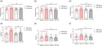
The intervention effect was found in 1-back accuracy (F (1,32) = 4.521, p = 0.042, FDR adjusted p value = 0.050, η 2 = 0.139, BF10 = 3.040) and 2-back accuracy (F (1,32) = 4.802, p = 0.037, FDR adjusted p value = 0.056, η 2 = 0.146, BF10 = 1.320), indicating that the n-back accuracy in iTBS group was higher than the sham group in the 1-back and 2-back condition. A significant intervention effect was found in 3-back accuracy (F (1,32) = 16.710, p = 0.001, FDR adjusted p value = 0.006, η 2 = 0.382, BF10 = 11.772), suggesting that iTBS group performed significantly higher accuracy at Day 15th relative to the sham group (Fig. 2). Further analysis demonstrated that at all three levels of n-back, iTBS group produced increased accuracy in day 15th relative to baseline, (1-back: FDR adjusted p value <0.01; 2-back: FDR adjusted p value <0.01; 3-back: FDR adjusted p value <0.001), while the performance of sham group remains stable at lower cognitive load condition (1-back, 2-back) but showed a significant reduction on accuracy at 3-back level (FDR adjusted p value <0.05) (Fig. 2A–C).
Accuracy and d-prime of three levels N-back task for two groups at baseline and day 15th. (A) N-back accuracy in1-back task, (B) N-back accuracy in 2-back task, (C) N-back accuracy in 3-back task, (D)1-back d-prime. (E) 2-back d-prime. (F) 3-back d-prime. d’: d-prime. *Represented p < 0.05.

Sensitivity index d’ (d-prime) reflects participants discrimination ability. The intervention effect was found in 2-back condition (F (1,32) = 7.427, p = 0.011, FDR adjusted p value = 0.022, η 2 = 0.210, BF10 = 1.382) and 3-back condition (F (1,32) = 16.870, p = 0.011, FDR adjusted p value = 0.001, η 2 = 0.385, BF10 = 6.847) but not in 1-back condition (F (1,32) = 4.165, p = 0.051, FDR adjusted p value = 0.051, η 2 = 0.129, BF10 = 3.731), indicating iTBS group displayed higher d′ score relative to a sham group at day 15th. Further analysis indicated that the iTBS group on day 15th displayed an increased d′ score compared with baseline (1-back: FDR adjusted p value <0.001; 2-back: FDR adjusted p value <0.01; 3-back: FDR adjusted p value <0.001) at all three-level, but a not sham group, performing a better detection ability that can discriminate signal from noise in three levels of N-back task (Fig. 2D–F). More details can be found in sTable 1.
For reaction time analysis, the cognitive load main effect was observed (FDR adjusted p value <0.001), revealing that increased cognitive load accompanied increased RTs. No session and group main effects were found in three levels of the n-back task. The interaction effect also displayed insignificance (sFigure 1).
Global changes in resting-state EEG power
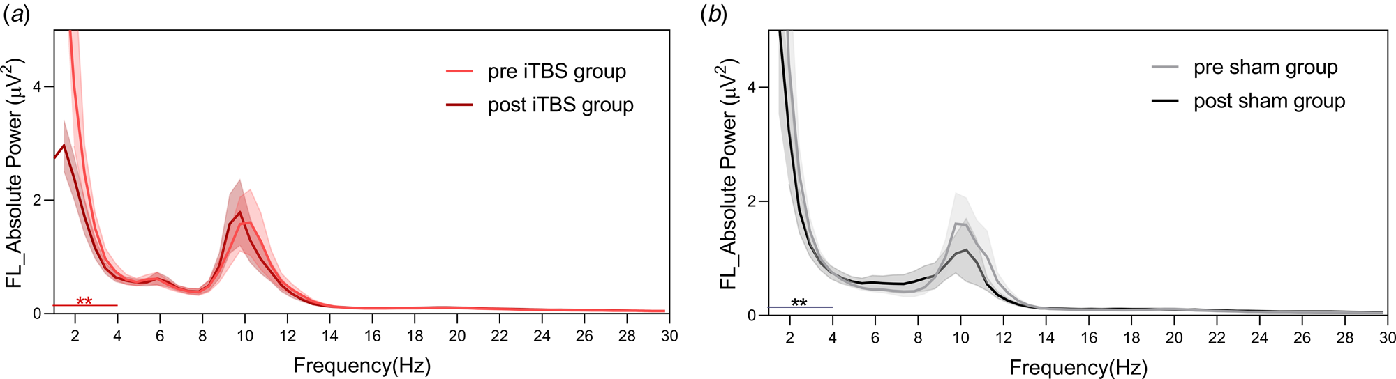
The overall pattern of absolute power was illustrated in Fig. 3. Absolute EEG power was obtained from the FL, CL, PL, and CC subregions in the iTBS group (N = 15) and the sham group (N = 13). sFigure 3 showed the scalp topography of the two groups in baseline and post-treatment conditions in each band.
Absolute EEG power at FL in the iTBS group and sham group. (A) the absolute EEG power at the left-frontal subregion (FL) in the iTBS group. (B) the absolute EEG power at the left-frontal subregion (FL) in the sham group. Thin lines indicate the SEM across subjects. Red and black lines above the X-axes indicate the frequency at which differentiated between two sessions. * Represent FDR adjusted p value<0.01, ** represent FDR adjusted p value <0.01.

An RMANOVA was conducted to measure the differences of four power bands (delta, theta, alpha, beta) in sessions (baseline, post-treatment) and groups (iTBS, sham) in subregions (CL, FL, PL, CC) separately. We found the main session effect in the delta band at FL, PL and CC subregions (FL: F(1,26) = 32.495, FDR adjusted p value <0.01, η 2 = 0.556, BF10 = 2137.369; PL: F(1,26) = 10.066, FDR adjusted p value <0.01, η 2 = 0.279, BF10 = 12.734; CC: F(1,26) = 13.022, FDR adjusted p value <0.01, η 2 = 0.334, BF10 = 29.965) and marginal significance at CL(F(1,26) = 9.823, FDR adjusted p value = 0.084, η 2 = 0.274, BF10 = 8.364).
There was no interaction effect between sessions and groups. The further analysis indicated a significant reduction at delta band in post-treatment condition in iTBS group and sham group (iTBS: t = 3.968, p = 0.001, d = 1.025, BF10 = 29.002; sham: t = 4.264, p = 0.001, d = 1.183, BF10 = 36.567) relative to baseline at left-frontal region (baseline: iTBS group: M = 4.51 s.d. = 2.685; sham: M = 5.30 s.d. = 3.046; post-treatment: iTBS: M = 1.77 s.d. = 0.950; sham: M = 2.76 s.d. = 2.251). While only iTBS group displayed a significant reduction in post-treatment (CL: t = 2.457, p = 0.028, d = 0.635, BF10 = 2.446; CC: t = 2.886, p = 0.012, d = 0.745, BF10 = 4.808; PL: t = 2.677, p = 0.018, d = 0.691, BF10 = 3.443) compared with baseline (Baseline: CL: M = 2.95 s.d. = 3.872; CC: M = 10.71 s.d. = 11.775; PL: M = 7.14 s.d. = 8.416; Sham: CL: M = 0.53 s.d. = 0.295; CC: M = 1.87 s.d. = 1.047; PL: M = 1.21 s.d. = 1.505) at central, left-central, and left-parietal regions (sFigure 2).
Association between WM change and delta power
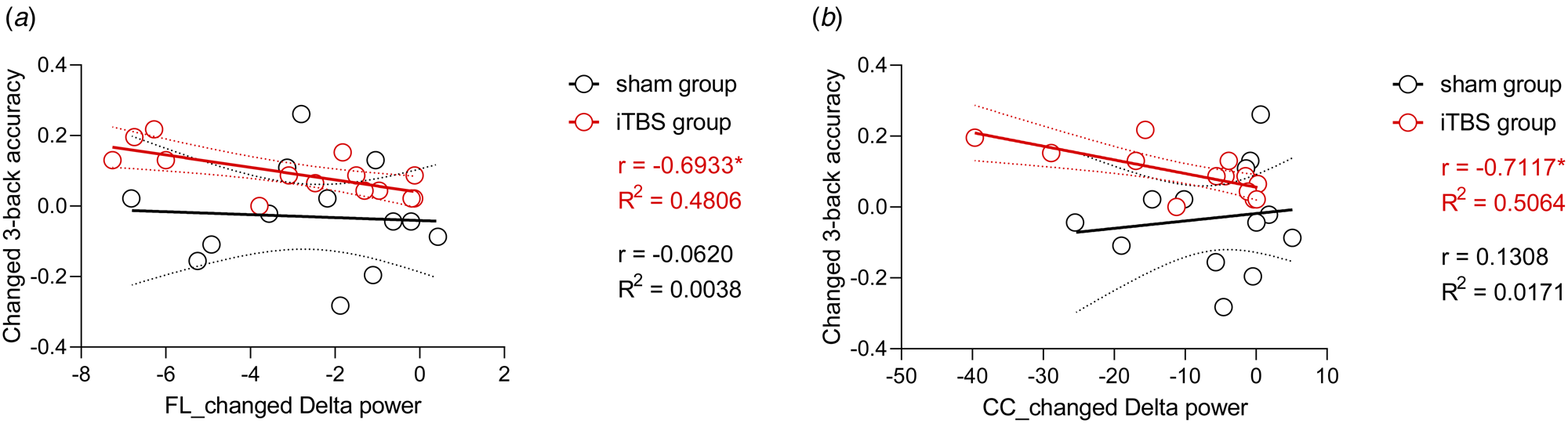
After Pearson's correlation analysis, we found a significant correlation between increased 3-back accuracy and decreased delta power in iTBS group at FL, CL, PL and CC subregions rather than sham group (active iTBS:FL: r = −0.6933, p < 0.01, BF10 = 59.219; CL: r = −0.6043, p < 0.05, BF10 = 14.028; PL: r = −0.7744, p < 0.01, BF10 = 45.818; CC: r = −0.7117, p < 0.01, BF10 = 54.107; Sham group: FL: r = −0.062, p = 0.869, BF10 = 0.345; CL: r = 0.275, p = 0.586, BF10 = 0.391; PL: r = 0.143, p = 0.838, BF10 = 0.348; CC: r = 0.131, p = 0.853, BF10 = 0.346) (Fig. 4, sFigure 4). We also found significant differences in the correlation coefficients between two groups in CL (z = −2.3968, p = 0.017), PL (z = −2.7440, p = 0.006), and CC (z = −2.7440, p = 0.006). However, only a marginal significance was observed in the FL (z = −1.8502, p = 0.064).
Correlation between changed delta power and changed N-back indexes in FL and CC.(A) correlation between changes in 3-back accuracy and changes in Delta power at left-frontal. (B) correlation between percent changes in 3-back accuracy and changes in Delta power at central. The red words represent the r-value and effect size for the iTBS group and the black words represent the r-value and effect size for the sham group.

Discussion
This is, to our knowledge, the first study to report WM improvement following twice-daily iTBS intervention to the left DLPFC in MUD patients, as well as identifying EEG signatures associated with iTBS memory response. We observed a significant improvement in WM performance following active iTBS, including enhanced accuracy and discrimination ability, together with faster reaction time. iTBS resulted in resting-state EEG power reduction in the delta band over central and left frontal electrodes. Our exploratory analysis also identified the correlation between delta power change and WM improvements.
In the current study, the spatial WM performance was improved after 20 sessions of iTBS intervention. In healthy subjects, a single session of iTBS improved WM up to 40 min following stimulation (Hoy et al., Reference Hoy, Bailey, Michael, Fitzgibbon, Rogasch, Saeki and Fitzgerald2016), and the effects could be optimized to individualized frequencies or number of pulses in iTBS protocol parameters (Chung et al., Reference Chung, Sullivan, Rogasch, Hoy, Bailey, Cash and Fitzgerald2019; Chung, Rogasch, Hoy, & Fitzgerald, Reference Chung, Rogasch, Hoy and Fitzgerald2018). Notably, a single session of iTBS did not improve WM in Parkinson's disease (PD) patients (Hill et al., Reference Hill, McModie, Fung, Hoy, Chung and Bertram2020), which may be related to impaired fronto-striatal circuity, which is important for WM, in these subjects (Morgante, Espay, Gunraj, Lang, & Chen, Reference Morgante, Espay, Gunraj, Lang and Chen2006). Another possibility is that a single session cannot induce detectable cognitive changes in PD patients, repeated sessions are required (Hill et al., Reference Hill, McModie, Fung, Hoy, Chung and Bertram2020). That is, repeated sessions of iTBS are expected to potentiate cortical functioning in DLPFC and connected network circuits and improve cognitive function. Our results are in line with the premise of accumulated dosage effects for multiple daily sessions of TMS/TBS treatments, as evidenced in trajectory analyses from depression patients (Kaster et al., Reference Kaster, Downar, Vila-Rodriguez, Thorpe, Feffer, Noda and Blumberger2019). Indeed, the trajectory of responses tracked the number of sessions rather than pulses (Schulze et al., Reference Schulze, Feffer, Lozano, Giacobbe, Daskalakis, Blumberger and Downar2018), implying that the inter-session interval is key to cortical plasticity responses and behavioral changes. Prolonged TBS protocol evoked different cortical plasticity than classical 600 pulses (Gamboa, Antal, Moliadze, & Paulus, Reference Gamboa, Antal, Moliadze and Paulus2010; Suppa et al., Reference Suppa, Huang, Funke, Ridding, Cheeran, Di Lazzaro and Rothwell2016), suggesting that more pulses within a single session might worsen the expected outcome. Collectively, these lines of evidence support the application of daily multiple session-iTBS in clinical practice, rather than a single long session using a prolonged protocol. Our twice-daily iTBS protocol was well-tolerated, and these data warrant large clinical trials in the future investigation of iTBS for cognitive enhancement.
Resting-state EEG analyses revealed a decreased delta power in the left frontal regions after repeated iTBS intervention. The underlying neural basis for these changes requires additional investigation. It is possible that such EEG changes relate to altered dopaminergic neurotransmission. For example, it was found that prefrontal cortical stimulation restored dopaminergic transmission in caudate (Strafella, Paus, Barrett, & Dagher, Reference Strafella, Paus, Barrett and Dagher2001), a substantial neural substrate underlying WM (e.g. mediating information transfer between prefrontal areas and parietal cortex) (Rieckmann, Karlsson, Fischer, & Bäckman, Reference Rieckmann, Karlsson, Fischer and Bäckman2011). Dopamine acts as one critical modulator for WM capacity (Vijayraghavan, Wang, Birnbaum, Williams, & Arnsten, Reference Vijayraghavan, Wang, Birnbaum, Williams and Arnsten2007), accompanied by theta oscillation changes in this process (Eckart, Fuentemilla, Bauch, & Bunzeck, Reference Eckart, Fuentemilla, Bauch and Bunzeck2014). Altered power for delta, beta, and alpha bands was observed in alcohol, meth, cocaine, and heroin addictions (King et al., Reference King, Herning, Gorelick and Cadet2000; Newton et al., Reference Newton, Cook, Kalechstein, Duran, Monroy, Ling and Leuchter2003; Rass et al., Reference Rass, Ahn and O'Donnell2016; Son et al., Reference Son, Choi, Lee, Park, Lim, Lee and Kwon2015). Our study reported the decreased delta power with iTBS treatment, which is consistent with previous rTMS treatment studies (Kalechstein et al., Reference Kalechstein, De la Garza, Newton, Green, Cook and Leuchter2009; Pripfl, Tomova, Riecansky, & Lamm, Reference Pripfl, Tomova, Riecansky and Lamm2014). Notably, the correlation between changes in 3-back accuracy and decreased delta power at FL, CL, PL, and CC subregions were identified in the iTBS group, but not in the sham-treated group. Although the comparison of correlations only showed marginal significance at FL between the two groups, which may be due to the sample size limitation as the significant differences were observed in other subregions. Therefore, these findings may suggest that delta power changes may be associated with treatment response. It has been suggested that delta band power recorded with EEG during WM can discriminate between low, medium, and high WM load (Zarjam, Epps, & Chen, Reference Zarjam, Epps and Chen2011), and task-related delta power was detected to increase in more difficult WM tasks compared with easier one (Harmony, Reference Harmony2013). In addition, at the resting state, the increased delta power has been related to a deviation from optimal brain homeostasis − reflecting the chaotic brain activity and deficient inhibition ability (Knyazev, Reference Knyazev2012). Moreover, combing resting EEG and functional magnetic resonance imaging (fMRI), activity in the function network of WM was observed to negatively correlate with delta power (Jann, Kottlow, Dierks, Boesch, & Koenig, Reference Jann, Kottlow, Dierks, Boesch and Koenig2010). Further causality could be established in animal studies where optogenetic stimulation at delta frequency indeed caused WM deficits in a rat model of schizophrenia (Duan et al., Reference Duan, Varela, Zhang, Shen, Xiong, Wilson and Lisman2015). Furthermore, dementia individuals displayed increased delta power at the left prefrontal regions, which further negatively correlated to WM performance (Fonseca, Tedrus, Prandi, Almeida, & Furlanetto, Reference Fonseca, Tedrus, Prandi, Almeida and Furlanetto2011). Taken together, high-level delta power in MA patients might be an important biomarker for WM deficits of the patients, and the restoration of delta power through rTMS observed in our study suggests a promising way to recover WM functions in the patient group.
Besides the involvement of PFC, the posterior parietal cortex (PPC) also plays a pivotal role in WM (Curtis, Reference Curtis2006; D'Esposito et al., Reference D'Esposito, Detre, Alsop, Shin, Atlas and Grossman1995; Harding, Yücel, Harrison, Pantelis, & Breakspear, Reference Harding, Yücel, Harrison, Pantelis and Breakspear2015; Wang, Itthipuripat, & Ku, Reference Wang, Itthipuripat and Ku2019), especially in verbal WM (Jonides et al., Reference Jonides, Schumacher, Smith, Koeppe, Awh, Reuter-Lorenz and Willis1998). PPC also demonstrated changes in activity following DLPFC stimulation and was further associated with changes in WM performance (Wu et al., Reference Wu, Tseng, Chang, Pai, Hsu, Lin and Juan2014). We also observe changes in brain activity in the parietal region, which reflects co-activation of the frontal−parietal networks during WM processes. In the future, it will be important to dissect the contribution of PFC v. PPC in WM recovery for MA patients.
This study has some limitations. First, the number of subjects is limited in each group. The limited sample size could bias the effect size (Loken & Gelman, Reference Loken and Gelman2017; Vasishth, Mertzen, Jäger, & Gelman, Reference Vasishth, Mertzen, Jäger and Gelman2018). Although we conducted Bayesian analysis to quantify evidence for the current results, research based on a larger sample was required to further generalize the conclusion. Second, EEG during the WM task was not recorded, and only resting EEG was analyzed as a signature. Last but not least, it would be important to investigate if baseline delta power or first treatment-induced changes could predict the final treatment response trajectory in MA patients, which could not be accessed in the current study.
In conclusion, the analyses revealed that twice-daily iTBS at L-DLPFC intervention could enhance WM in MUD subjects, and the changes are correlated to decreased delta power at frontal and parietal regions. Future studies will be required to replicate these findings, and to explore the potential efficiency of resting EEG signature as treatment biomarkers.
Supplementary material
The supplementary material for this article can be found at https://doi.org/10.1017/S003329172100430X.
Data
The datasets generated during and/or analyzed during the current study are available from the corresponding author on reasonable request.
Acknowledgements
We thank Jing Shen, Xi He, Hangbin Zhang, Ziqi Liu, Xue Ma, Yuxuan Zhang for their help during data collection and management.
Author contributions
YZ and TFY designed the experiment; YZ performed the study; YZ, YK, JS, ZJD, and TFY analyzed the results and wrote the paper together. All authors have read and approved the final version of the manuscript.
Financial support
The study was supported by Guangdong grant’ Key technologies for the treatment of brain disorders' (No. 2018B030331001), NSFC grants [81822017, 31771215, 32171082], Medicine and Engineering Interdisciplinary Research Fund of Shanghai Jiao Tong University (ZH2018ZDA30), Shenzhen-Hong Kong Institute of Brain Science - Shenzhen Fundamental Research Institutions (NYKFKT20190020), The Science and Technology Commission of Shanghai Municipality (18JC1420304, 19ZR1416700).
Conflict of interest
None declared for all authors.
Ethical standards
The authors assert that all procedures contributing to this work comply with the ethical standards of the relevant national and institutional committees on human experimentation and with the Helsinki Declaration of 1975, as revised in 2000.