Introduction
The genus Skrjabinolecithum (Waretrematinae, Srivastava, 1937) was first proposed by Belous (Reference Belous1954) based on the type species S. spasskii from the intestine of redlip mullet Liza haematocheila (Temminck et Schlegel, 1845) (old name: Mugil soiuy) from Razdolnaya River (Suifun), Primorsky Region, Russia. Overstreet & Curran (Reference Overstreet, Curran, Gibson, Jones and Bray2005) revised the family Haploporidae and included in Skrjabinolecithum indicum (Zhukov, 1972), S. vitellosum (Martin, 1973), S. lobolecithum (Martin, 1973) and S. bengalensis (Madhavi, 1979), which infect marine and estuarine fishes in the eastern hemisphere. Besprozvannykh et al. (Reference Besprozvannykh, Atopkin, Ngo, Beloded, Ermolenko, Ha and Tang2015b) published new morphological and molecular data on S. spasskii collected from L. haematocheila and Mugil cephalus Linnaeus, 1758 from the Primorsky Region, Russia, and from Vietnamese M. cephalus. A number of diagnostic features for S. spasskii were revised, and intraspecific molecular variation of this species was detected. Here, we provide morphometric and molecular data for a new species of Skrjabinolecithum from the intestines of L. haematocheila and M. cephalus from the Primorsky Region, Russia.
Materials and methods
Collection of trematodes
Trematodes were collected during parasitological investigations of L. haematocheila and M. cephalus fish in estuaries of the Kievka River (42°52ʹN, 133°39ʹE) and the Razdolnaya River (43°20ʹN, 131°47ʹE) in the Primorsky Region, Russia. Following removal from the intestine, worms from each fish species were rinsed in distilled water, killed in hot distilled water and preserved in 70% ethanol. Following fixation, flukes were transferred to 96% ethanol and whole mounts were stained in alum carmine, dehydrated in an ethanol series, cleared in xylene and mounted in Canada balsam. All sizes are given in millimetres (mm).
DNA extraction, amplification and sequencing
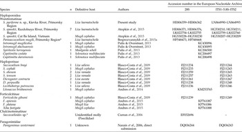
Four adult specimens of S. pyriforme n. sp. from L. haematocheila collected from the Kievka River of the Primorsky Region were used for molecular analysis (table 1). Total DNA was extracted from flukes using a ‘hot shot’ technique (Truett, Reference Truett and Kieleczawa2006).
List of taxa used for molecular analysis.

*Nucleotide sequences used only for p-distance calculation.
28S ribosomal DNA (rDNA) was amplified with the following primers: DIG12 (5ʹ-AAG CAT ATC ACT AAG CGG-3ʹ) and 1500R (5ʹ-GCT ATC CTG AGG GAA ACT TCG-3ʹ) (Tkach et al., Reference Tkach, Littlewood, Olson, Kinsella and Swiderski2003) at an annealing temperature of 55°C. A ribosomal ITS1–5.8S–ITS2 fragment was amplified with primers BD1 (5ʹ-GTC GTA ACA AGG TTT CCG TA-3ʹ) and BD2 (5ʹ-TAT GCT TAA ATT CAG CGG GT-3ʹ) (Luton et al., Reference Luton, Walker and Blair1992) with an annealing temperature of 54°C. Negative and positive controls using both primer pairs were included.
Polymerase chain reaction (PCR) products were directly sequenced using an ABI Big Dye Terminator v. 3.1 Cycle Sequencing Kit (Applied Biosystems, Waltham, Massachusetts, USA), as recommended by the manufacturer, with the internal sequencing primers described by Tkach et al. (Reference Tkach, Littlewood, Olson, Kinsella and Swiderski2003) for 28S rDNA and by Luton et al. (Reference Luton, Walker and Blair1992) for ITS. Products of sequencing were analysed using an ABI 3130 genetic analyser at the Institute of Biology and Soil Sciences, Far Eastern Branch of Russian Academy of Sciences (FEB RAS). Sequences were submitted to the European Nucleotide Archive (ENA).
Alignments and the phylogenetic analysis
Ribosomal DNA sequences were assembled with SeqScape v. 2.6 software, provided by Applied Biosystems. Alignments and estimations of the number of variable sites and sequence differences were performed using the MEGA 6.0 software (Tamura et al., Reference Tamura, Stecher, Peterson, Filipski and Kumar2013). The values of genetic p-distances were calculated for two ribosomal DNA fragments separately. Phylogenetic relationships were obtained using a concatenated dataset of partial sequences of the 28S rRNA gene and ITS2 rDNA. Phylogenetic analysis was performed using the Bayesian algorithm with the MrBayes v. 3.1.2 software (Huelsenbeck et al., Reference Huelsenbeck, Ronquist, Nielsen and Bollback2001). The best nucleotide substitution model, the general time reversible model (Tavare, Reference Tavare1986) with estimates of invariant sites and gamma-distributed among-site variation (GTR + I + G), was estimated using jModeltest v. 2.1.5 software (Darriba et al., Reference Darriba, Taboada, Doallo and Posada2012). Bayesian analysis was performed using 10,000,000 generations with two independent runs. Summary parameters and the phylogenetic tree were calculated with burn-in of 2,500,000 generations. The significance of the phylogenetic relationships was estimated using posterior probabilities (Huelsenbeck et al., Reference Huelsenbeck, Ronquist, Nielsen and Bollback2001).
Results
Skrjabinolecithum pyriforme n. sp.
Taxonomic summary
Type host
Liza haematocheila (Temminck et Schlegel).
Other host
Mugil cephalus L.
Type locality
Kievka River (43°52ʹN, 133°39ʹE), Primorsky Region (southern Far East, Russia).
Other locality
Razdolnaya River (43°20ʹN, 131°47ʹE), Primorsky Region.
Site
Intestine.
Intensity of infection
Liza haematocheila: 4–27 worms per fish; M. cephalus: 3–6 worms per fish.
Material examined
Fifty specimens. Worms were found in the intestines of 9 of the 27 specimens of L. haematocheila and in 2 of the 7 specimens of M. cephalus.
Type deposited
Holotype No. Tr-68, paratypes No. Tr-69-73. This material is held at the parasitological collection of the Zoological Museum (Institute of Biology and Soil Science, Far East Branch of the Russian Academy of Sciences, Vladivostok, Russia), e-mail: petrova@ibss.dvo.ru. Deposited: 29 July 2010.
Etymology
The name of the species refers to the pyriform shape of the body.
Description
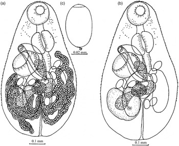
Based on 15 specimens (fig. 1, table 2).
Skrjabinolecithum pyriforme n. sp.: (a) ventral view of holotype, (b) ventral view of holotype without vitellarium, (c) egg.

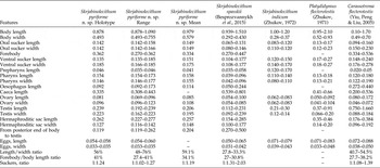
Morphometric comparison of Skrjabinolecithum pyriforme n. sp. with similar species.

Adult worms
Body pyriform, spined from anterior end to posterior third. Eye-spots in anterior third of body, diffuse. Oral sucker subterminal; prepharynx short; pharynx round, equal to size of oral sucker. Oesophagus longer than prepharynx; caeca wide, reach posterior margin of testis. Ventral sucker equal or slightly larger than oral sucker. Testis single, V-shaped, far posterior to ventral sucker. External seminal vesicle well-developed, may reach level of testis. Hermaphroditic sac sacciform, dorsal to ventral sucker and partly covered by it, with internal seminal vesicle and few prostatic cells. Posterior margin of hermaphroditic sac at level of posterior margin of ventral sucker. Genital pore dextral to median line of body, anterior to ventral sucker, with muscle sphincters at the posterior end of the hermaphroditic sac. Ovary round or irregular form, adjacent to anterior margin of testis. Mehlis' gland composed of large cells. Vitellarium, thread-like, consists of small, round follicles, in close contact with each other. Vitelline fields from ventral sucker to posterior end of body, merge at median line of body and cover ovary, testis and caeca. Uterus short, between hermaphroditic sac and anterior border of testis, with 7–22 eggs. Eggs light-yellow, operculated, with knob, unembryonated. Excretory bladder I-shaped.
Miracidium
The development of miracidia takes place only in water and finishes (at a temperature of 21–22°C) with hatching on days 8–9. Miracidium with eye-spot, span 11 h.
Molecular data
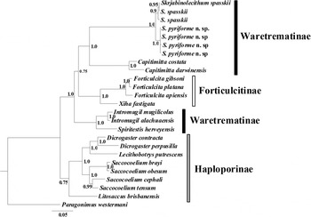
For S. pyriforme n. sp., totals of 1243 and 1294 alignable characters were available for analysis in the 28S rRNA gene and ITS1–5.8S–ITS2 rDNA fragment datasets, respectively. Three variable sites were detected within the ITS2 fragment. The 28S rRNA gene fragment was conservative. Alignment of the concatenated nucleotide sequence dataset was trimmed by adding ribosomal DNA sequences for Haploporidae species obtained from GenBank for phylogenetic analysis. Thus, for Bayesian analysis 1525 bp (608 bp and 917 bp fragments of the 28S rRNA gene and ITS1–5.8S–ITS2 rDNA, respectively) were available (fig. 2).
Phylogenetic relationships of the family Haploporidae obtained with the Bayesian algorithm based on a concatenated nucleotide sequence dataset of ITS2 rDNA and partial 28S rRNA gene. Nodal numbers: posterior probabilities that indicate statistical support of phylogenetic relationships.

Remarks
Specimens of S. pyriforme n. sp. resemble representatives of the type species, S. spasskii (in the description of Besprozvannykh et al., Reference Besprozvannykh, Atopkin, Ngo, Beloded, Ermolenko, Ha and Tang2015b) in a number of morphological characteristics: thread-like vitelline fields, V-shaped testis and unembryonated eggs in the uterus. Skrjabinolecithum pyriforme n. sp. differs from S. vitellosum and S. lobolecithum by the presence of two caeca. Skrjabinolecithum vitellosum and S. lobolecithum have a single caecum. Besides that, S. pyriforme n. sp. differs from S. spasskii and S. indicum species in body width, sizes of oral sucker, pharynx and eggs, and ratio of length and width of the body (table 2). The new species differs from S. bengalensis in having a threefold smaller body size, V-shaped testis and thread-like vitelline (Madhavi, Reference Madhavi1979). Specimens of S. pyriforme n. sp. were similar in most morphological and metric features to Platydidymus flecterotestis, which was first described by Zhukov (Reference Zhukov1971) in Leiocassis brashnikowi (Berg, 1907) and L. haematocheila from the Liao River. Overstreet & Curran (Reference Overstreet, Curran, Gibson, Jones and Bray2005) noted that the generic name ‘Hapalotrema’ is nomen praeoccupatum, studied a number of specimens of worms collected by Zhukov and included them in a new genus, Platydidymus, as type species P. flecterotestis (Waretrematinae). Yiu et al. (Reference Yiu, Peng and Liu2005), on the basis of a study of the morphology of worms found in Liza carinatus from Taiwan, transferred Hapalotrema flecterotestis to Carassotrema as C. flecterotestis (Waretrematinae). Skrjabinolecithum pyriforme n. sp. is most similar morphologically to the worms presented in the publication of Zhukov (Reference Zhukov1971). In both cases, these worms have a thread-like vitellarium and V-shaped testis. However, specimens of S. pyriforme n. sp. differ from the worms described by Zhukov in having lower maximal sizes of body length, and lower sizes of testis and eggs (table 2). Specimens of S. pyriforme n. sp. differ from C. flecterotestis, as described by Yiu et al. (Reference Yiu, Peng and Liu2005), by the same metric features (table 2). In descriptions of C. flecterotestis and P. flecterotestis, the vitellarium consists of separate elongated follicles (Overstreet & Curran, Reference Overstreet, Curran, Gibson, Jones and Bray2005; Yiu et al., Reference Yiu, Peng and Liu2005). Besides that, species of these two genera differ from S. pyriforme n. sp. by the presence of miracidia in eggs in the uterus (Overstreet & Curran, Reference Overstreet, Curran, Gibson, Jones and Bray2005). Trematodes identified in mullet fish from the Primorsky Region and described in the present paper, therefore, cannot be attributed to any of the species described previously by Zhukov (Reference Zhukov1971), Yiu et al. (Reference Yiu, Peng and Liu2005) or Overstreet & Curran (Reference Overstreet, Curran, Gibson, Jones and Bray2005) on the basis of morphological features.
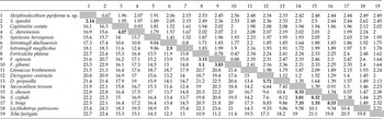
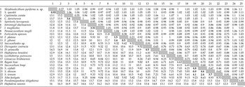
Sequence differentiation, as estimated through p-distance calculation (tables 3 and 4) of S. pyriforme n. sp. and S. spasskii, was 0.93 ± 0.27% and 2.14 ± 0.67% according to 28S rDNA and ITS rDNA sequence data, respectively. These values correspond with the interspecific level of genetic differentiation estimated by ribosomal DNA fragments, obtained earlier for other representatives of Haploporidae (Blasco-Costa et al., Reference Blasco-Costa, Balbuena, Kostadinova and Olson2009; Pulis et al., Reference Pulis and Overstreet2013). For example, genetic differentiation within Saccocoelium ranged from 0.9 ± 0.31% to 4.8 ± 0.68% by 28S rDNA and from 1.55 ± 0.57% to 8.33 ± 1.39% by ITS rDNA sequence data, respectively (tables 3 and 4). Within Waretrematinae, genetic differentiation of S. pyriforme n. sp. ranged from 11.1 ± 0.94% (Parasaccocoelium mugili) to 13.7 ± 1.05% (Capitimitta darwinensis) by 28S rDNA, and from 15.6 ± 1.91% (Spiritestis herveyensis) to 18.1 ± 2.15% (Intromugil mugilicolus) by ITS rDNA sequence data. The same values of genetic differentiation were obtained after comparative analysis of S. spasskii relative to other Waretrematinae species (tables 3 and 4).
Genetic differentiation (below the diagonal, %) and standard error (above the diagonal, %) of species of the family Haploporidae based on nucleotide sequences of ITS2 rDNA.

Genetic differentiation (below the diagonal, %) and standard error (above the diagonal, %) of species of the family Haploporidae based on nucleotide sequences of partial 28S rRNA gene.

Phylogenetic tree topology (fig. 2) showed close relationships of S. pyriforme n. sp. to S. spasskii. These species formed a clade closely related to the genus Capitimitta of the subfamily Waretrematinae. However, I. mugilicolus, I. alachuaensis and S. herveyensis, which are present in the subfamily Waretrematinae (Pulis et al., Reference Pulis, Fayton, Curran and Overstreet2013) are highly differentiated from the [Skrjabinolecithum/Capitimitta] clade, suggesting polyphyly of the subfamily Waretrematinae.
Financial support
This study was supported by grant No. 16-34-00222 of the Russian Foundation for Basic Research.
Conflict of interest
None.