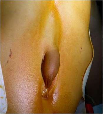
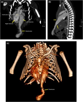
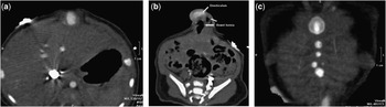
Pentalogy of Cantrell is a rare congenital cardiac disease. The full spectrum consists of five anomalies, such as a deficiency of the anterior diaphragm, a midline supraumbilical abdominal wall defect, a defect in the diaphragmatic pericardium, various congenital intracardiac abnormalities, and a defect of the lower sternum. Only a few patients with the full spectrum have been described. In up to 75% of cases, genetic anomalies or additional cardiac and somatic abnormalities accompany the diagnosis of pentalogy of Cantrell.Reference van Hoorn, Moonen, Huysentruyt, Ernest van Heurn, Offermans and Twan Mulder1, Reference Zhan, Qun-Jun, Ze-Wei, Li-Yang and Liang-long2 Early diagnosis and management are mandatory in order to avoid complications mainly linked with the cardiac and abdominal anomalies. A 3-month-old baby was referred to our hospital with a clinically pulsatile supraumbilical mass (Fig 1). Ultrasound and multi-slice computed tomography scan showed a left ventricular diverticulum and a large ventricular septal defect. Computed tomography showed further anomalies. Frontal and sagittal reconstructed images from the scan data show a large ventricular septal defect with overriding aorta and a left ventricular diverticulum (Fig 2a and b). A three-dimensional view of the cardiac diverticulum and the sternum was also available from the scan data (Fig 2c). Transverse sections of the abdomen show diastasis of the rectus abdominis muscles and bowel hernia adjacent to the cardiac diverticulum (Fig 3a and b). A reconstructed frontal view of the thorax wall shows a rather short sternum with normal ossification (Fig 3c). These findings were consistent with pentalogy of Cantrell. All findings were confirmed at surgery and early surgical correction of the cardiac malformations was successful.
Clinically pulsatile supraumbilical mass before surgery.

(a and b) Frontal and sagittal reconstructions show a left ventricular diverticulum and a large ventricular septal defect with overriding aorta. The sagittal view shows the anterior diaphragmatic and pericardial defect (arrows). (c) A three-dimensional view of the diverticulum and the sternum.

(a and b) Transverse views of the abdomen show disatasis of the rectus abdominis muscles and bowel hernia adjacent to the cardiac diverticulum. (c) Frontal reconstruction shows a rather short sternum with normal ossification.